Published online Jun 16, 2021. doi: 10.12998/wjcc.v9.i17.4262
Peer-review started: December 21, 2020
First decision: March 27, 2021
Revised: April 1, 2021
Accepted: April 23, 2021
Article in press: April 23, 2021
Published online: June 16, 2021
Processing time: 156 Days and 0.7 Hours
Osseous hemangiomas, especially those located in the manubrium, are rare benign tumors. In a review of the literature, only three case reports of sternal hemangioma were found. A precise diagnosis is difficult because of their nonspecific findings on computed tomography (CT)/magnetic resonance imaging (MRI).
An 88-year-old woman was suffering from a progressively enlarging mass in the manubrium. Chest CT images showed an osteolytic and expansile lesion with cortical destruction. Vascular malformation was suspected after CT-guided biopsy. On the dynamic MRI scans, the mass showed a bright signal on the T2-weighted image, peripheral nodular enhancement on the early-phase images and progressive centripetal fill-in on the delayed-phase images. Cavernous heman
This uncommon case demonstrates the possible characteristic features of manubrium cavernous hemangioma on dynamic MRI scans; knowledge about these features may prevent patients from developing catastrophic complications, such as rupture or internal hemorrhage, caused by biopsy or surgery.
Core Tip: Osseous hemangiomas, especially those located in the manubrium, are rare benign tumors. We presented a case suffering from a progressively enlarging mass in the manubrium. Chest computed tomography images showed aggressive appearance and the dynamic magnetic resonance imaging scans revealed progressive centripetal fill-in on the delayed-phase images. The diagnosis of cavernous hemangioma was confirmed after surgery. This uncommon case demonstrates the possible characteristic features of manubrium cavernous hemangioma on dynamic MRI scans.
- Citation: Lin TT, Hsu HH, Lee SC, Peng YJ, Ko KH. Dynamic magnetic resonance imaging features of cavernous hemangioma in the manubrium: A case report. World J Clin Cases 2021; 9(17): 4262-4267
- URL: https://www.wjgnet.com/2307-8960/full/v9/i17/4262.htm
- DOI: https://dx.doi.org/10.12998/wjcc.v9.i17.4262
The imaging findings of osseous hemangioma are nonspecific. This tumor appears as a localized or expanding osteolytic mass with fine spiculations and is difficult to differentiate from other malignancies or neoplasms[1]. The enhancement pattern of osseous hemangioma is variable and depends on the components of the tumor. If the lesion has abundant fat content, it shows slight enhancement. Conversely, if the lesion has little fat content, elevated contrast enhancement is observed[2]. In this article, we report a case of a cavernous hemangioma in the manubrium that presented with a possible characteristic enhancement pattern on dynamic magnetic resonance imaging (MRI).
A woman, aged 88 years, visited Tri-Service General Hospital after finding an enlarging mass in the manubrium for 6 years.
The patient was suffering from progressive chest pain in the manubrium for about 3 mo.
The patient had a free previous medical history.
The patient had a free previous personal and family history.
A protruding mass was identified at the sternal region.
The laboratory results were within normal limits.
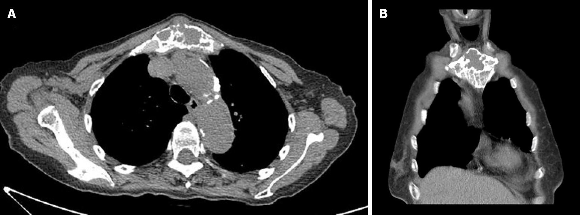
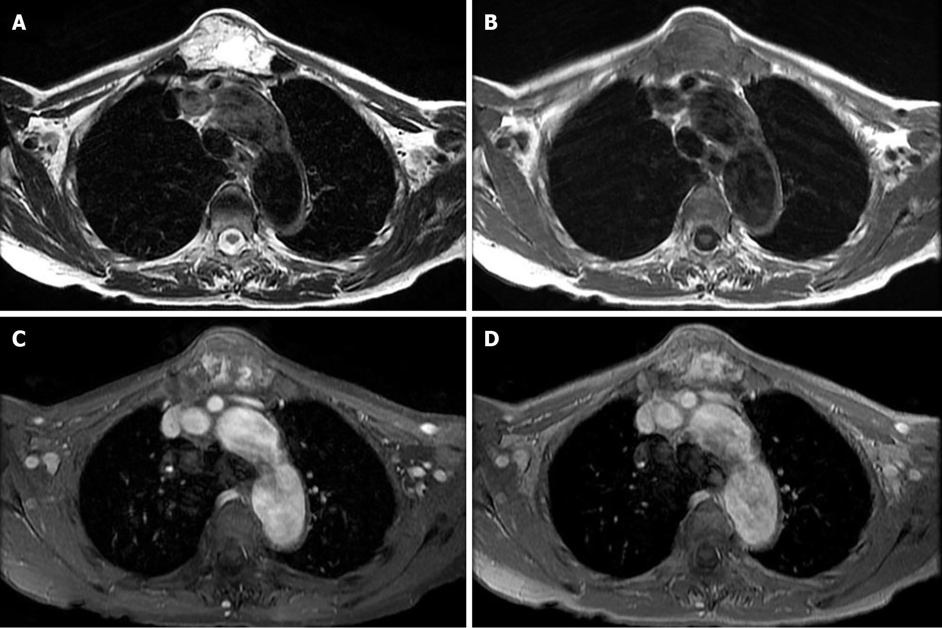
Computed tomography demonstrated an aggressive, expansile and osteolytic lesion in the manubrium with cortical destruction (Figure 1). The technetium-99m MDP scintigraphy was performed to identify potential metastasis and showed moderately increased uptake in the right-sided sternal manubrium but did not detect other lesions. Therefore, dynamic contrast-enhanced MRI was performed to evaluate the nature of the tumor and revealed hyperintensity on T2-weighted sequences, isointensity on T1-weighted images and mild peripheral enhancement with more centripetal fill-in on the delayed-phase images acquired with dynamic sequences (Figure 2). A sternal hemangioma was first considered based on its enhancement pattern.
Multidisciplinary team recommended that she need sternotomy for resection of the manubrium tumor in consideration of the symptomatic and progressively enlarged mass.
The final diagnosis was confirmed to be cavernous hemangioma in the manubrium.
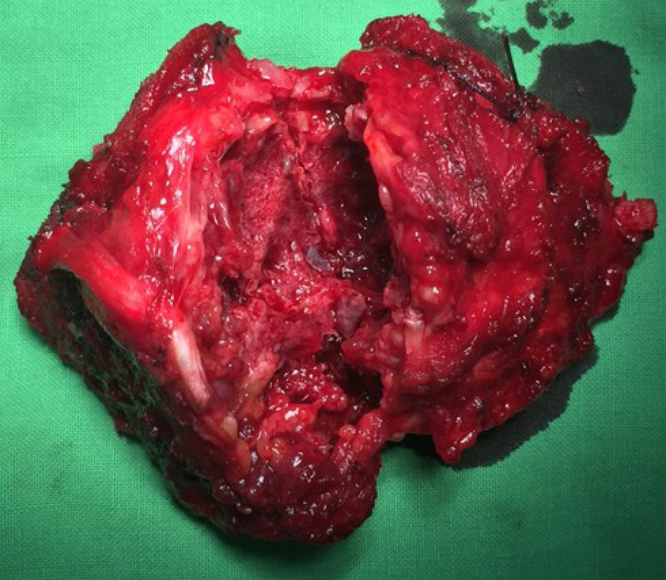
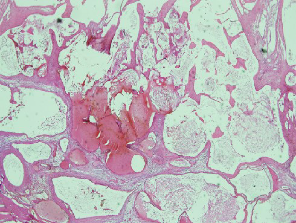
The patient underwent sternotomy for resection of the manubrium tumor, and a plate and polypropylene mesh were applied to the postresectional space to repair the defect of the sternal tumor. The resected tumor was approximately 4 cm × 3 cm × 4 cm in size and had a hypervascular appearance (Figure 3). The cross-sections showed sponge-like tumor components within the bone tissue. The histologic findings of the mass showed large, dilated, blood-filled vessels lined by flattened endothelium with a haphazard arrangement within the sternal manubrium tissue (Figure 4). The immunohistochemical stains revealed positive on endothelial cell makers (CD34, CD31). The diagnosis of cavernous hemangioma was confirmed.
The patient presented no evidence of chest pain and no signs of recurrence at the 1-year follow-up.
Primary neoplasms in the sternal region are rare, and malignancies should always be considered the first possibility for sternal osteolytic lesions. Benign neoplasms include osteochondroma, osteoma, hemangioma, fibrous dysplasia, and Langerhan’s cell histiocytosis[1]. Osseous hemangiomas are rare among these benign tumors, accounting for approximately 1% of bone tumors, and occur in the skull, rib, and vertebral columns[1,3]. The computed tomography (CT) image may appear with “spoke wheel trabeculations” or a “lattice-like pattern”[4], but these patterns were not present in our case. The MRI findings of osseous hemangioma depend on its different compositions. Typically, hemangiomas present with high signal intensity on T2-weighted sequences and intermediate signal intensity on T1-weighted sequences due to its vascularity. If the amount of lipomatous soft tissue of the hemangioma increases, the signal intensity will increase on T1-weighted and T2-weighted sequences[5,6]. The use of an intravenous gadolinium contrast agent to image hemangiomas demonstrates various enhancement patterns. Some hemangiomas present with a ‘‘bunch of grapes’’ appearance on T2- weighted sequences due to cavernous vascular spaces[6]. In addition, thrombi in hemangioma will show low signal foci within high-signal lesions on T2-weighed sequences[7]. In a review of the literature, only three case reports of sternal hemangioma were found. Medalion et al[8] described a sternal hemangioma in a 30-year-old woman who had a 2-year history of chest pain. The mass was located in the lower part of the sternum and invaded the sternal cortex on MRI scans. Onat et al[9] presented another case about a 32-year-old woman with anterior chest pain and an enlarging mass. A contrast-enhanced CT scan display an expanding mass and focal destruction of the sternal cortex. The MRI scan was not performed in this patient. Therefore, the dynamic features of MRI were rarely reported in osseous hemangiomas because of the rarity of this tumor.
Klotz et al[10] described three histological subtypes: capillary hemangioma, cavernous hemangioma and sclerosing hemangioma of the liver. Among these three subtypes, cavernous hemangioma is the most common. The enhancement pattern is classically nodular, with peripheral enhancement and progressive and complete centripetal filling. Yamashita et al[11] explained that the size of the vascular space is related to the hemodynamics of cavernous hemangiomas. The peripheral areas with a smaller vascular size were enhanced early. The gradual fill-in area had a larger vascular size, and slower enhancement was observed. The central area is seldom enhanced because of fibrosis, bleeding or thrombosis. When vascular malformation is found on histopathology and a destructive lytic pattern is observed on the CT images, osseous angiosarcoma should be considered. The common symptoms of angiosarcoma are painful swollen lesions that are often combined with pathological fractures. On MRI, these tumors display hypointensity on T1-weighted images, heterogeneity on T2-weighted images and irregular contrast enhancement. High-grade tumors may invade regional soft tissues, and approximately 66% of cases reveal distant metastases during diagnosis[12]. In our case, the dynamic MRI scans revealed typical imaging features in the sternum, which were similar to those of cavernous hepatic hemangiomas. This differential diagnosis was considered before the operation. After resection surgery, the diagnosis was confirmed.
In conclusion, osseous hemangiomas are uncommon benign tumors of the sternum, and imaging findings may demonstrate aggressive osteolytic lesions. Dynamic MRI studies may help distinguish osseous hemangiomas from other malignancies when the lesion reveals classical features of peripheral nodular enhancement on early-phase images and progressive centripetal fill-in on delayed-phase images. An early precise diagnosis of hemangioma may prevent patients from developing catastrophic complications, such as rupture or internal hemorrhage, caused by biopsy or surgery.
The authors are grateful to Professors Hsian-He Hsu, Dr. Kai-Hsiung Ko for their assistance in diagnosis and reviewing this manuscript. In addition, we are thankful to Shih-Chun Lee, who were involved in the patient’s care and Yi-Jen Peng for path-ological interpretation. We also thank the patient and her family for their cooperation and permission.
Manuscript source: Unsolicited manuscript
Specialty type: Radiology, nuclear medicine and medical imaging
Country/Territory of origin: Taiwan
Peer-review report’s scientific quality classification
Grade A (Excellent): 0
Grade B (Very good): 0
Grade C (Good): C
Grade D (Fair): 0
Grade E (Poor): 0
P-Reviewer: Liu HL S-Editor: Gong ZM L-Editor: A P-Editor: Liu JH
| 1. | Singh A, Chandrashekhara SH, Triveni GS, Kumar P. Imaging in Sternal Tumours: A Pictorial Review. Pol J Radiol. 2017;82:448-456. [RCA] [PubMed] [DOI] [Full Text] [Full Text (PDF)] [Cited by in Crossref: 6] [Cited by in RCA: 9] [Article Influence: 1.1] [Reference Citation Analysis (0)] |
| 2. | Qaseem Y, Bermo M, Matesan M, Behnia F, Verma N, Elojeimy S. A case of multiple aggressive osseous hemangiomas on bone scan. Radiol Case Rep. 2020;15:226-229. [RCA] [PubMed] [DOI] [Full Text] [Full Text (PDF)] [Cited by in Crossref: 1] [Cited by in RCA: 1] [Article Influence: 0.2] [Reference Citation Analysis (0)] |
| 3. | Clements RH, Turnage RB, Tyndal EC. Hemangioma of the rib: a rare diagnosis. Am Surg. 1998;64:1027-1029. [PubMed] |
| 4. | Yamada K, Whitbeck MG Jr, Numaguchi Y, Shrier DA, Tanaka H. Symptomatic vertebral hemangioma: atypical spoke-wheel trabeculation pattern. Radiat Med. 1997;15:239-241. [PubMed] |
| 5. | Ching BC, Wong JS, Tan MH, Jara-Lazaro AR. The many faces of intraosseous haemangioma: a diagnostic headache. Singapore Med J. 2009;50:e195-e198. [PubMed] |
| 6. | Li W, Zou F, Dai M, Zhang B, Nie T. A rare case of pure primary hemangioma of the scapula: A case report. Oncol Lett. 2015;10:2265-2268. [RCA] [PubMed] [DOI] [Full Text] [Cited by in Crossref: 2] [Cited by in RCA: 2] [Article Influence: 0.2] [Reference Citation Analysis (0)] |
| 7. | Vilanova JC, Barceló J, Smirniotopoulos JG, Pérez-Andrés R, Villalón M, Miró J, Martin F, Capellades J, Ros PR. Hemangioma from head to toe: MR imaging with pathologic correlation. Radiographics. 2004;24:367-385. [RCA] [PubMed] [DOI] [Full Text] [Cited by in Crossref: 157] [Cited by in RCA: 121] [Article Influence: 5.8] [Reference Citation Analysis (0)] |
| 8. | Medalion B, Bar I, Neuman R, Shargal Y, Merin G. Sternal hemangioma: a rare tumor. J Thorac Cardiovasc Surg. 1996;112:1402-1403. [RCA] [PubMed] [DOI] [Full Text] [Cited by in Crossref: 3] [Cited by in RCA: 3] [Article Influence: 0.1] [Reference Citation Analysis (0)] |
| 9. | Onat S, Ulku R, Avci A, Mizrak B, Ozcelik C. Hemangioma of the sternum. Ann Thorac Surg. 2008;86:1974-1976. [RCA] [PubMed] [DOI] [Full Text] [Cited by in Crossref: 8] [Cited by in RCA: 8] [Article Influence: 0.5] [Reference Citation Analysis (0)] |
| 10. | Klotz T, Montoriol PF, Da Ines D, Petitcolin V, Joubert-Zakeyh J, Garcier JM. Hepatic haemangioma: common and uncommon imaging features. Diagn Interv Imaging. 2013;94:849-859. [RCA] [PubMed] [DOI] [Full Text] [Cited by in Crossref: 77] [Cited by in RCA: 71] [Article Influence: 5.9] [Reference Citation Analysis (1)] |
| 11. | Yamashita Y, Ogata I, Urata J, Takahashi M. Cavernous hemangioma of the liver: pathologic correlation with dynamic CT findings. Radiology. 1997;203:121-125. [RCA] [PubMed] [DOI] [Full Text] [Cited by in Crossref: 119] [Cited by in RCA: 105] [Article Influence: 3.8] [Reference Citation Analysis (0)] |
| 12. | Gaballah AH, Jensen CT, Palmquist S, Pickhardt PJ, Duran A, Broering G, Elsayes KM. Angiosarcoma: clinical and imaging features from head to toe. Br J Radiol. 2017;90:20170039. [RCA] [PubMed] [DOI] [Full Text] [Cited by in Crossref: 94] [Cited by in RCA: 150] [Article Influence: 18.8] [Reference Citation Analysis (0)] |


