Published online Jan 6, 2021. doi: 10.12998/wjcc.v9.i1.218
Peer-review started: July 28, 2020
First decision: November 3, 2020
Revised: November 5, 2020
Accepted: November 21, 2020
Article in press: November 21, 2020
Published online: January 6, 2021
Processing time: 157 Days and 7.9 Hours
Papillary thyroid cancer (PTC) has good prognosis so that the local recurrence or distant metastasis can occur later on the lifetime follow up. In this study, we report recurrence of PTC in subcutaneous area combined with lymph node metastasis. A suspicion of needle tract implantation after core needle biopsy was found.
A 66-year-old female patients who underwent right thyroid lobectomy for PTC complained of palpable nodule on anterior neck area. The location of the palpable nodule was not associated with her postoperative scar. After excision of the skin tumor, it was diagnosed as recurrence of PTC. Furthermore, results of subsequent imaging showed lymph node metastasis on her right cervical area. According to the previous medical records, the patient received core needle biopsy through the neck of the patient midline and hematoma was noted after the procedure. The time interval from the first diagnosis to local recurrence or metastasis to the skin and lymph nodes was ten years. As treatment, the patient underwent lymph node dissection in the right and completion thyroidectomy for radioisotope treatment.
Needle tract implantation can occur after core needle biopsy. Further studies are needed to compare core-needle biopsy and fine-needle aspiration.
Core Tip: Local recurrence of papillary thyroid cancer can occur in the late period. Clinicians should pay attention to needle tract implantation related core needle biopsy.
- Citation: Kim YH, Choi IH, Lee JE, Kim Z, Han SW, Hur SM, Lee J. Late recurrence of papillary thyroid cancer from needle tract implantation after core needle biopsy: A case report. World J Clin Cases 2021; 9(1): 218-223
- URL: https://www.wjgnet.com/2307-8960/full/v9/i1/218.htm
- DOI: https://dx.doi.org/10.12998/wjcc.v9.i1.218
Papillary thyroid carcinoma (PTC) has good prognosis and the survivals have been assessed as 10 years, which is exceeding 90%. Smaller tumors can be incidentally found by ultrasound before evident symptoms emerged, PTCs less than 1 cm has excellent prognosis. The recurrence rate are higher in young age but the outcome is better than the older patients[1]. It has been known that the most common site of distant metastasis is lung, followed by bones and multiple organ involvement[2].
Fine needle aspiration cytology (FNAC) is widely used for the diagnosis of PTC. It is considered safe and effective due to the low chance of fatal complications. Needle tract implantation (NTI) from FNAC was reported in less than 0.2% of the cases[3]. However, NTI from core needle biopsy (CNB) has not yet been investigated. In a retrospective analysis of 11745 PTC patients, 9.1% of NTI patients showed local recurrence, while 40.9% had distant metastasis during follow-up[4].
The authors found late skin recurrence combined with lymph node metastasis of PTC, presumably from NTI after CNB.
A 66-year-old female patient with the chief complaint of an anterior neck mass sought consult from our surgical department.
The mass was located midline to the right side. It was first noticed a few months ago. She claimed that it rapidly grew one month prior to consultation.
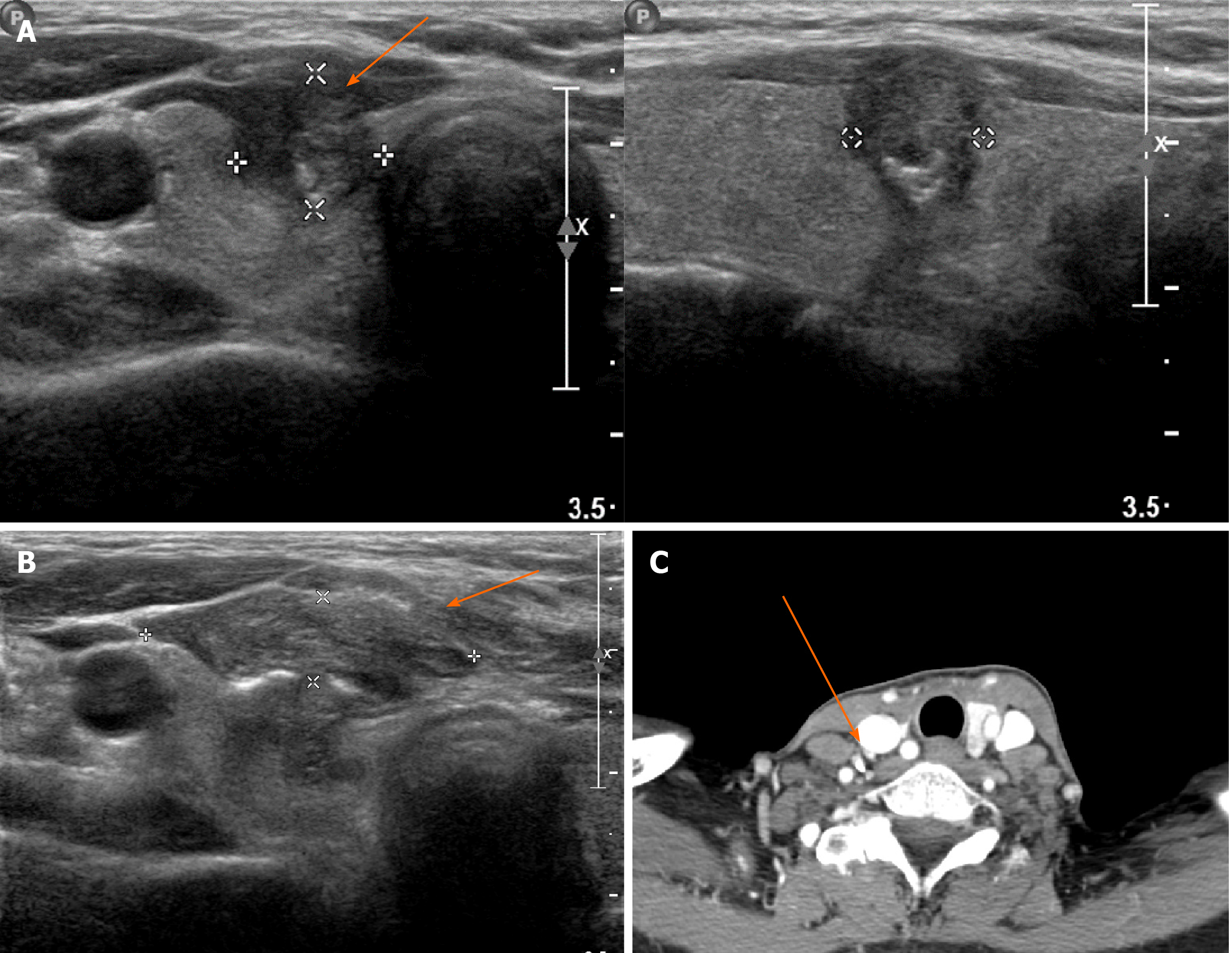
She had a 4-cm scar from her previous right thyroid lobectomy ten years ago. At her initial visit for the thyroidectomy, an irregular ill-defined mixed hypoechogenic nodule with internal calcification was found on the mid pole of the right thyroid (Figure 1A). FNAC along with CNB using a 20-gauge Franseen needle was performed at the time of diagnosis, there found a hematoma after procedure (Figure 1B). A pathologic report revealed an 8-mm papillary carcinoma with focal extension to perithyroidal soft tissue without resection margin involvement and lymph nodal metastasis. The patient was lost to follow-up for 115 months until she noted a soft tissue mass on anterior neck.
She was taking medication for hypertension and hyperlipidemia few years ago, except that there were no other diseases diagnosed.
She did not have any other family members that have thyroid cancer, or other type of cancers.
The 1.4 cm × 0.65 cm nodule was palpated 5 mm away from previous operative scar. The nodule was not, in any way or form, connected to the previous scar. The mass was located subcutaneously, slightly movable, and tense. Excisional biopsy without FNAC or CNB was performed under local anesthesia with an elliptical excision using previous scar. After excision, the mass was revealed to be a papillary carcinoma on soft tissue. Her previous medical records were reviewed.
On the laboratory test before the second surgery, thyroid hormone test including TSH, free T4, T3 were within normal range. Serum thyroglobulin level was 13.8 ng/mL (3.5-77 ng/mL), and the antibody to thyroglobulin was 11.9 IU/mL (0-115 IU/mL).
Additional imaging studies were preformed to find out another metastatic lesion. A suspicious metastatic lymph node measured 5 mm on level IV was observed on the right cervical area through ultrasound and computed tomography imaging (Figure 1C). Furthermore, FNAC confirmed the presence of a metastatic lymph node. Thyroglobulin level were greater than 500 ng/mL in the aspirate.
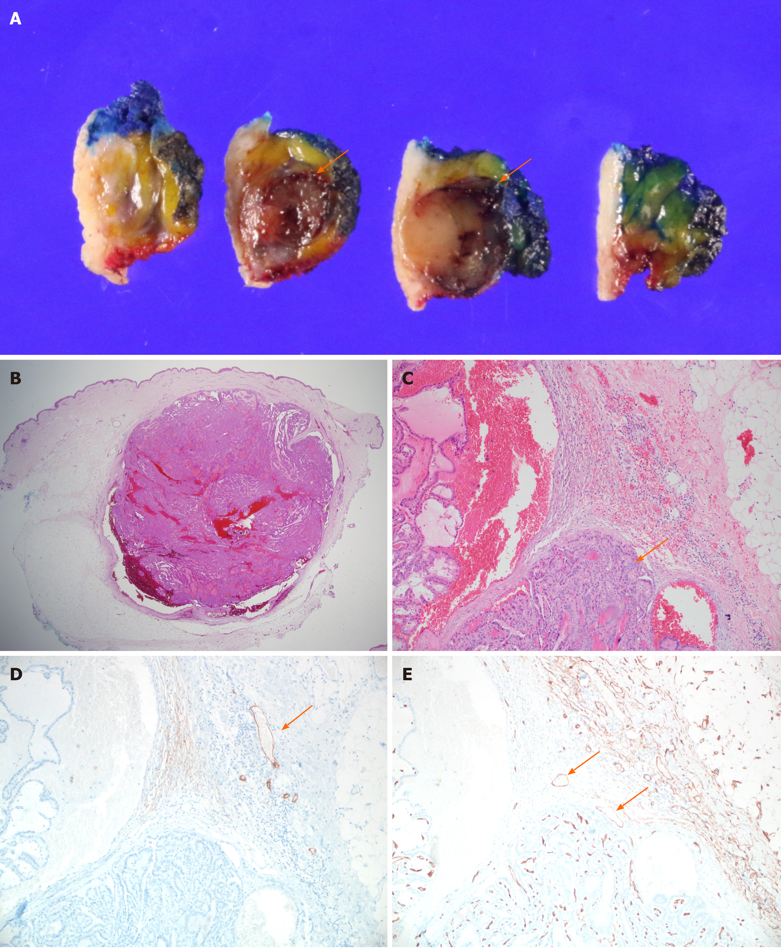
During the gross examination for excisional biopsy of skin and soft tissue, the specimen reveals a round light yellow to brown solid soft mass without necrosis in the superficial subcutaneous layer, measuring 1.4 cm × 1.0cm, Figure 2A. On microscopic examination, it shows a relatively well defined round solid mass in subcutaenous. It reveals neither lymph nodal architecture nor residual thyroid tissue in the submitted specimen, Figure 2B. The mass is composed of multiple papillary architecture showing nuclear enlargement, nuclear groove and inclusion which is shown in typical papillary thyroid carcinoma, Figure 2C. The tumor reveals neither lymphatic nor perineural invasion using histologic features. And the results of immunohistochemical stainings, Figure 2D and E. Using the deeper cut section, the tumor reveals no lymph node architecture.
Completion thyroidectomy with modified radical neck dissection on the right cervical lymph node was performed. There were no further malignant findings on the remnant thyroid tissue. Lymph node metastasis was found in 3 out of 19 nodes. The maximal size of lymph node metastasis is 1.2 mm without extranodal soft tissue extension. There were no immediate surgical complications. Due to the patient’s clinical history and pathologic findings on the excisional biopsy (Figure 2), the anterior neck mass was suspected to be a local recurrence of her initial tumor, rather than metastasis to the skin.
Radioisotope treatment was provided after surgery (100 mCi).
After the radioisotope treatment, serum thyroglobulin level maintained less than 0.1 ng/mL after 10 mo after surgery.
We found a late recurrence of subcutaneous NTI after needle biopsy in PTC patients combined with lymph node metastasis. No significant findings suggested whether the nodule was NTI or metastasis. Cutaneous or intramuscular metastasis of thyroid cancer is rare[5]. Therefore, the direction of biopsy needle tract, seedings in linear fashion, seedings not accompanied by lymphoid or neurovascular tissue, and its presence away from the initial surgical incision, led us to a diagnosis of NTI, rather than soft tissue metastasis. NTI may be a manifestation of an underlying disease, particularly lymph node metastasis as presented in this case.
CNB has applied to thyroid nodule evaluation because of some inconclusive results from FNAC. Its routine use is not recommended by several guidelines because of limited evidence so far[6,7]. CNB use is accepted as complementary modality after FNAC, compared to repeated FNAC. Pain, hematoma, edema, hoarseness, and infection are common complications of CNB. It can be safely performed by an expert and under ultrasonography guidance.
The cumulative incidence of NTI after FNAC of PTC was reportedly 0.1% after five years and 0.3% after ten years[4]. The time interval between FNAC and NTI in PTC was reported to range from six months to seven years[8]. In this case, our patient exhibited symptoms at a relatively later time period of almost 10 years (115 mo). NTI can occur in any type of cancer, but it has been accepted as the benefit overrides the harm[9,10]. One of the factors related to NTI reported in other cancer types is needle diameter, suggesting that a similar pattern can occur during CNB in the thyroid. Although, it has not been established in any form of thyroid cancer.
Evaluation of 26 NTI patients showed that old age, lymph node metastasis, and extrathyroidal extension were related to NTI[11]. In this case, the patient was above 55 years old and had aggressive feature of extrathyroidal extension. For FNAC, there are a few tips from experts to avoid NTI such as removing negative pressure during needle withdrawal, removing sternothyroid muscle during thyroidectomy, or using ultrasound guidance to prevent seeding on the posterior part of thyroid[8]. Currently, there is no evidence to avoid NTI in CNB.
We found a post-biopsy hematoma due to CNB. The incidence of hematoma was found to be greater in CNB than in FNAC[12], regardless of the nodule size, nodule composition, malignancy suspicion by ultrasound, or vascularity. There is no direct evidence that post-biopsy hematoma is related to NTI. Rather, a hypothesis that hematoma can prevent the healing of the needle tract suggests its action as a pool for disseminating tumor cells into the surrounding tissue.
The role of CNB can be less effective in other differentiated thyroid carcinomas[13,14]. In this case, although NTI may act as a clue to diagnose the patient with lymph node metastasis, using the FNAC and CNB simultaneously on initial diagnosis should be refrained when the ultrasound results shows a nodule highly suspicious for PTC.
To directly compare the occurrence of NTI between FNAC and CNB, a larger population and a longer observation time is required. A late recurrence of PTC in a CNB site suggests that clinicians must carefully choose diagnostic method for PTC patients. Furthermore, signs of post-biopsy hematoma in NTI should be more thoroughly investigated in the future.
Manuscript source: Unsolicited manuscript
Specialty type: Oncology
Country/Territory of origin: South Korea
Peer-review report’s scientific quality classification
Grade A (Excellent): 0
Grade B (Very good): 0
Grade C (Good): C, C, C
Grade D (Fair): 0
Grade E (Poor): 0
P-Reviewer: Casella C, Handra-Luca A, Munoz M S-Editor: Zhang H L-Editor: A P-Editor: Li JH
| 1. | Gilliland FD, Hunt WC, Morris DM, Key CR. Prognostic factors for thyroid carcinoma. A population-based study of 15,698 cases from the Surveillance, Epidemiology and End Results (SEER) program 1973-1991. Cancer. 1997;79:564-573. [RCA] [PubMed] [DOI] [Full Text] [Cited by in RCA: 2] [Reference Citation Analysis (0)] |
| 2. | Ruegemer JJ, Hay ID, Bergstralh EJ, Ryan JJ, Offord KP, Gorman CA. Distant metastases in differentiated thyroid carcinoma: a multivariate analysis of prognostic variables. J Clin Endocrinol Metab. 1988;67:501-508. [RCA] [PubMed] [DOI] [Full Text] [Cited by in Crossref: 266] [Cited by in RCA: 232] [Article Influence: 6.3] [Reference Citation Analysis (0)] |
| 3. | Ito Y, Tomoda C, Uruno T, Takamura Y, Miya A, Kobayashi K, Matsuzuka F, Kuma K, Miyauchi A. Needle tract implantation of papillary thyroid carcinoma after fine-needle aspiration biopsy. World J Surg. 2005;29:1544-1549. [RCA] [PubMed] [DOI] [Full Text] [Cited by in Crossref: 41] [Cited by in RCA: 43] [Article Influence: 2.3] [Reference Citation Analysis (0)] |
| 4. | Hayashi T, Hirokawa M, Higuchi M, Kudo T, Ito Y, Miyauchi A. Needle Tract Implantation Following Fine-Needle Aspiration of Thyroid Cancer. World J Surg. 2020;44:378-384. [RCA] [PubMed] [DOI] [Full Text] [Cited by in Crossref: 10] [Cited by in RCA: 11] [Article Influence: 2.2] [Reference Citation Analysis (0)] |
| 5. | Alwaheeb S, Ghazarian D, Boerner SL, Asa SL. Cutaneous manifestations of thyroid cancer: a report of four cases and review of the literature. J Clin Pathol. 2004;57:435-438. [RCA] [PubMed] [DOI] [Full Text] [Cited by in Crossref: 53] [Cited by in RCA: 51] [Article Influence: 2.4] [Reference Citation Analysis (0)] |
| 6. | Haugen BR, Alexander EK, Bible KC, Doherty GM, Mandel SJ, Nikiforov YE, Pacini F, Randolph GW, Sawka AM, Schlumberger M, Schuff KG, Sherman SI, Sosa JA, Steward DL, Tuttle RM, Wartofsky L. 2015 American Thyroid Association Management Guidelines for Adult Patients with Thyroid Nodules and Differentiated Thyroid Cancer: The American Thyroid Association Guidelines Task Force on Thyroid Nodules and Differentiated Thyroid Cancer. Thyroid. 2016;26:1-133. [RCA] [PubMed] [DOI] [Full Text] [Cited by in Crossref: 10769] [Cited by in RCA: 9672] [Article Influence: 1074.7] [Reference Citation Analysis (1)] |
| 7. | Na DG, Baek JH, Jung SL, Kim JH, Sung JY, Kim KS, Lee JH, Shin JH, Choi YJ, Ha EJ, Lim HK, Kim SJ, Hahn SY, Lee KH, Choi YJ, Youn I, Kim YJ, Ahn HS, Ryu JH, Baek SM, Sim JS, Jung CK, Lee JH; Korean Society of Thyroid Radiology (KSThR) and Korean Society of Radiology. Core Needle Biopsy of the Thyroid: 2016 Consensus Statement and Recommendations from Korean Society of Thyroid Radiology. Korean J Radiol. 2017;18:217-237. [RCA] [PubMed] [DOI] [Full Text] [Full Text (PDF)] [Cited by in Crossref: 98] [Cited by in RCA: 117] [Article Influence: 14.6] [Reference Citation Analysis (1)] |
| 8. | Polyzos SA, Anastasilakis AD. A systematic review of cases reporting needle tract seeding following thyroid fine needle biopsy. World J Surg. 2010;34:844-851. [RCA] [PubMed] [DOI] [Full Text] [Cited by in Crossref: 42] [Cited by in RCA: 35] [Article Influence: 2.3] [Reference Citation Analysis (0)] |
| 9. | Gobien RP. Aspiration biopsy of the solitary thyroid nodule. Radiol Clin North Am. 1979;17:543-554. [PubMed] |
| 10. | Hales MS, Hsu FS. Needle tract implantation of papillary carcinoma of the thyroid following aspiration biopsy. Acta Cytol. 1990;34:801-804. [PubMed] |
| 11. | Ito Y, Hirokawa M, Higashiyama T, Takamura Y, Kobayashi K, Miya A, Miyauchi A. Clinical significance and prognostic impact of subcutaneous or intrastrap muscular recurrence of papillary thyroid carcinoma. J Thyroid Res. 2012;2012:819797. [RCA] [PubMed] [DOI] [Full Text] [Full Text (PDF)] [Cited by in Crossref: 3] [Cited by in RCA: 5] [Article Influence: 0.4] [Reference Citation Analysis (0)] |
| 12. | Chae IH, Kim EK, Moon HJ, Yoon JH, Park VY, Kwak JY. Ultrasound-guided fine needle aspiration versus core needle biopsy: comparison of post-biopsy hematoma rates and risk factors. Endocrine. 2017;57:108-114. [RCA] [PubMed] [DOI] [Full Text] [Cited by in Crossref: 11] [Cited by in RCA: 11] [Article Influence: 1.4] [Reference Citation Analysis (0)] |
| 13. | Renshaw AA, Pinnar N. Comparison of thyroid fine-needle aspiration and core needle biopsy. Am J Clin Pathol. 2007;128:370-374. [RCA] [PubMed] [DOI] [Full Text] [Cited by in Crossref: 105] [Cited by in RCA: 100] [Article Influence: 5.6] [Reference Citation Analysis (0)] |
| 14. | Yeon JS, Baek JH, Lim HK, Ha EJ, Kim JK, Song DE, Kim TY, Lee JH. Thyroid nodules with initially nondiagnostic cytologic results: the role of core-needle biopsy. Radiology. 2013;268:274-280. [RCA] [PubMed] [DOI] [Full Text] [Cited by in Crossref: 97] [Cited by in RCA: 103] [Article Influence: 8.6] [Reference Citation Analysis (0)] |


