Published online Jan 6, 2020. doi: 10.12998/wjcc.v8.i1.168
Peer-review started: October 19, 2019
First decision: November 29, 2019
Revised: December 4, 2019
Accepted: December 6, 2019
Article in press: December 6, 2019
Published online: January 6, 2020
Processing time: 79 Days and 22.6 Hours
The technique of percutaneous endoscopic lumbar discectomy (PELD) as a transforaminal approach has been used to treat highly migrated lower lumbar disc herniations. However, due to the different anatomic characteristics of the upper lumbar spine, conventional transforaminal PELD may fail to remove the highly migrated upper lumbar disc nucleus pulposus. Therefore, the purpose of this study was to describe a novel surgical technique, two-level PELD, for the treatment of highly migrated upper lumbar disc herniations and to report its related clinical outcomes.
A 60-year-old male presented with a complaint of pain at his lower back and right lower limb. The patient received 3 mo of conservative treatments but the symptoms were not alleviated. Physical examination revealed a positive femoral nerve stretch test and a negative straight leg raise test for the right leg, and preoperative visual analog scale (VAS) score for the lower back was 6 points and for the right leg was 8 points. Magnetic resonance imaging (MRI) demonstrated L2-L3 disc herniation on the right side and the herniated nucleus pulposus migrated to the upper margin of L2 vertebral body. According to physical examination and imaging findings, surgery was the primary consideration. Therefore, the patient underwent surgical treatment with two-level PELD. The pain symptom was relieved and the VAS score for back and thigh pain was one point postoperatively. The patient was asymptomatic and follow-up MRI scan 1 year after operation revealed no residual nucleus pulposus.
Two-level PELD as a transforaminal approach can be a safe and effective procedure for highly migrated upper lumbar disc herniation.
Core tip: Conventional open surgery has been considered a gold standard procedure for highly migrated upper lumbar disc herniations. However, conventional open surgery needs to remove extensive lamina and facet joint, which may induce iatrogenic instability. In this study, we creatively introduced the two-level percutaneous endoscopic lumbar discectomy for highly migrated upper lumbar disc herniations, enabling us to completely remove the highly migrated nucleus pulposus and reduce the incidence of surgical complications. Therefore, two-level percutaneous endoscopic lumbar discectomy is a safe and effective procedure for highly migrated upper lumbar disc herniations.
- Citation: Wu XB, Li ZH, Yang YF, Gu X. Two-level percutaneous endoscopic lumbar discectomy for highly migrated upper lumbar disc herniation: A case report. World J Clin Cases 2020; 8(1): 168-174
- URL: https://www.wjgnet.com/2307-8960/full/v8/i1/168.htm
- DOI: https://dx.doi.org/10.12998/wjcc.v8.i1.168
Upper lumbar disc herniations, including L1-L2 and L2-L3 level herniations, are less common than lower lumbar disc herniations, and the incidence is no more than 2% of all lumbar disc herniations[1-3]. Conventional open surgery has been considered a gold standard procedure for upper lumbar disc herniations when conservative treatments fail to relieve the symptoms[4]. However, conventional open surgery needs to extensively resect the lamina and facet joints, which may result in iatrogenic instability and back pain after surgery[4-7]. In addition, compared with lower lumbar disc herniations, upper lumbar disc herniations always have less favorable outcomes after open surgery due to the specific anatomic characteristics, such as narrow spinal canal and lamina window of the upper lumbar spine[2,8]. Therefore, the choice of surgical approach becomes an important issue.
Recently, the technique of percutaneous endoscopic lumbar discectomy (PELD) has been widely used for the treatment of upper lumbar disc herniations and its clinical efficacy has been validated by previous studies[9,10]. However, the indications of PELD are limited to non-migrated upper lumbar disc herniations due to the anatomic barriers. It is very difficult to use PELD as a transforaminal approach to remove highly migrated upper lumbar disc nucleus pulposus.
To improve the clinical outcomes, we introduced the two-level PELD technique for the treatment of highly migrated lower lumbar disc herniations[11]; however, its clinical efficacy to the highly migrated upper lumbar disc herniations is unclear. Therefore, the purpose of this study was to describe the surgical procedures and clinical outcomes of two-level PELD for the treatment of highly migrated upper lumbar disc herniations.
A 60-year-old male patient presented with a complaint of pain at his lower back and right lower limb, mainly around the groin and the front of the thigh.
Patient’s symptoms started 3 mo ago with no other concomitant symptoms.
The patient had no previous medical history.
Preoperative physical examinations demonstrated a positive femoral nerve stretch test and a negative straight leg raise test for the right leg. Muscle force and feelings in the right lower limb, as well as bilateral knee reflexes and Achilles tendon reflexes were normal.
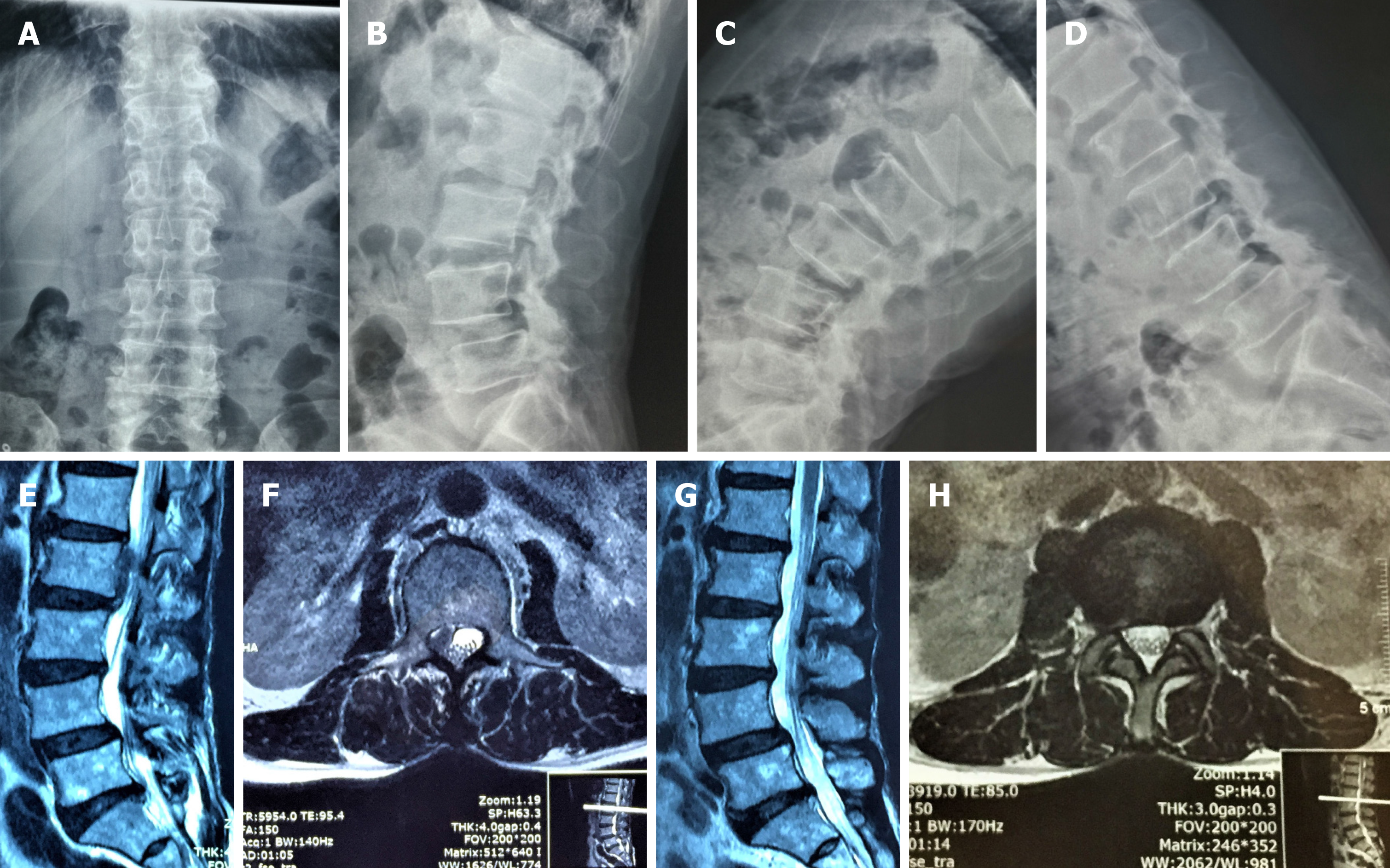
Magnetic resonance imaging (MRI) showed L2-L3 disc herniation on the right side and the herniated nucleus pulposus migrated to the upper margin of L2 vertebral body. A dynamic imaging x-ray indicated instability of the L4-L5 disc space (Figure 1A-F).
The patient was finally diagnosed as lumbar disc herniation (L2-L3), combined with lumbar spondylolisthesis (L4-L5).
The patient obtained conservative treatments by using physiotherapy and symptomatic treatment for 3 mo, but the symptoms persisted. After obtaining an accurate diagnosis, we decided to remove the highly migrated nucleus pulposus with the two-level PELD technique.
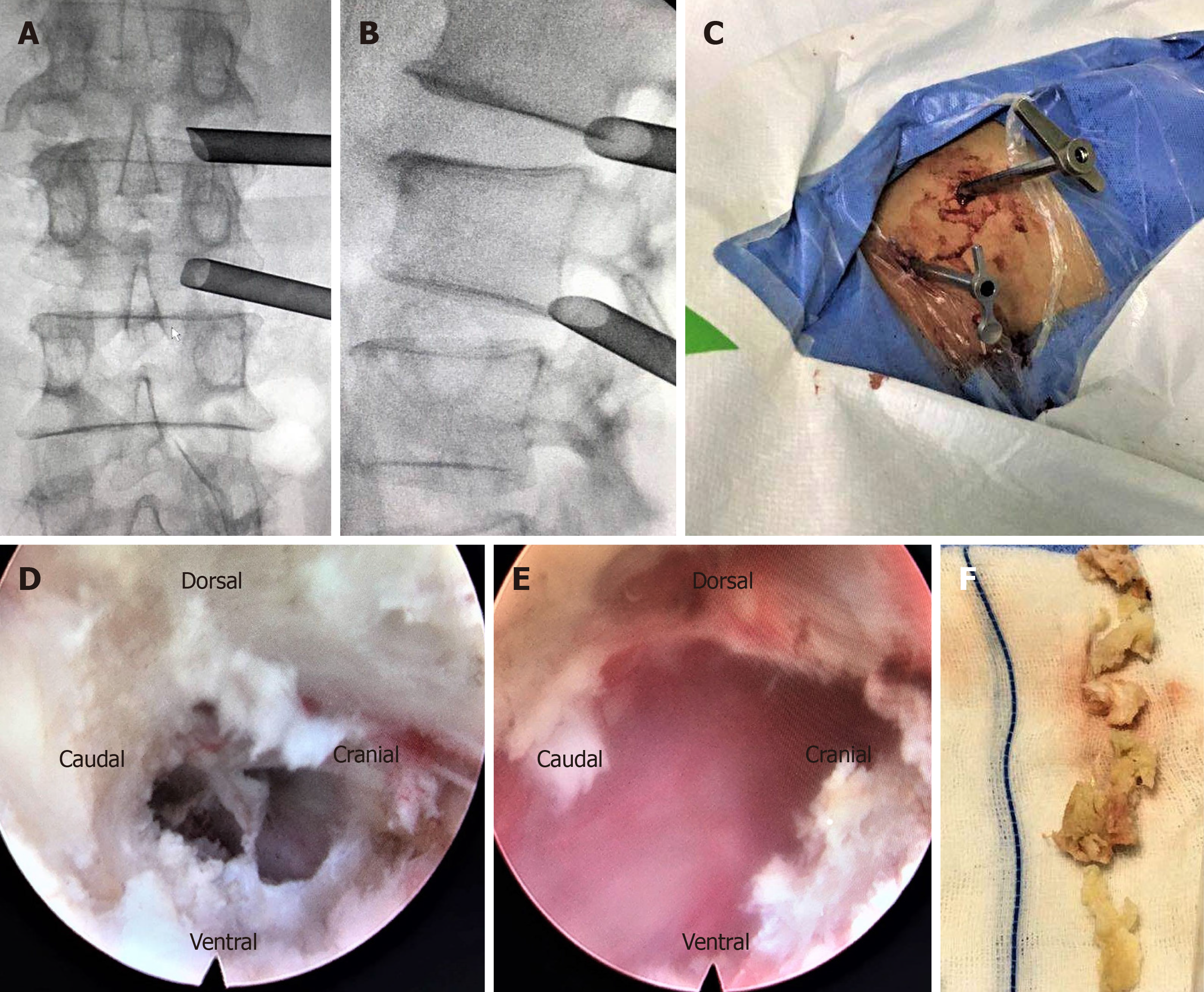
Specifically, PELD with an outside-in approach was performed according to the standard procedure, which has been detailed in our previous study[11]. The patient was positioned in the prone position on a radiolucent operating table and C-arm fluoroscopy was used to confirm the target segment. We marked the lumbar spinous process, L1, L2, and L3 pedicles, intervertebral space, and target position according to preoperative localization. The surgical puncture point was 7 cm from the midline for the L1/2 and 8 cm for L2/3 segments according to safety puncture distance based on the cross-section measurement of preoperative MRI. The puncture path was deviated to the cranial direction for the L2/3 segment and deviated to the caudal direction for the L1/2 segment. Routine disinfection and shop towels were used. These procedures were performed under local anesthesia with 1% lidocaine solution, with continuous feedback permitted from the patient during the whole surgical procedure (Supplementary material). After local anesthesia was achieved around the puncture pathway, an 18-gauge needle was inserted under fluoroscopic guidance through the L2/3 right intervertebral foramen. The target point of the needle was the medial pedicle line on the anteroposterior view and the central of the intervertebral space on the lateral view. Subsequently, the needle was replaced by a guidewire and an 8-mm skin incision was made around it. Sequential dilators of increasing diameter were placed along the guide wire to widen the soft tissue channel and a guide rod was inserted. A 7.5-mm-diameter working channel was directly placed into the intervertebral foramen, and the intraoperative fluoroscopy showed that the working channel was completely placed diagonally in the intervertebral foramen. Then a spinal endoscope was inserted through the working channel and the soft tissue was gradually separated with a flexible bipolar radiofrequency probe. After revealing the position of the dural sac, the working channel was rotated upward along the dural sac to expose the migrated nucleus pulposus tissue. The migrated nucleus pulposus tissues could be removed with a straight forceps or curved forceps under direct vision and explored to the axilar of L2 nerve root. After removing the migrated nucleus pulposus, the endoscope was pulled out and the working channel was kept in place to prevent the nucleus pulposus from shifting downward (Figure 2A-C).
The anesthesia and puncture operation of the L1-L2 segments were the same as L2-L3 segments. The working channel was completely placed in the L1-L2 intervertebral foramen and its oblique face deviated to the caudal direction confirmed by intra-operative fluoroscopy. The soft tissue was isolated and the residual nucleus pulposus was found below the nerve root. Curved forceps were used to remove the free nucleus pulposus tissues until there were no compressions of the nerve root. After decompression of the L1-L2 intervertebral space, we re-entered the L2-L3 working channel to check if any nucleus shifted away. Finally, we confirmed that there were no further remnants via the two-level working channels and the incision was sutured after the working channel was removed (Figure 2D-F).
The preoperative visual analog scale (VAS) score for lower back was 6 points and that for the right leg was 8 points. The postoperative pain symptoms of the lower back and leg were significantly relieved and the VAS score for back and leg pain was one point. Physical examinations revealed that the femoral nerve stretch test was negative. The back and leg pain symptoms completely disappeared after 1-year of follow-up and no residual nucleus pulposus was found by MRI examination (Figure 1G-H).
The definition of upper lumbar disc herniations remains controversial. Some authors consider upper lumbar disc herniations to be at L1-L2 and L2-L3, but others have expanded the definition to L3-L4. However, Sanderson et al[2] found that the anatomic characteristics of L3-L4 were similar to the lower lumbar spine and the postoperative outcomes for lumbar disc herniations at the L3-L4 level were significantly better than those occurring at L1-L2 and L2-L3. Therefore, the upper lumbar disc herniations should be defined as herniations occurring at the L1-L2 and L2-L3[1]. In addition, the herniated nucleus pulposus of upper lumbar rarely migrates due to the anatomic barriers such as large dural sac, smaller epidural space, and vascular structures[12]. Therefore, the unique anatomical characteristics are the major reasons for the low incidence of highly migrated disc herniations.
Upper lumbar disc herniations are associated with more severe clinical symptoms due to the complicated nerve structures such as the spinal cone[3]. Compared with lower lumbar disc herniations, it is difficult to accurately diagnose the disc herniation by only symptoms and signs (e.g., deep tendon reflex and manual muscle testing), because the nerve root in the upper lumbar spine does not innervate any specific muscles[10]. Therefore, early diagnosis is most important which may benefit to the patient’s postoperative recovery.
Due to the severe clinical symptoms, surgical treatment is necessary and conventional microdiscectomy surgery is the gold standard procedure for upper lumbar disc herniations[4]. During the process of microdiscectomy, extensive lamina and facet joint need to be resected in order to remove the migrated nucleus pulposus, which may locate at the inferior margin of upper pedicle, pedicle, and the upper margin of the upper pedicle[7]. However, wide laminectomy may induce iatrogenic instability and adjacent vertebral disease[13]. In this case, x-ray revealed instability of the L4/5 segment. Therefore, it would be inappropriate to conduct laminectomy unless screws are added, but it would increase the cost for the patient.
Compared with traditional open surgery, many randomized controlled trials have confirmed that the clinical outcomes of PELD technique for lower lumbar disc herniation are effective in selected patients, such as those with non-migrated disc herniations[14-17]. Furthermore, the PELD technique has many merits, e.g., shorter length of hospital stay, less lumbosacral muscle dissection[14,16,18]. Therefore, the PELD technique has become a popular technique for the treatment of lower lumbar disc herniation. Recently, to improve the clinical efficacy and reduce the incidence of iatrogenic instability, some scholars have introduced PELD for upper lumbar disc herniation and demonstrated the safety and effectiveness of this technique[9,10,19].
However, there are still limitations for traditional PELD techniques applied in the treatment of highly migrated upper lumbar disc herniations. Evidence has shown that PELD as a transforaminal approach cannot provide sufficient exposure due to anatomic barriers, such as short and fixed nerve roots and narrow spinal canal[5,6,20], which may increase the incidence of nucleus pulposus residue and dural injury. In addition, implementation of the PELD as an interlaminar approach is also limited due to the relatively narrow window and low interlaminar gap for the highly migrated nucleus pulposus, and thus, this technique is only applicable to lower non-migrated and migrated lumbar disc herniations. By contrast, the two-level PELD technique is able to provide adequate surgical vision through two working channels, which can alleviate complications after surgery.
Xin et al[21] introduced PELD through translaminar osseous channel for highly migrated upper lumbar disc herniations and obtained good clinical outcomes. However, this procedure has some limitations. First, without the cover of the yellow ligament, dural tears and cauda equine injury can occur during the process of enlarging the bottom of the bony tunnel. Second, nucleus pulposus residues may occur due to special anatomical structures. Third, this technique limits the management of central disc herniation. Therefore, the application of this approach is limited. In this study, we first introduced two-level transforaminal PELD for highly migrated upper lumbar disc herniations and achieved favorable clinical outcomes. The preoperative VAS scores for back and leg pain were 7 and 8 points, respectively, which completely disappeared 12 mo after surgery and no residual nucleus pulposus was found by MRI examination.
The most common complication of PELD for highly migrated disc herniations is nucleus pulposus residues. According to a literature review, it has been reported that approximately 5%-13% of patients have incomplete nucleus pulposus removal, which need reoperation[22,23]. However, there have been no reports on the incidence of nucleus pulposus residues of PELD for highly migrated upper lumbar disc herniations. The nucleus pulposus residue is associated with the characteristic of migrated nucleus pulposus and anatomical structure. First, the highly migrated nucleus pulposus are usually multi-fragmented. Kim et al[23] reported that multi-fragmented nucleus pulposus were found in 19 of 53 patients. In the current case, we found that the migrated nucleus pulposus was composed of several parts. Therefore, those fragmented herniations could not be completely removed just by grasping the proximal part of the herniation. In addition, due to the narrow epidural space and complex nerve tissues, the working channel could not be rotated freely to check whether there was residual nucleus pulposus. In contrast, through the two-level PELD technique, we were able to check whether the migrated nucleus pulposus had been completely removed through two different directions. In addition, we could also remove the residual nucleus pulposus through the other working channel. Therefore, the two-level PELD technique is of great benefit to reduce the incidence of postoperative nucleus pulposus residue.
The two-level PELD as a transforaminal approach is a safe and effective procedure for highly migrated upper lumbar disc herniations.
Manuscript source: Unsolicited Manuscript
Specialty type: Medicine, research and experimental
Country of origin: China
Peer-review report classification
Grade A (Excellent): 0
Grade B (Very good): 0
Grade C (Good): C
Grade D (Fair): 0
Grade E (Poor): 0
P-Reviewer: Kung WM S-Editor: Wang YQ L-Editor: Filipodia E-Editor: Xing YX
| 1. | Kim DS, Lee JK, Jang JW, Ko BS, Lee JH, Kim SH. Clinical features and treatments of upper lumbar disc herniations. J Korean Neurosurg Soc. 2010;48:119-124. [RCA] [PubMed] [DOI] [Full Text] [Cited by in Crossref: 33] [Cited by in RCA: 31] [Article Influence: 2.1] [Reference Citation Analysis (0)] |
| 2. | Sanderson SP, Houten J, Errico T, Forshaw D, Bauman J, Cooper PR. The unique characteristics of "upper" lumbar disc herniations. Neurosurgery. 2004;55:385-9; discussion 389. [RCA] [PubMed] [DOI] [Full Text] [Cited by in Crossref: 58] [Cited by in RCA: 62] [Article Influence: 3.0] [Reference Citation Analysis (0)] |
| 3. | Albert TJ, Balderston RA, Heller JG, Herkowitz HN, Garfin SR, Tomany K, An HS, Simeone FA. Upper lumbar disc herniations. J Spinal Disord. 1993;6:351-359. [RCA] [PubMed] [DOI] [Full Text] [Cited by in Crossref: 60] [Cited by in RCA: 54] [Article Influence: 1.7] [Reference Citation Analysis (0)] |
| 4. | Iwasaki M, Akino M, Hida K, Yano S, Aoyama T, Saito H, Iwasaki Y. Clinical and radiographic characteristics of upper lumbar disc herniation: ten-year microsurgical experience. Neurol Med Chir (Tokyo). 2011;51:423-426. [RCA] [PubMed] [DOI] [Full Text] [Cited by in Crossref: 12] [Cited by in RCA: 12] [Article Influence: 0.9] [Reference Citation Analysis (0)] |
| 5. | Jha RT, Syed HR, Catalino M, Sandhu FA. Contralateral Approach for Minimally Invasive Treatment of Upper Lumbar Intervertebral Disc Herniation: Technical Note and Case Series. World Neurosurg. 2017;100:583-589. [RCA] [PubMed] [DOI] [Full Text] [Cited by in Crossref: 8] [Cited by in RCA: 8] [Article Influence: 1.0] [Reference Citation Analysis (0)] |
| 6. | Karaaslan B, Aslan A, Börcek AÖ, Kaymaz M. Clinical and surgical outcomes of upper lumbar disc herniations: a retrospective study. Turk J Med Sci. 2017;47:1157-1160. [RCA] [PubMed] [DOI] [Full Text] [Cited by in Crossref: 7] [Cited by in RCA: 7] [Article Influence: 0.9] [Reference Citation Analysis (0)] |
| 7. | Son S, Lee SG, Kim WK, Ahn Y. Advantages of a Microsurgical Translaminar Approach (Keyhole Laminotomy) for Upper Lumbar Disc Herniation. World Neurosurg. 2018;119:e16-e22. [RCA] [PubMed] [DOI] [Full Text] [Cited by in Crossref: 9] [Cited by in RCA: 15] [Article Influence: 2.1] [Reference Citation Analysis (0)] |
| 8. | Ido K, Shimizu K, Tada H, Matsuda Y, Shikata J, Nakamura T. Considerations for surgical treatment of patients with upper lumbar disc herniations. J Spinal Disord. 1998;11:75-79. [RCA] [PubMed] [DOI] [Full Text] [Cited by in Crossref: 16] [Cited by in RCA: 16] [Article Influence: 0.6] [Reference Citation Analysis (0)] |
| 9. | Ahn Y, Lee SH, Lee JH, Kim JU, Liu WC. Transforaminal percutaneous endoscopic lumbar discectomy for upper lumbar disc herniation: clinical outcome, prognostic factors, and technical consideration. Acta Neurochir (Wien). 2009;151:199-206. [RCA] [PubMed] [DOI] [Full Text] [Cited by in Crossref: 75] [Cited by in RCA: 82] [Article Influence: 5.1] [Reference Citation Analysis (0)] |
| 10. | Wu J, Zhang C, Zheng W, Hong CS, Li C, Zhou Y. Analysis of the Characteristics and Clinical Outcomes of Percutaneous Endoscopic Lumbar Discectomy for Upper Lumbar Disc Herniation. World Neurosurg. 2016;92:142-147. [RCA] [PubMed] [DOI] [Full Text] [Cited by in Crossref: 14] [Cited by in RCA: 17] [Article Influence: 1.9] [Reference Citation Analysis (0)] |
| 11. | Wu X, Fan G, Gu X, Guan X, He S. Surgical Outcome of Two-Level Transforaminal Percutaneous Endoscopic Lumbar Discectomy for Far-Migrated Disc Herniation. Biomed Res Int. 2016;2016:4924013. [RCA] [PubMed] [DOI] [Full Text] [Full Text (PDF)] [Cited by in Crossref: 22] [Cited by in RCA: 18] [Article Influence: 2.0] [Reference Citation Analysis (0)] |
| 12. | Wiltse LL, Fonseca AS, Amster J, Dimartino P, Ravessoud FA. Relationship of the dura, Hofmann's ligaments, Batson's plexus, and a fibrovascular membrane lying on the posterior surface of the vertebral bodies and attaching to the deep layer of the posterior longitudinal ligament. An anatomical, radiologic, and clinical study. Spine (Phila Pa 1976). 1993;18:1030-1043. [RCA] [PubMed] [DOI] [Full Text] [Cited by in Crossref: 134] [Cited by in RCA: 112] [Article Influence: 3.5] [Reference Citation Analysis (0)] |
| 13. | Guha D, Heary RF, Shamji MF. Iatrogenic spondylolisthesis following laminectomy for degenerative lumbar stenosis: systematic review and current concepts. Neurosurg Focus. 2015;39:E9. [RCA] [PubMed] [DOI] [Full Text] [Cited by in Crossref: 63] [Cited by in RCA: 86] [Article Influence: 9.6] [Reference Citation Analysis (0)] |
| 14. | Ahn SS, Kim SH, Kim DW, Lee BH. Comparison of Outcomes of Percutaneous Endoscopic Lumbar Discectomy and Open Lumbar Microdiscectomy for Young Adults: A Retrospective Matched Cohort Study. World Neurosurg. 2016;86:250-258. [RCA] [PubMed] [DOI] [Full Text] [Cited by in Crossref: 74] [Cited by in RCA: 92] [Article Influence: 9.2] [Reference Citation Analysis (0)] |
| 15. | Lee SH, Chung SE, Ahn Y, Kim TH, Park JY, Shin SW. Comparative radiologic evaluation of percutaneous endoscopic lumbar discectomy and open microdiscectomy: a matched cohort analysis. Mt Sinai J Med. 2006;73:795-801. [PubMed] |
| 16. | Ruan W, Feng F, Liu Z, Xie J, Cai L, Ping A. Comparison of percutaneous endoscopic lumbar discectomy versus open lumbar microdiscectomy for lumbar disc herniation: A meta-analysis. Int J Surg. 2016;31:86-92. [RCA] [PubMed] [DOI] [Full Text] [Cited by in Crossref: 174] [Cited by in RCA: 153] [Article Influence: 17.0] [Reference Citation Analysis (0)] |
| 17. | Qin R, Liu B, Hao J, Zhou P, Yao Y, Zhang F, Chen X. Percutaneous Endoscopic Lumbar Discectomy Versus Posterior Open Lumbar Microdiscectomy for the Treatment of Symptomatic Lumbar Disc Herniation: A Systemic Review and Meta-Analysis. World Neurosurg. 2018;120:352-362. [RCA] [PubMed] [DOI] [Full Text] [Cited by in Crossref: 38] [Cited by in RCA: 74] [Article Influence: 10.6] [Reference Citation Analysis (0)] |
| 18. | Pan L, Zhang P, Yin Q. Comparison of tissue damages caused by endoscopic lumbar discectomy and traditional lumbar discectomy: a randomised controlled trial. Int J Surg. 2014;12:534-537. [RCA] [PubMed] [DOI] [Full Text] [Cited by in Crossref: 58] [Cited by in RCA: 67] [Article Influence: 6.1] [Reference Citation Analysis (0)] |
| 19. | Shin MH, Bae JS, Cho HL, Jang IT. Extradiscal Epiduroscopic Percutaneous Endoscopic Discectomy for Upper Lumbar Disc Herniation A Technical Note. Clin Spine Surg. 2019;32:98-103. [RCA] [PubMed] [DOI] [Full Text] [Cited by in Crossref: 9] [Cited by in RCA: 9] [Article Influence: 1.5] [Reference Citation Analysis (0)] |
| 20. | Bae J, Lee SH, Shin SH, Seo JS, Kim KH, Jang JS. Radiological analysis of upper lumbar disc herniation and spinopelvic sagittal alignment. Eur Spine J. 2016;25:1382-1388. [RCA] [PubMed] [DOI] [Full Text] [Cited by in Crossref: 20] [Cited by in RCA: 27] [Article Influence: 3.0] [Reference Citation Analysis (0)] |
| 21. | Xin Z, Liao W, Ao J, Qin J, Chen F, Ye Z, Cai Y. A Modified Translaminar Osseous Channel-Assisted Percutaneous Endoscopic Lumbar Discectomy for Highly Migrated and Sequestrated Disc Herniations of the Upper Lumbar: Clinical Outcomes, Surgical Indications, and Technical Considerations. Biomed Res Int. 2017;2017:3069575. [RCA] [PubMed] [DOI] [Full Text] [Full Text (PDF)] [Cited by in Crossref: 9] [Cited by in RCA: 13] [Article Influence: 1.6] [Reference Citation Analysis (0)] |
| 22. | Choi G, Lee SH, Lokhande P, Kong BJ, Shim CS, Jung B, Kim JS. Percutaneous endoscopic approach for highly migrated intracanal disc herniations by foraminoplastic technique using rigid working channel endoscope. Spine (Phila Pa 1976). 2008;33:E508-E515. [RCA] [PubMed] [DOI] [Full Text] [Cited by in Crossref: 145] [Cited by in RCA: 150] [Article Influence: 8.8] [Reference Citation Analysis (0)] |
| 23. | Kim HS, Ju CI, Kim SW, Kim JG. Endoscopic transforaminal suprapedicular approach in high grade inferior migrated lumbar disc herniation. J Korean Neurosurg Soc. 2009;45:67-73. [RCA] [PubMed] [DOI] [Full Text] [Cited by in Crossref: 38] [Cited by in RCA: 44] [Article Influence: 2.8] [Reference Citation Analysis (0)] |


