Published online Nov 26, 2019. doi: 10.12998/wjcc.v7.i22.3778
Peer-review started: September 12, 2019
First decision: September 23, 2019
Revised: October 10, 2019
Accepted: October 15, 2019
Article in press: October 15, 2019
Published online: November 26, 2019
Processing time: 74 Days and 15.4 Hours
Epidermoid cysts can be found at any location in the human body. However, perianal epidermoid cysts are extremely rare and only a few cases have been reported. As far as we know, there is no special literature on the value of contrast-enhanced computed tomography (CT) for the diagnosis of perianal epidermoid cysts.
A 60-year-old male patient presented to the department of general surgery of PLA Strategic Support Force Characteristic Medical Center with the chief complaint of a mass in the perianal region gradually expanding for more than 30 years and perianal discomfort upon sitting for a preceding period of 2 mo. Physical examination revealed a painless mass in the left perianal region. Contrast-enhanced CT was used for preoperative diagnosis. The patient was treated by total mass excision under epidural anesthesia. Postoperative pathological examination revealed the presence of a perianal epidermoid cyst. The patient showed a satisfactory recovery during the 6-month follow-up period.
Contrast-enhanced CT may be a beneficial, useful, and convenient approach for assistance for preoperative diagnosis and surgical decision-making for patients with perianal epidermoid cysts.
Core tip: Perianal epidermoid cysts are extremely rare and there is no special literature on the value of contrast-enhanced computed tomography (CT) for the diagnosis of perianal epidermoid cysts. This case report describes the value of contrast-enhanced CT for the diagnosis and surgical decision-making of patients with perianal epidermoid cysts and highlights that it may be a beneficial, useful, and convenient approach for these patients.
- Citation: Sun PM, Yang HM, Zhao Y, Yang JW, Yan HF, Liu JX, Sun HW, Cui Y. Contrast-enhanced computed tomography findings of a huge perianal epidermoid cyst: A case report. World J Clin Cases 2019; 7(22): 3778-3783
- URL: https://www.wjgnet.com/2307-8960/full/v7/i22/3778.htm
- DOI: https://dx.doi.org/10.12998/wjcc.v7.i22.3778
Epidermoid cysts usually are benign cysts that are commonly found in the skin, including on the face, scalp, neck, and trunk[1]. However, perianal epidermoid cysts are very rare and a few cases have been reported on[2]. Perianal epidermoid cysts do not exhibit any unique clinical symptoms during the early stages and may be misdiagnosed as other anal diseases, due to pain or other symptoms[3]. Since a perianal epidermoid cyst may get infected and inflamed, surgical resection is recommended. Preoperative diagnosis is very important for the selection of a suitable surgical method. Although there is some literature on the use of ultrasound and magnetic resonance imaging for the location of perianal/presacral epidermoid cysts[2,4,5], the value of the use of contrast-enhanced computed tomography (CT) for perianal epidermoid cysts is not reported. Here we present a case of perianal epidermoid cyst, for which contrast-enhanced CT was successfully used for preoperative diagnosis and surgical decision-making.
A 60-year-old male patient was admitted to the department of general surgery of PLA Strategic Support Force Characteristic Medical Center, with a complaint of a mass gradually expanding for more than 30 years and perianal discomfort upon sitting, which had persisted for 2 mo.
The mass was painless and grew slowly. There were no signs of redness, swelling, or heat of local skin and no anal swelling, itching, bearing down, or difficulty during defecation.
He had a history of hypertension for over 10 years but had no other previous medical issues.
The patient had no history of smoking, alcohol abuse, or illicit drug use and lacked a family history of other diseases.
Physical examination revealed a painless, mobile mass, with a clear margin, at 3 to 6 points of the lithotomy position, 2 cm over the anal verge. The top of the mass was exposed to the skin, measuring 6 cm, and the bottom of the mass could not be palpated clearly. Preoperative digital anal examination and anoscopy showed a normal rectal mucosa and the mass could be palpated through the left sidewall of the rectum.
Laboratory tests, including tumor markers, were normal.
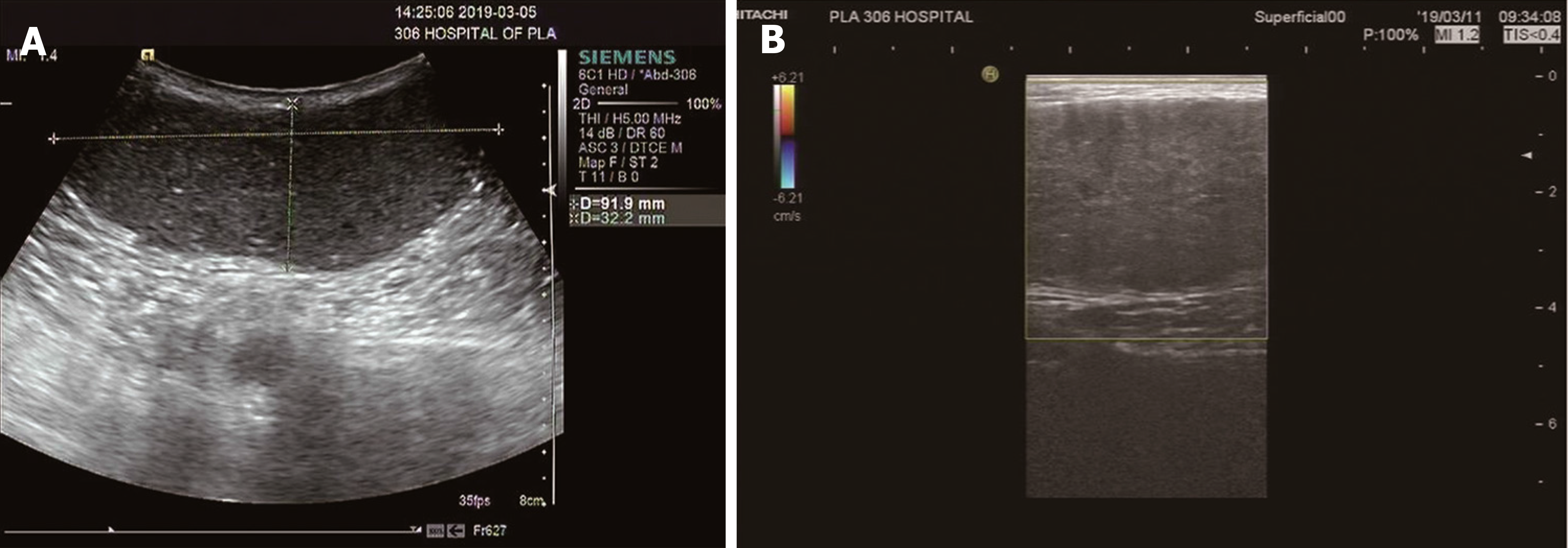
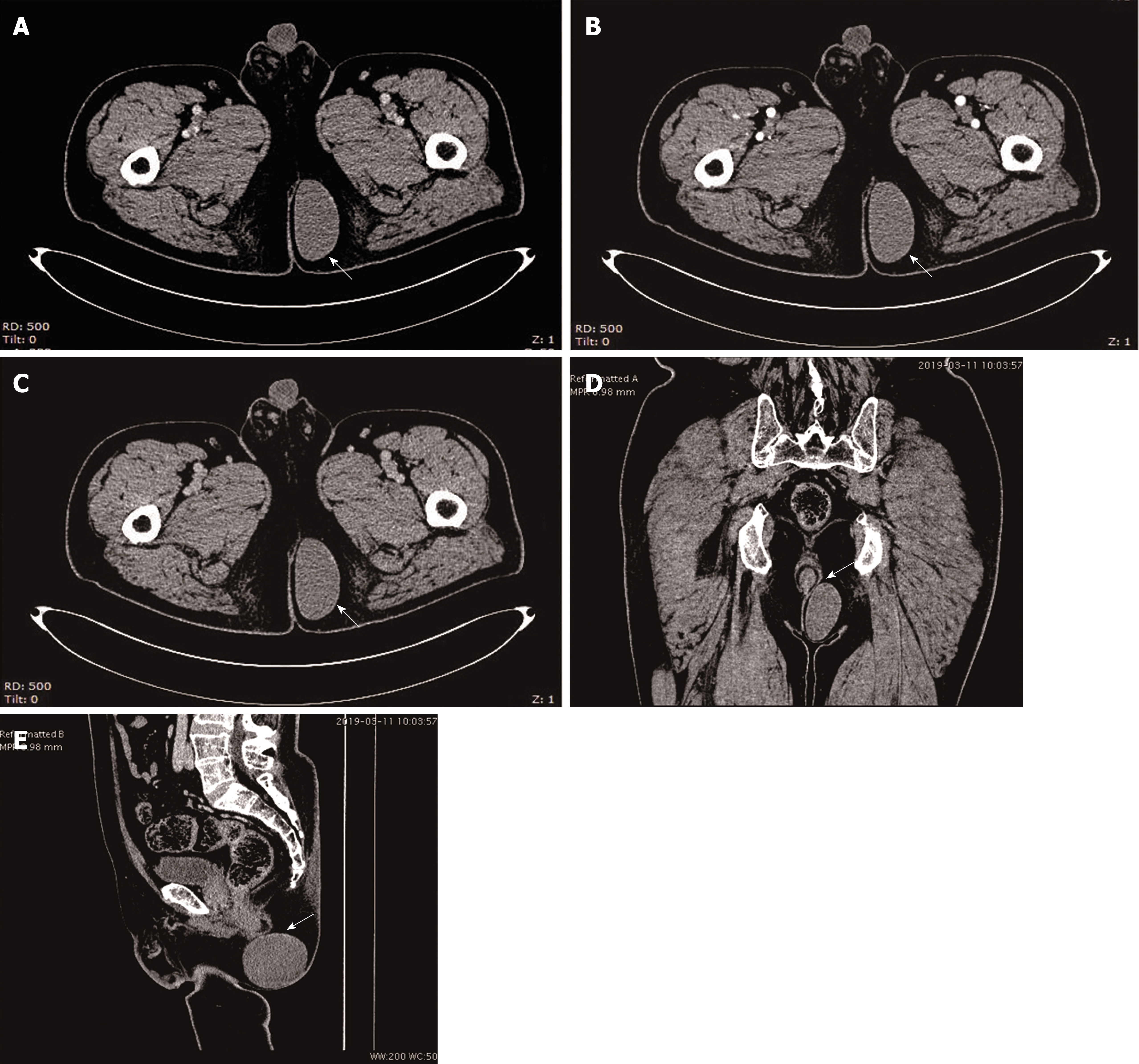
Ultrasound showed a hypoechoic mass, measuring 9.2 cm × 3.7 cm, with a clear margin, regular morphology, and no obvious blood flow (Figure 1). Contrast-enhanced CT revealed a cystic low-density mass with a smooth edge. The upper right edge of the mass was very close to the anal canal (Figure 2D and 2E). The wall of the cyst was not enhanced in the arterial phase, venous phase, and delayed phase and there was no blood vessel in and around the mass (Figure 2A, 2B and 2C), while the CT density was 40 HU.
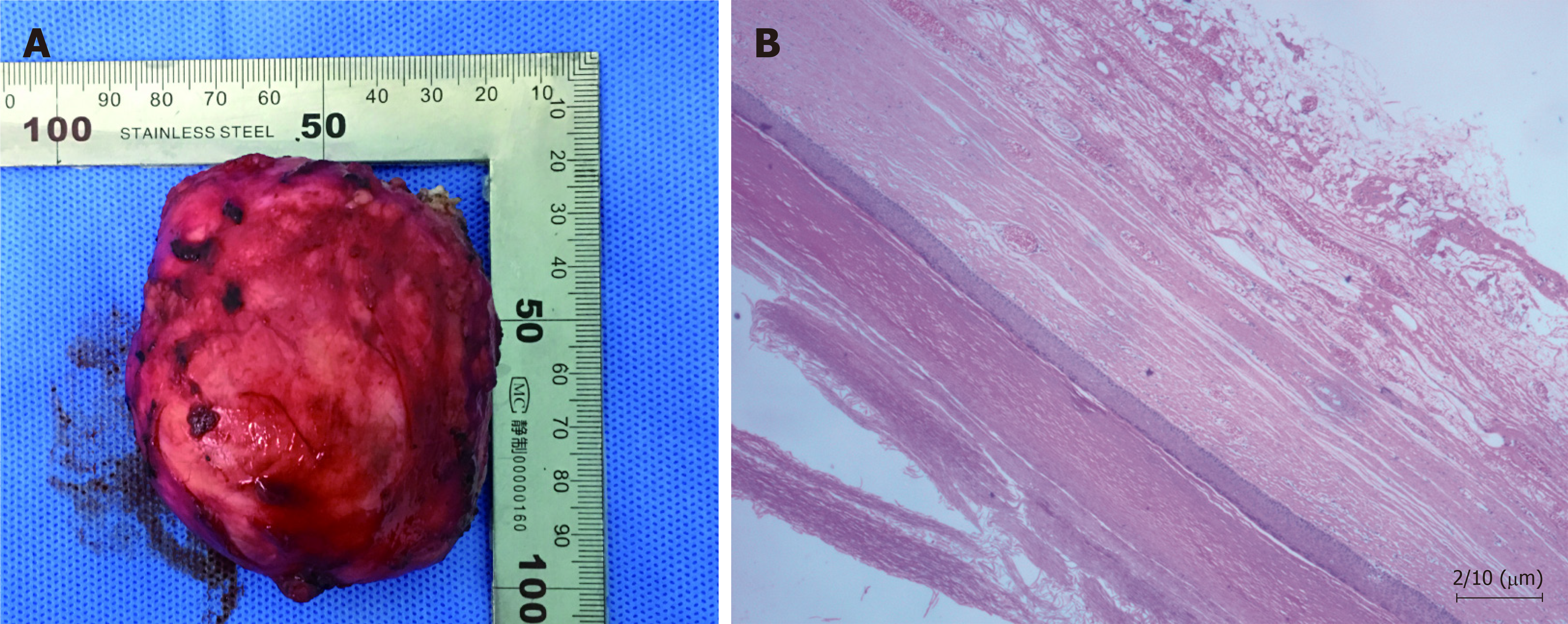
The pathologic diagnosis showed an epidermoid cyst. The specimen was a greyish-red cystic mass, which contained greyish-white substance in the capsule. Microscopic and histopathological examinations revealed that the cyst wall was 40-60 μm thick and lined with stratified squamous epithelia, which had distinct granular layers. The cavity of the cyst was filled with layered uniform red-stained keratin (Figure 3).
The patient was taken to the operating room for mass excision as the preoperative diagnosis was a perianal mass. The surgical exploration showed a greyish-red subcutaneous mass with an intact capsule, tough and flexible, which measured about 10 cm. The mass was completely resected after being carefully separated from surrounding tissues. Digital anal examination and anoscopy showed that there was no fistula between the surgical area and the anal canal, after the resection.
The patient was discharged on postoperative day 5 without complications and showed a satisfactory recovery, as observed during the 6-mo follow-up period.
Epidermoid cysts are extremely common and can occur in any hair-containing area, and are more frequently found in men than in women (2:1), within the age range of 30-40 years[6]. This disease is caused by the ectodermal cells that remain in tissues from embryonic development developing into cysts, as a result of inflammation around a pilosebaceous follicle, or deep implantation of the epidermis by a blunt or penetrating injury or surgery[3]. The wall of the epidermoid cyst is mainly composed of stratified squamous epithelium[7], and does not contain a skin attachment structure in the dermis, and can become weak when the cyst extends. The cysts are generally filled with keratin, scales, fat, and cholesterol. Usually the cysts are slow-growing and asymptomatic. Pain and tenderness may be felt if the cysts are infected and inflamed, or develop into a malignancy, which is rare[1].
Perianal epidermoid cysts are difficult to diagnose during the early stages, because there are no unique clinical symptoms. The symptoms of the cysts vary according to their size, location, and infected status. When increasing in size, the epidermoid cyst will oppress the anal sphincter or/and rectum, inducing perianal discomfort[2], such as in this case. If the cysts are infected and inflamed, symptoms including the pain and tenderness of the perianal region may be reported. When the cysts rupture after infection, a purulent secretion can be found in the perianal region. Therefore, perianal epidermoid cysts are often misdiagnosed as perianal abscesses and/or anal fistulas and undergo unsuitable operation, leading to postoperative recurrence.
The preoperative diagnosis of perianal epidermoid cysts is very important for the selection of a suitable surgical method and may prevent intraoperative and postoperative complications. In our case, contrast-enhanced CT was used for preoperative diagnosis and surgical decision-making. Contrast-enhanced CT has the advantages of high resolution, fast scanning speed, and competitive price, and it can clearly indicate the size, location, density, and margin of the epidermoid cysts, as well as its relationship with surrounding tissues[8]. Contrast-enhanced CT is significantly valuable in showing the blood supply of the cysts. A typical imaging feature of perianal epidermoid cysts is a subcutaneous low density cystic mass, round or elliptical, with a smooth edge, being either a single cyst or polycystic. The density of cysts differs according to keratin and cholesterol content, as well as proportion, calcification, and bleeding[9]. The wall of the cysts is thin and no or slight enhancement is seen in contrast-enhanced CT examination. When the cysts are infected and form granulomatous structures, rim enhancement may be present around the mass[10]. Furthermore, contrast-enhanced CT can clearly show the location of perianal epidermoid cysts, and the relationship between the cyst and the anal sphincter and rectum, as well as the blood supply to the cyst or around the cyst. This imaging information is of significant value for guiding the surgery. As in our case, contrast-enhanced CT clearly shows the texture, density, edge, and the wall of the mass with no enhancement and whether there are blood vessels in and around the mass. The upper right edge of the mass can be very close to the anal canal, suggesting that surgery should be carried out while being very careful of this area to avoid damage of the anal sphincter and rectum.
Wide surgical resection is recommended for the treatment of epidermoid cysts[11], because there is some risk of recurrence. Suitable first surgical treatment is very important for epidermoid cysts because an incorrect or insufficient treatment could compromise the chances of complete surgical excision and increase the risk of recurrence, as well as cause repeated operations, prolonged morbidity, and serious complications[7]. Surgery should remove the cysts completely and avoid rupture and residue of the cystic wall, as much as possible. For perianal epidermoid cysts, it is important to explore the relationship between the cysts and the anus during surgery to avoid damage to the anal sphincter and rectum. The prognosis and outcome of these lesions are excellent, with a postoperative recurrence rate of only 3%[12], which generally occurs locally.
Contrast-enhanced CT examination could show the morphology, location, and blood supply of the perianal epidermoid cysts and would be a beneficial, useful, and convenient approach for preoperative diagnosis and surgical decision-making.
We thank our coworkers of the departments of radiology, laboratory, electrocardiogram, and operating room for their invaluable technical help.
Manuscript source: Unsolicited manuscript
Specialty type: Medicine, Research and Experimental
Country of origin: China
Peer-review report classification
Grade A (Excellent): 0
Grade B (Very good): B, B
Grade C (Good): 0
Grade D (Fair): D
Grade E (Poor): 0
P-Reviewer: Gonoi W, Sersar SI, Zavras N S-Editor: Dou Y L-Editor: Wang TQ E-Editor: Liu MY
| 1. | Faltaous AA, Leigh EC, Ray P, Wolbert TT. A Rare Transformation of Epidermoid Cyst into Squamous Cell Carcinoma: A Case Report with Literature Review. Am J Case Rep. 2019;20:1141-1143. [RCA] [PubMed] [DOI] [Full Text] [Full Text (PDF)] [Cited by in Crossref: 8] [Cited by in RCA: 21] [Article Influence: 3.5] [Reference Citation Analysis (0)] |
| 2. | Nicolay S, De Schepper A, Pouillon M. Epidermal inclusion cyst of the perianal region. JBR-BTR. 2014;97:166-167. [PubMed] |
| 3. | Sritharan K, Ghani Y, Thompson H. An unusual encounter of an epidermoid cyst. BMJ Case Rep. 2014;2014. [RCA] [PubMed] [DOI] [Full Text] [Cited by in Crossref: 2] [Cited by in RCA: 2] [Article Influence: 0.2] [Reference Citation Analysis (0)] |
| 4. | Turkay R, Caymaz I, Yildiz B, Livaoglu A, Turkey B, Bakir B. A rare case of epidermoid cyst of perineum: Diffusion-weighted MRI and ultrasonography findings. Radiol Case Rep. 2015;8:593. [RCA] [PubMed] [DOI] [Full Text] [Full Text (PDF)] [Cited by in Crossref: 6] [Cited by in RCA: 6] [Article Influence: 0.6] [Reference Citation Analysis (0)] |
| 5. | Halefoglu AM, Sen EY. Precoccygeal epidermal inclusion cyst: ultrasound and MR imaging features. JBR-BTR. 2012;95:294-296. [PubMed] |
| 6. | Saeed U, Mazhar N. Epidermoid cyst of perineum: a rare case in a young female. BJR Case Rep. 2016;3:20150352. [RCA] [PubMed] [DOI] [Full Text] [Full Text (PDF)] [Cited by in Crossref: 3] [Cited by in RCA: 3] [Article Influence: 0.3] [Reference Citation Analysis (0)] |
| 7. | Jain V, Misra S, Tiwari S, Rahul K, Jain H. Recurrent Perianal Sinus in Young Girl Due To Pre-sacral Epidermoid Cyst. Ann Med Health Sci Res. 2013;3:458-460. [RCA] [PubMed] [DOI] [Full Text] [Full Text (PDF)] [Cited by in Crossref: 2] [Cited by in RCA: 3] [Article Influence: 0.3] [Reference Citation Analysis (0)] |
| 8. | Cianci P, Tartaglia N, Altamura A, Fersini A, Vovola F, Sanguedolce F, Ambrosi A, Neri V. A recurrent epidermoid cyst of the spleen: report of a case and literature review. World J Surg Oncol. 2016;14:98. [RCA] [PubMed] [DOI] [Full Text] [Full Text (PDF)] [Cited by in Crossref: 28] [Cited by in RCA: 23] [Article Influence: 2.6] [Reference Citation Analysis (0)] |
| 9. | Dahan H, Arrivé L, Wendum D, Docou le Pointe H, Djouhri H, Tubiana JM. Retrorectal developmental cysts in adults: clinical and radiologic-histopathologic review, differential diagnosis, and treatment. Radiographics. 2001;21:575-584. [RCA] [PubMed] [DOI] [Full Text] [Cited by in Crossref: 153] [Cited by in RCA: 153] [Article Influence: 6.4] [Reference Citation Analysis (0)] |
| 10. | Riojas CM, Hahn CD, Johnson EK. Presacral epidermoid cyst in a male: a case report and literature review. J Surg Educ. 2010;67:227-232. [RCA] [PubMed] [DOI] [Full Text] [Cited by in Crossref: 12] [Cited by in RCA: 14] [Article Influence: 0.9] [Reference Citation Analysis (0)] |
| 11. | Kesici U, Sakman G, Mataraci E. Retrorectal/Presacral epidermoid cyst: report of a case. Eurasian J Med. 2013;45:207-210. [RCA] [PubMed] [DOI] [Full Text] [Cited by in Crossref: 9] [Cited by in RCA: 10] [Article Influence: 1.0] [Reference Citation Analysis (0)] |
| 12. | Houdek MT, Warneke JA, Pollard CM, Lindgren EA, Taljanovic MS. Giant epidermal cyst of the gluteal region. Radiol Case Rep. 2015;5:476. [RCA] [PubMed] [DOI] [Full Text] [Full Text (PDF)] [Cited by in Crossref: 13] [Cited by in RCA: 16] [Article Influence: 1.6] [Reference Citation Analysis (0)] |


