Published online Nov 6, 2019. doi: 10.12998/wjcc.v7.i21.3639
Peer-review started: May 8, 2019
First decision: September 9, 2019
Revised: September 23, 2019
Accepted: September 25, 2019
Article in press: September 25, 2019
Published online: November 6, 2019
Processing time: 186 Days and 23.8 Hours
We report a rare case of numbness in the right hand, finally diagnosed as bilateral common carotid artery common trunk with aberrant right subclavian artery combined with right subclavian steal syndrome and explain the cause of these diseases.
The patient was a 65-year-old woman. She complained of dizziness, numbness and weakness of the right hand for 6 mo. She was diagnosed with bilateral common carotid artery common trunk with aberrant right subclavian artery combined with right subclavian steal syndrome by ultrasound, enhanced computed tomography, computed tomography angiography and other examinations. Considering the surgical risks, the patient refused the aberrant right subclavian artery stent implantation and was discharged. We hypothesize that these two kinds of deformity and right subclavian steal syndrome may not occur by accident and result from multiple malformations.
Bilateral common carotid artery common trunk with aberrant right subclavian artery combined with right subclavian steal syndrome is rare. This case reminds interventional radiologists of the possibility of these abnormalities before surgery.
Core tip: Bilateral common carotid artery common trunk with aberrant right subclavian artery combined with right subclavian steal syndrome is rare. No other similar domestic or foreign cases have been described. We hypothesized that the occurrence of blood steal syndrome may not be accidental and results from multiple malformations. This case reminds surgeons and interventional radiologists to be aware of the possibility of these abnormalities in patients with aortic arch abnormalities before surgery.
- Citation: Sun YY, Zhang GM, Zhang YB, Du X, Su ML. Bilateral common carotid artery common trunk with aberrant right subclavian artery combined with right subclavian steal syndrome: A case report. World J Clin Cases 2019; 7(21): 3639-3648
- URL: https://www.wjgnet.com/2307-8960/full/v7/i21/3639.htm
- DOI: https://dx.doi.org/10.12998/wjcc.v7.i21.3639
There are many anatomical variations of the aortic arch and its branches, which are related to race and environment[1]. Anatomically, this is related to the complex process of embryonic development, that is, the imbalance of the developmental speed of the aortic arch during the embryonic period[2,3]. When variations in the aortic arch and its branches affect anatomy or hemodynamics, patients often develop symptoms[4].
We report a case of a patient who complained of numbness and weakness in her right hand. After ultrasonography, digital subtraction angiography, enhanced computed tomography (CT), CT angiography (CTA) and other examinations, she was finally diagnosed with bilateral common carotid artery common trunk with aberrant right subclavian artery combined with right subclavian steal syndrome. The coexistence of these two deformities is rare when combined with the presence of right subclavian steal syndrome. No other similar domestic or foreign cases have been described. We hypothesize that these two kinds of deformities and right subclavian steal syndrome may not occur by accident and result from multiple malformations.
The patient was a 65-year-old woman. Her chief complaints were dizziness, numbness in the right hand and weakness for 6 mo.
Dizziness occurred > 6 mo ago without obvious inducement, lasted for 30 min, without fainting, chest tightness, chest pain and abdominal pain. The patient was treated in a community hospital for pain and other discomfort and was considered to have a history of hypertension. Two months ago, the blood pressure difference between the two upper arms was > 40 mmHg. In the outpatient department of the hospital, the relevant examination was completed to confirm severe stenosis of the subclavian artery. When she was admitted to the hospital, recent mood, diet, sleep pattern and urine test were normal, and body weight showed no significant changes.
She had a history of hypertension for > 10 yr and chronic hepatitis B for > 40 yr. She denied history of hepatitis, tuberculosis, typhoid fever and other infectious diseases, blood transfusion, trauma, poisoning and surgery.
No other members of her family had a similar medical history. She denied a family history of hereditary diseases, cancer, liver disease, infectious diseases and inflammatory diseases.
The patient came to our hospital for treatment for dizziness and numbness and weakness of the right hand for 6 mo. Physical examination and blood pressure measurement showed a blood pressure difference of 40 mmHg between the upper arms on both sides. It was significantly lower in the right upper arm.
Laboratory examination results were: N-terminal pro-brain natriuretic peptide 135.1 pg/mL (reference range 0-125 pg/mL); uric acid 378 μmol/L (reference range 142-339 μmol/L); endogenous creatinine clearance rate 66 mL/min × 1.73 m2 (reference range 80-120 mL/min × 1.73m2).
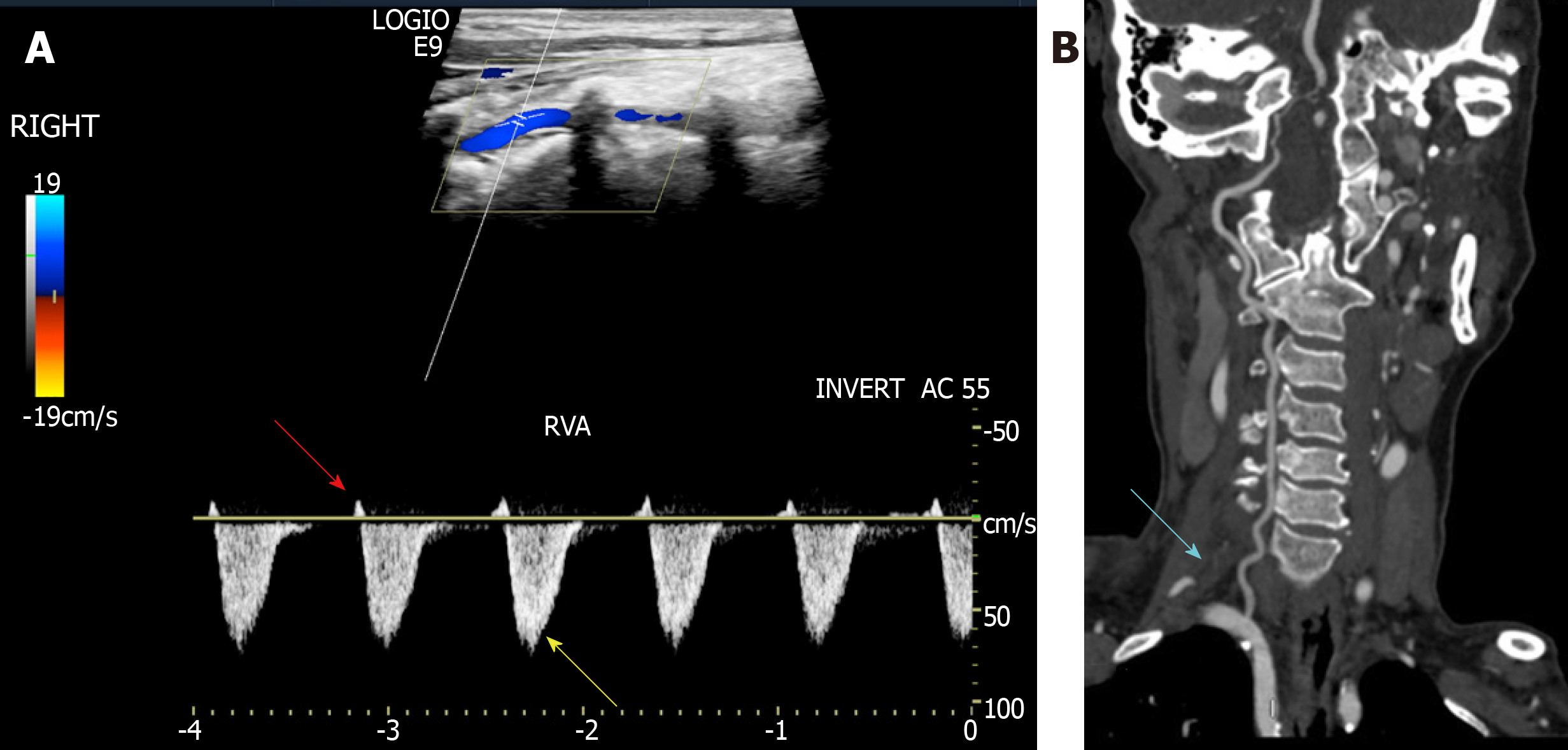
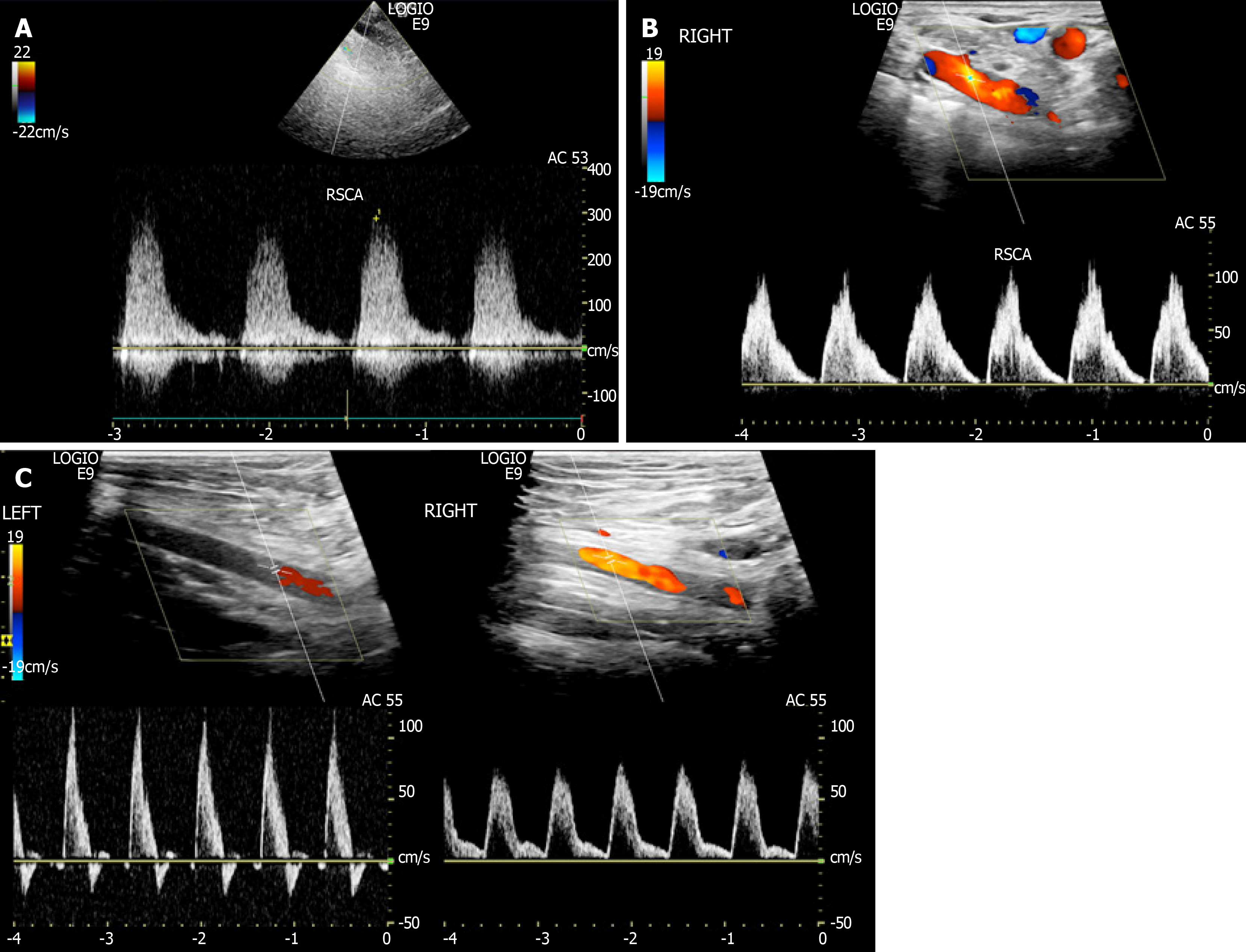
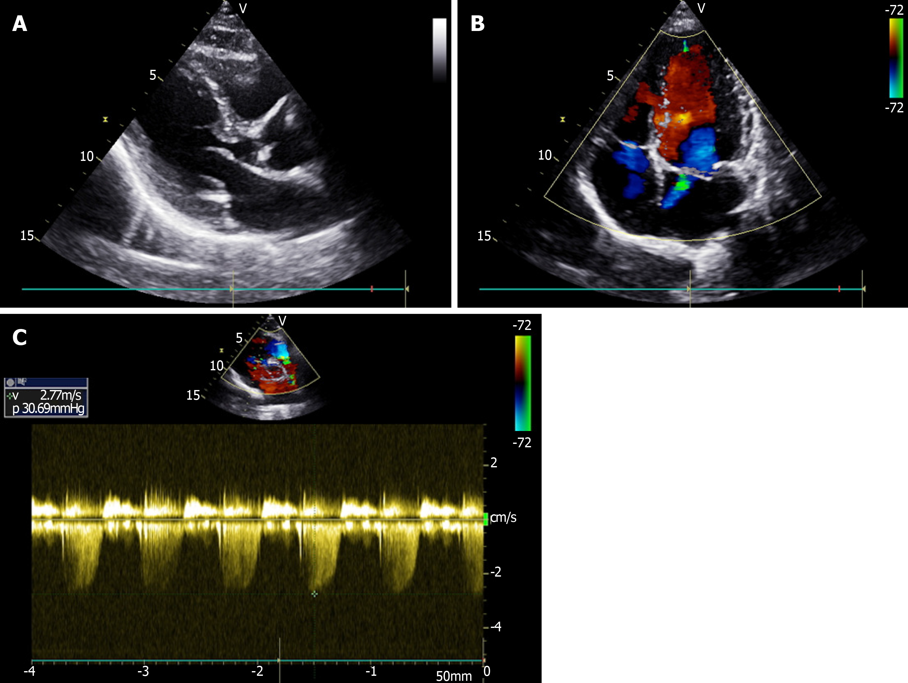
Ultrasound showed anomalous origin of the right subclavian artery and type II right subclavian steal syndrome. The blood flow in the right vertebral artery showed a weak positive contractile peak in early systole, followed by a large contractile wave in late systole below baseline and the diastolic blood flow disappeared (Figure 1A). She had right vertebral artery dysplasia (Figure 1B). The initial segment of the right subclavian artery showed 80%–99% stenosis. The blood flow velocity at the area of stenosis was up to 289 cm/s (Figure 2A) and was reduced at the distal segment of the stenosis, presenting as a single-phase wave change (Figure 2B). The right axillary artery and the following arteries showed blood deficiency compared with the healthy left side, and the flow velocity of the right axillary artery was significantly reduced and showed unidirectional changes (Figure 2C). Echocardiography showed calcification of the posterior mitral valve ring (Figure 3A) and mild regurgitation of the mitral and tricuspid valves (Figure 3B and 3C).
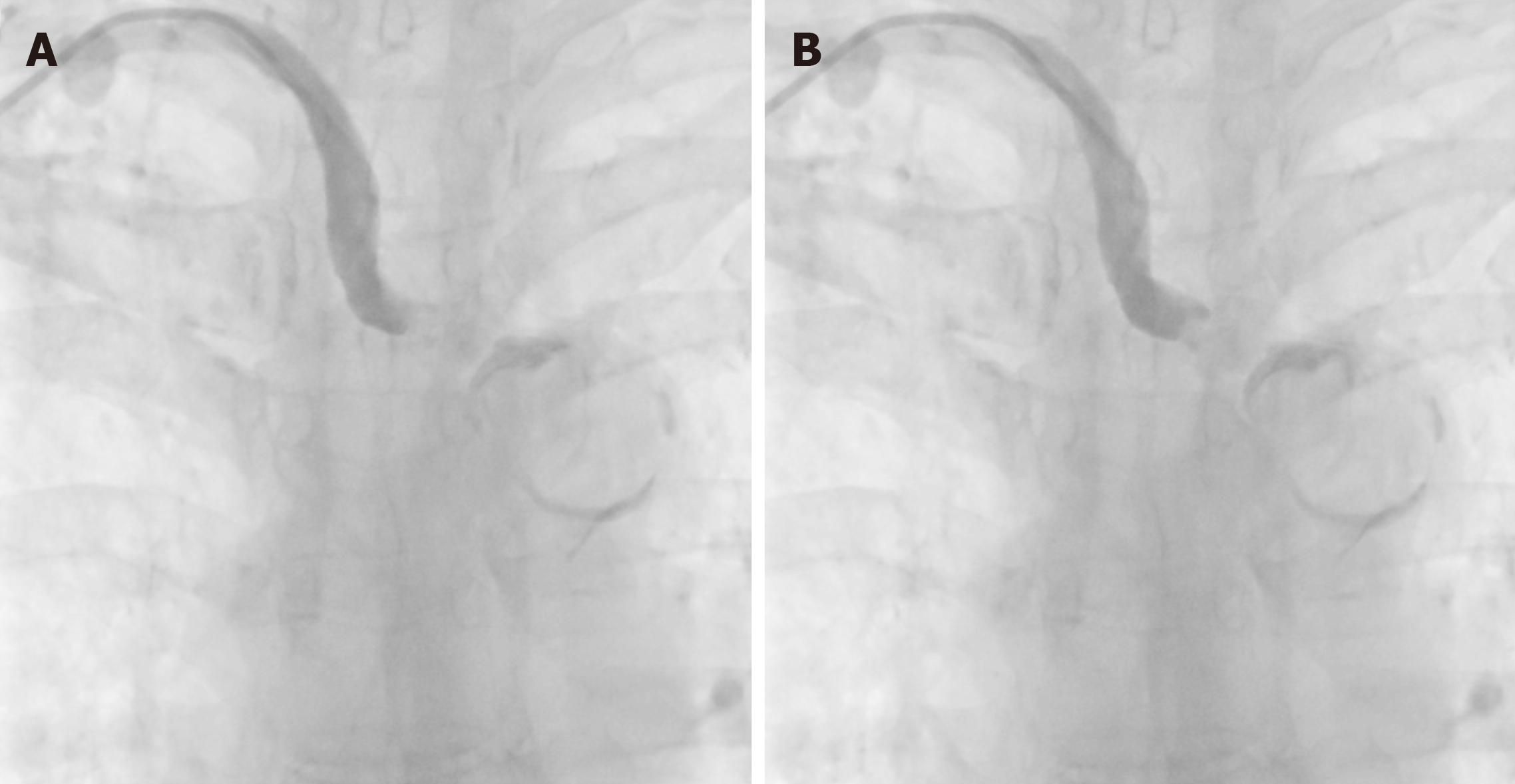
In order to confirm and treat the right subclavian steal syndrome, digital subtraction angiography was performed. However, after successful puncture of the right radial artery, the direction of the catheter guide wire was different from that in normal patients and combined with severe right subclavian artery stenosis, it displayed as arterial occlusion (Figure 4). If the direction of the catheter guide wire was blindly adjusted only according to experience, the blood vessel might be damaged, so the doctors discontinued the continuous angiography. Considering the surgical risks, the patient refused the aberrant right subclavian artery stent implantation and was discharged.
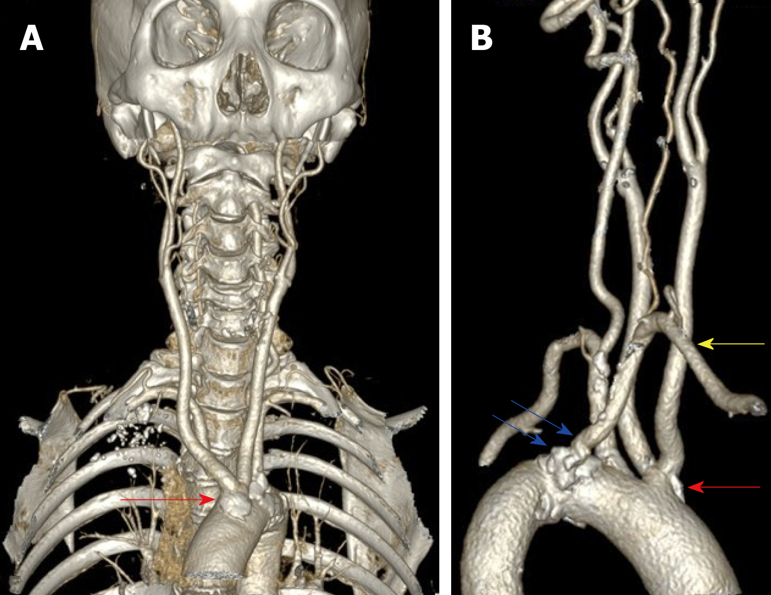
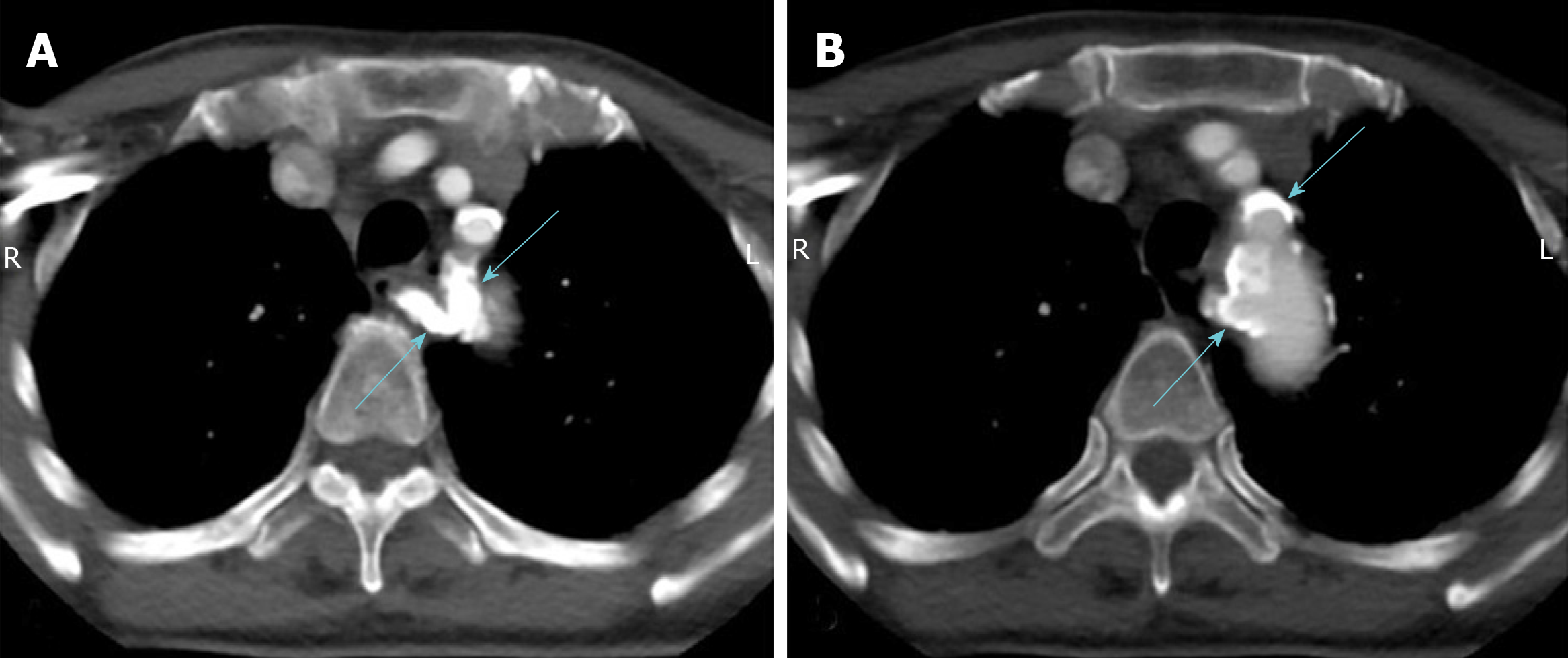
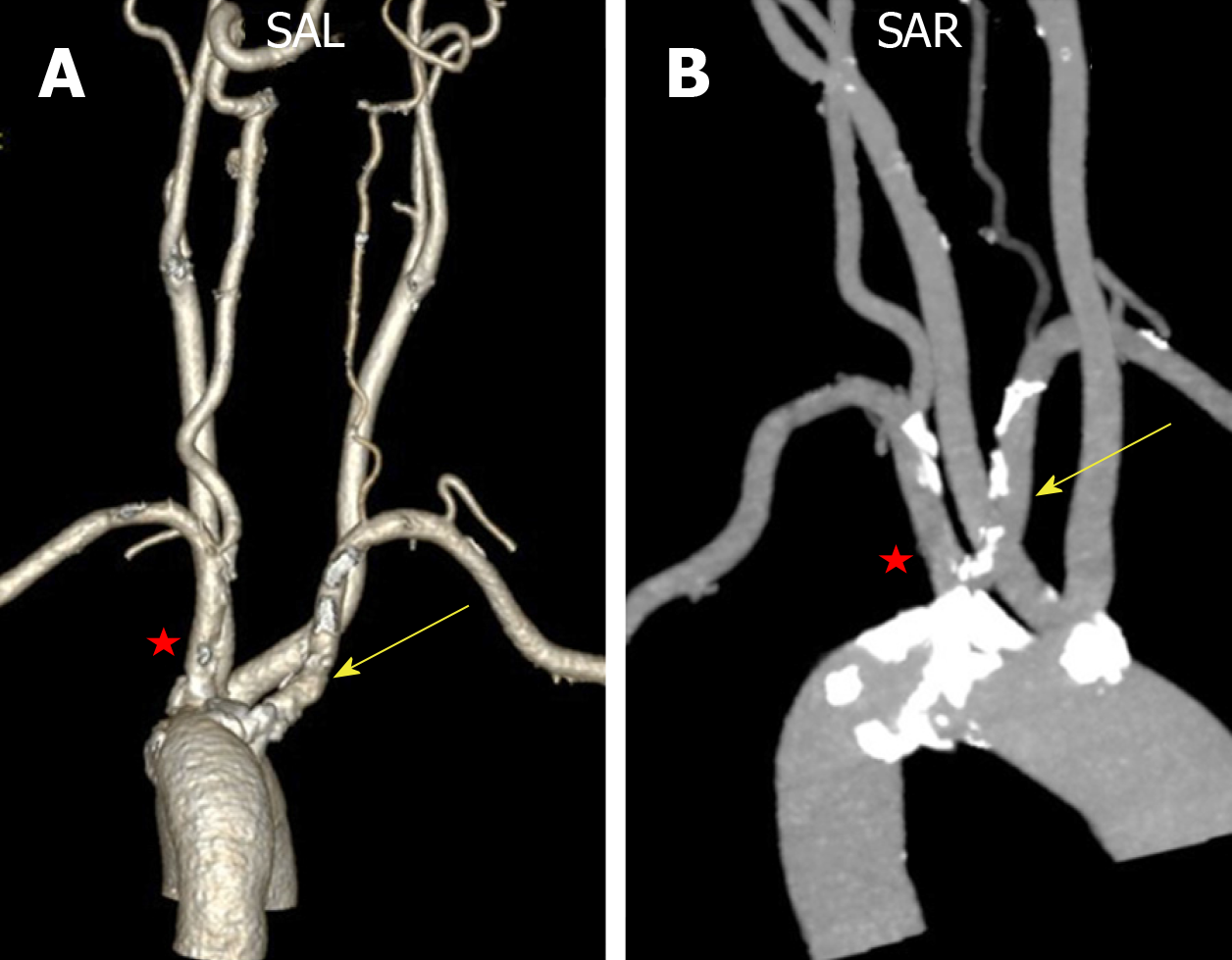
Ten days later, the patient underwent enhanced CT and CTA examination, which showed that the common trunk of the bilateral common carotid artery arose from the aortic arch (Figure 5A) and aberrant right subclavian artery (Figure 5B). A large number of calcified plaques were found at the beginning of the aberrant right subclavian artery and the common trunk of the bilateral common carotid artery (Figure 5B). The right vertebral artery was slender (Figure 1B). All cardiologists met to discuss the patient, analyzed the causes of cerebral angiography failure and lessons learnt from it.
The common trunk of bilateral common carotid artery arose from the aortic arch and the aberrant right subclavian artery. Type II right subclavian steal syndrome and right vertebral artery dysplasia. Right subclavian artery stenosis at the initial segment (80%-99%). Ischemia change in the right axillary artery.
The patient underwent digital subtraction angiography. However, after the successful puncture of right radial artery, the direction of the catheter guide wire appeared different from that in “normal” patients, and severe right subclavian artery stenosis was displayed as arterial occlusion. If the direction of the catheter guide wire was blindly adjusted only according to examiner’s experience, the blood vessel might be damaged, so the angiography was discontinued. Considering the surgical risks, the patient refused the aberrant right subclavian artery stent implantation and was discharged.
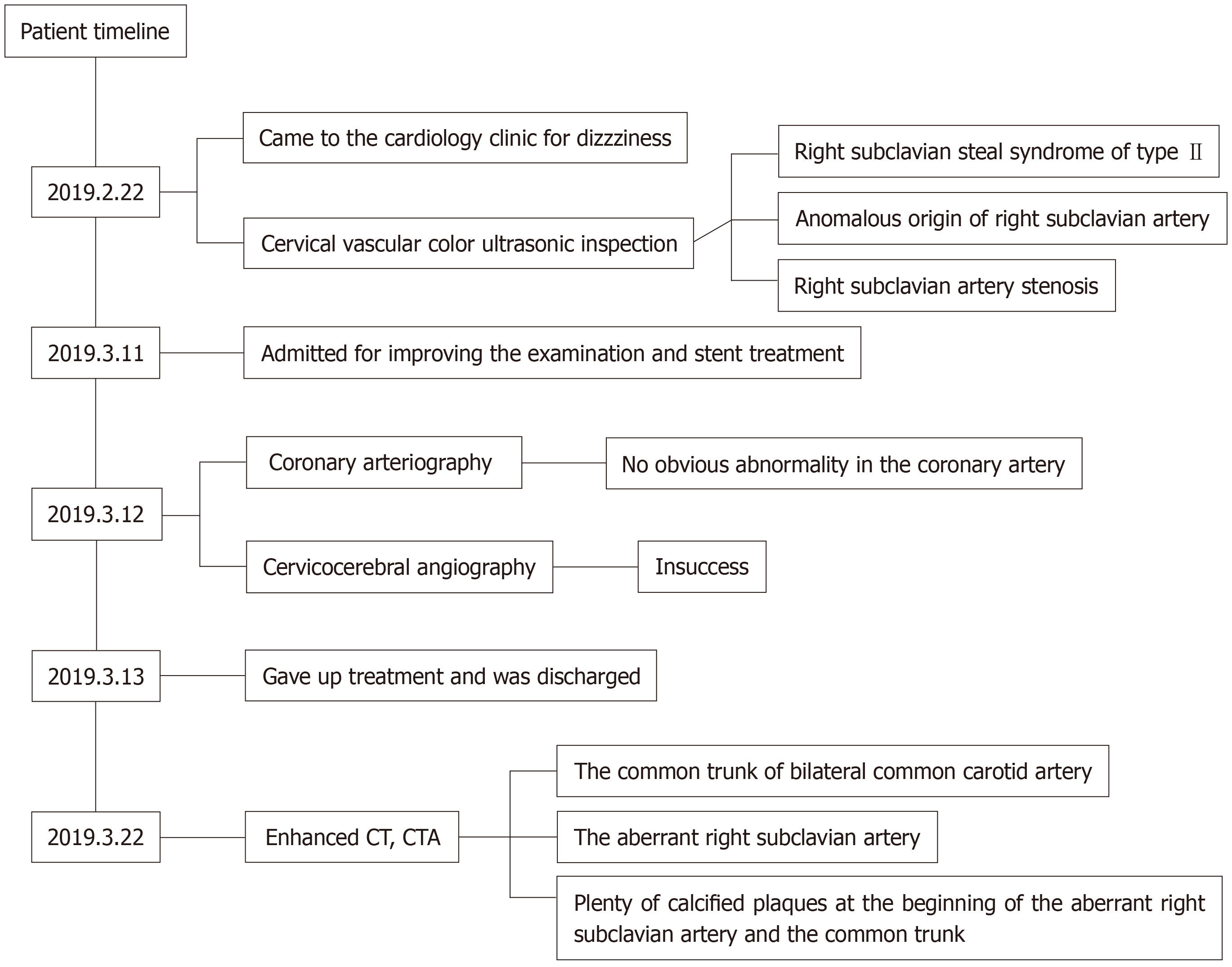
The patient underwent enhanced CT and CTA 10 d after discharge, and a final diagnosis was established. All cardiologists met to discuss the patient, analyzed the causes of cerebral angiography failure and the lessons learnt from this case. Timeline of patient diagnosis, treatment and follow-up (Figure 6).
Vertebrates have six pairs of arterial arches in the embryonic stage, and the third, fourth and sixth pairs of arterial arches are important for the formation of human blood vessels[5-7]. During the embryonic period, under normal development, degeneration and atrophy occur between the tail of the arterial arch and the right subclavian artery, and finally the arterial arch sends out three main blood vessels, namely the brachiocephalic trunk, left common carotid artery and left subclavian artery, from right to left[8,9]. When degenerative disruption occurs between the right common carotid artery and right subclavian artery, the aberrant right subclavian artery is formed[10]. The present case had a bilateral common carotid artery with aberrant right subclavian artery, which is a rare developmental deformity. This variation is caused by changes in the degenerative site of the fourth arterial arch during embryonic development, which leads to vascular malformations when the developmental process is blocked or mutation occurred[11].
The aberrant right subclavian artery may be located in the posterior esophageal pathway (80%–84%), between the trachea and esophagus (12.7%–15%) or in the anterior tracheal pathway (4.2%–5%)[12]. This anatomical variation is usually asymptomatic and therefore difficult to detect and does not require treatment. When blood vessels form an incomplete ring around them, they can compress the trachea and esophagus. Patients with dysphagia, dyspnea, chronic cough and other symptoms need surgical treatment[12,13]. Or as in this case, although there was no dysphagia, the right upper limb numbness and weakness and other symptoms also required surgical intervention, so it was necessary to reconstruct the continuity of the subclavian artery. In terms of differential diagnosis, it is easy to be confused with thoracic outlet syndrome in terms of symptoms alone, so detailed physical examination and vascular examination may help to distinguish the condition.
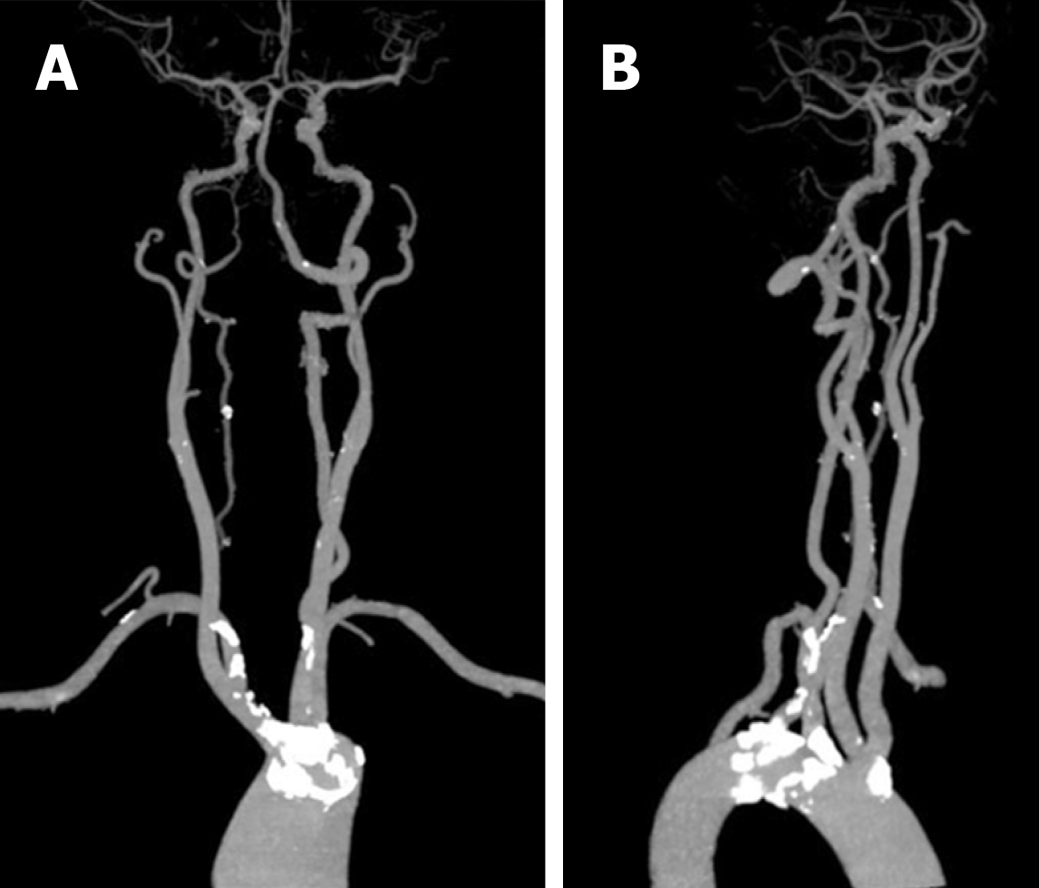
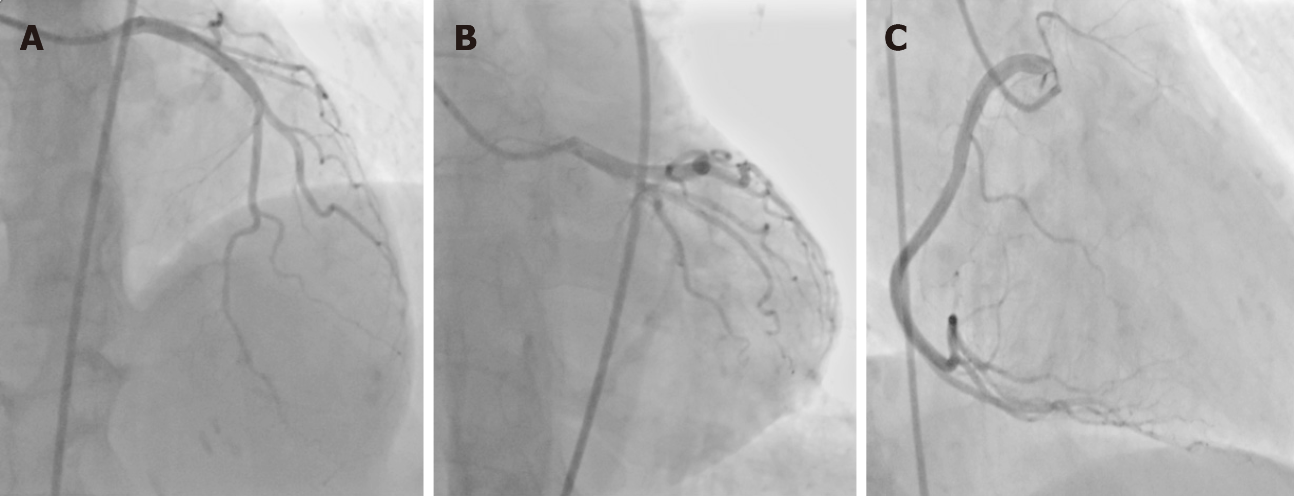
Are these two concomitant deformities associated with aberrant right subclavian steal syndrome? Is it related to the failure of cerebral angiography? First, CTA and enhanced CT showed that most of the artery wall calcification plaques were concentrated in the initial segment of the aortic branch artery, especially the aberrant right subclavian artery (Figure 7), which led to aberrant right subclavian steal syndrome. However, the patient’s aorta was in good condition, and CTA showed no stenosis in the thoracic aorta, abdominal aorta, common carotid artery and internal carotid artery, in addition to scattered small calcified plaques (Figure 8). In addition, coronary artery angiography showed no obvious stenosis in the left main, left anterior descending, left circumflex and right coronary arteries (Figure 9). Why did the aberrant right subclavian artery and common trunk of the bilateral common carotid artery show significant severe calcification with stenosis at the beginning under favorable vascular conditions? Compared with the severe stenosis of the aberrant right subclavian artery, the wall of the left subclavian artery was normal without stenosis. In fact, the bilateral subclavian arteries were close in origin and were almost symmetrical in shape with similar diameter (Figure 10).
Why were the two subclavian arteries with similar origins so different? After consulting the literature, we hypothesize that the reasons may be as follows. First, the aberrant right subclavian artery was congenital dysplasia. The arterial wall was weak in elasticity, especially at the beginning, and high blood pressure easily broke the intima[12-14]. Therefore, the blood flow impact was likely to cause intimal damage, plaque formation and then stenosis, which led to the blood steal syndrome. Second, the aberrant right subclavian artery arose directly from the aortic arch at an acute angle. According to the hemodynamic theory, there would be a greater shear force in the bloodstream[12,15]. The high blood flow rate in the aorta had a direct impact on the wall of the aberrant right subclavian artery, and the fragile and easily damaged wall caused by the congenital abnormality aggravated the damage. It was more prone to calcified plaque formation and stenosis than the other arteries were. Third, due to the coexistence of multiple morphological variants, hemodynamics were greatly changed[16,17]. Therefore, more plaques and stenosis were found at the initial segment of the abnormal vessels, while these at the beginning of the aberrant right subclavian artery were more significant. A large number of calcified plaques blocked entry of the guide wire, which was also a reason for the failure of cerebral angiography.
Bilateral common carotid artery common trunk with aberrant right subclavian artery is rare and combined with right subclavian steal syndrome is worthy of attention. By analyzing three possibilities, we hypothesized that the occurrence of blood steal syndrome of the aberrant right subclavian artery may not be accidental but rather a result from multiple malformations. This will be validated in future clinical studies. Surgeons and interventional radiologists should be aware of the possibility of these abnormalities in patients with aortic arch abnormalities before surgery; therefore, we may see more effective treatment being made available for patients in the future.
Manuscript source: Unsolicited manuscript
Specialty type: Medicine, Research and Experimental
Country of origin: China
Peer-review report classification
Grade A (Excellent): 0
Grade B (Very good): B
Grade C (Good): C
Grade D (Fair): 0
Grade E (Poor): 0
P-Reviewer: Akdeniz B, Kaypaklı O S-Editor: Zhang L L-Editor: Filipodia E-Editor: Qi LL
| 1. | Connolly PH, Meltzer AJ, Gallagher KA, Dayal R. A visceral aortic branch anomaly presenting with a common celiomesenteric-renal trunk. J Vasc Surg. 2013;57:1675. [RCA] [PubMed] [DOI] [Full Text] [Cited by in Crossref: 4] [Cited by in RCA: 4] [Article Influence: 0.3] [Reference Citation Analysis (0)] |
| 2. | Hara K, Yasuhara T, Maki M, Matsukawa N, Yu G, Xu L, Tambrallo L, Rodriguez NA, Stern DM, Yamashima T, Buccafusco JJ, Kawase T, Hess DC, Borlongan CV. Anomaly in aortic arch alters pathological outcome of transient global ischemia in Rhesus macaques. Brain Res. 2009;1286:185-191. [RCA] [PubMed] [DOI] [Full Text] [Full Text (PDF)] [Cited by in Crossref: 4] [Cited by in RCA: 4] [Article Influence: 0.3] [Reference Citation Analysis (0)] |
| 3. | Dumfarth J, Chou AS, Ziganshin BA, Bhandari R, Peterss S, Tranquilli M, Mojibian H, Fang H, Rizzo JA, Elefteriades JA. Atypical aortic arch branching variants: A novel marker for thoracic aortic disease. J Thorac Cardiovasc Surg. 2015;149:1586-1592. [RCA] [PubMed] [DOI] [Full Text] [Cited by in Crossref: 66] [Cited by in RCA: 86] [Article Influence: 8.6] [Reference Citation Analysis (0)] |
| 4. | Zhang J, Guileyardo JM, Roberts WC. Origin of the left subclavian artery as the first branch and origin of the right subclavian artery as the fourth branch of the aortic arch with crisscrossing posterior to the common carotid arteries. Proc (Bayl Univ Med Cent). 2016;29:423. [RCA] [PubMed] [DOI] [Full Text] [Cited by in Crossref: 2] [Cited by in RCA: 2] [Article Influence: 0.3] [Reference Citation Analysis (0)] |
| 5. | Cummings MS, Kuo BT, Ziada KM. A rare anomaly of the aortic arch: aberrant right subclavian artery associated with common carotid trunk. J Invasive Cardiol. 2011;23:E241-E243. [PubMed] |
| 6. | Cetin I, Varan B, Orün UA, Tokel K. Common trunks of the subclavian and the vertebral arteries: presentation of a new aortic arch anomaly. Ann Vasc Surg. 2009;23:142-143. [RCA] [PubMed] [DOI] [Full Text] [Cited by in Crossref: 3] [Cited by in RCA: 4] [Article Influence: 0.3] [Reference Citation Analysis (0)] |
| 7. | Yamashiro S, Kuniyoshi Y, Arakaki K, Inafuku H, Morishima Y, Kise Y. Total arch replacement with associated anomaly of the left vertebral artery. Ann Thorac Cardiovasc Surg. 2010;16:216-219. [PubMed] |
| 8. | Dhoble A, Dewar J, Szerlip M, Abidov A. Rare coronary anomaly: common origin of major coronary arteries from the right sinus of Valsalva and a small ramus branch originating from the left sinus of Valsalva. Int J Cardiol. 2012;158:e3-e4. [RCA] [PubMed] [DOI] [Full Text] [Cited by in Crossref: 2] [Cited by in RCA: 4] [Article Influence: 0.3] [Reference Citation Analysis (0)] |
| 9. | Prifti E, Crucean A, Bonacchi M, Bernabei M, Leacche M, Murzi B, Bartolozzi F, Vanini V. Postoperative outcome in patients with anomalous origin of one pulmonary artery branch from the aorta. Eur J Cardiothorac Surg. 2003;24:21-27. [RCA] [PubMed] [DOI] [Full Text] [Cited by in Crossref: 46] [Cited by in RCA: 40] [Article Influence: 1.8] [Reference Citation Analysis (0)] |
| 10. | Coskun KO, Bairaktaris A, Coskun ST, El Arousy M, AminParsa M, Koertke H, Koerfer R. Aortico-left ventricular tunnel, an unusual congenital cardiac anomaly in adult: application of a new operative technique. ASAIO J. 2006;52:e40-e42. [RCA] [PubMed] [DOI] [Full Text] [Cited by in Crossref: 2] [Cited by in RCA: 3] [Article Influence: 0.2] [Reference Citation Analysis (0)] |
| 11. | Roughneen PT, Al-Dossari GA. The Anomalous Coronary Artery in Aortic Valve Replacement: A Case For Caution. Ann Thorac Surg. 2016;102:e113-e115. [RCA] [PubMed] [DOI] [Full Text] [Cited by in Crossref: 1] [Cited by in RCA: 1] [Article Influence: 0.1] [Reference Citation Analysis (0)] |
| 12. | Myers PO, Fasel JH, Kalangos A, Gailloud P. Arteria lusoria: developmental anatomy, clinical, radiological and surgical aspects. Ann Cardiol Angeiol (Paris). 2010;59:147-154. [RCA] [PubMed] [DOI] [Full Text] [Cited by in Crossref: 77] [Cited by in RCA: 99] [Article Influence: 6.2] [Reference Citation Analysis (0)] |
| 13. | Kouchoukos NT, Masetti P. Aberrant subclavian artery and Kommerell aneurysm: surgical treatment with a standard approach. J Thorac Cardiovasc Surg. 2007;133:888-892. [RCA] [PubMed] [DOI] [Full Text] [Cited by in Crossref: 103] [Cited by in RCA: 112] [Article Influence: 6.2] [Reference Citation Analysis (0)] |
| 14. | Singh S, Grewal PD, Symons J, Ahmed A, Khosla S, Arora R. Adult-onset dysphagia lusoria secondary to a dissecting aberrant right subclavian artery associated with type B acute aortic dissection. Can J Cardiol. 2008;24:63-65. [RCA] [PubMed] [DOI] [Full Text] [Cited by in Crossref: 10] [Cited by in RCA: 15] [Article Influence: 0.9] [Reference Citation Analysis (0)] |
| 15. | Nasir A, Jadoon M, Ellis PK, Graham AN. Kommerell's diverticulum, risk factor for aortic dissection. J Card Surg. 2009;24:463. [RCA] [PubMed] [DOI] [Full Text] [Cited by in Crossref: 5] [Cited by in RCA: 5] [Article Influence: 0.3] [Reference Citation Analysis (0)] |
| 16. | Vistarini N, Aubert S, Gandjbakhch I, Bonnet N. Aberrant subclavian artery as origin of aortic dissection. Eur J Cardiothorac Surg. 2008;34:1109. [RCA] [PubMed] [DOI] [Full Text] [Cited by in Crossref: 6] [Cited by in RCA: 4] [Article Influence: 0.2] [Reference Citation Analysis (0)] |
| 17. | Goltz JP, Bastürk P, Hoppe H, Triller J, Kickuth R. Emergency and elective implantation of covered stent systems in iatrogenic arterial injuries. Rofo. 2011;183:618-630. [RCA] [PubMed] [DOI] [Full Text] [Cited by in Crossref: 10] [Cited by in RCA: 7] [Article Influence: 0.5] [Reference Citation Analysis (0)] |


