Published online Oct 26, 2019. doi: 10.12998/wjcc.v7.i20.3335
Peer-review started: June 19, 2019
First decision: September 9, 2019
Revised: September 21, 2019
Accepted: September 25, 2019
Article in press: September 25, 2019
Published online: October 26, 2019
Processing time: 134 Days and 16.4 Hours
Acute chest pain (ACP) is very common among patients presenting to emergency departments. Nevertheless, ACP caused by esophageal foreign body is relatively rarely reported.
A 56-year-old man suffering from chest pain (increased pain for the last 9 h) was admitted to our hospital on October 25, 2015. After undergoing physical examination and laboratory blood testing, he was diagnosed with acute anterior myocardial infarction. Consequently, the patient underwent emergency percutaneous coronary angiography; however, no myocardial infarction signs were observed. Later on, the patient experienced respiration failure and therefore was transferred to intensive care unit. Cardiac ultrasound showed pericardial effusion, which was considered as the cause of shock. He then underwent pericardium puncture drainage and the circulation temporarily improved. Nevertheless, persistent pericardial bleeding, unclear bleeding causes, and clot formation induced poor drainage led to worsening of cardiac tamponade symptoms. Consequently, the patient underwent emergency exploratory thoracotomy, which revealed a fish bone causing pericardial bleeding. The bone was removed, and the damaged blood vessels were mended. Eventually, the patient was discharged in good clinical condition.
For patients with chest pain, it is necessary to consider the possibility of foreign body in the esophagus or even in the heart. Careful history taking and the corresponding inspection can help to avoid unnecessary damage and safeguard patients from unnecessary pain.
Core tip: Chest pain is a symptom causing more attention by clinicians, and the common reasons include acute coronary events, aortic dissection, pulmonary embolism and so on, while that caused by esophageal foreign body is relatively rare. We present herein, a rare case of acute chest pain as the chief complaint that was diagnosed as acute myocardial infarction initially, then developed cardiac tamponade and severe shock, and was finally diagnosed as fish bone-induced myocardial injury by surgery. This case highlights the possibility of foreign body in the esophagus or even in the heart for patients with acute chest pain.
- Citation: Wang QQ, Hu Y, Zhu LF, Zhu WJ, Shen P. Fish bone-induced myocardial injury leading to a misdiagnosis of acute myocardial infarction: A case report. World J Clin Cases 2019; 7(20): 3335-3340
- URL: https://www.wjgnet.com/2307-8960/full/v7/i20/3335.htm
- DOI: https://dx.doi.org/10.12998/wjcc.v7.i20.3335
Chest pain is the early symptom of numerous life-threatening disease processes. The most common causes of acute chest pain (ACP) are acute coronary events (acute myocardial infarction or angina), aortic dissection, and pulmonary embolism. ACP caused by esophageal foreign body is relatively rare[1]. Most patients with ACP are accurately diagnosed and promptly treated based on the individual histories, blood biochemical tests, or imaging examination. Herein, we report the case of a patient with ACP as the chief complaint that was ultimately diagnosed as fish bone-induced myocardial injury.
A 56-year-old man suffering from chest pain presented at the Emergency Department of our hospital on October 25, 2015. The patient’s symptoms lasted for nine hours with persistent episodes of chest pain, which have especially worsened over the last 2 h.
The patient had no previous medical history.
None.
The patient’s temperature was 36.6 °C, heart rate was 120 bpm, respiratory rate was 23 breaths per minute, blood pressure was 97/70 mmHg, and the oxygen saturation in room air was 98%. Heart sounds were very faint and rales were heard in both lungs. All other signs were negative.
The patient’s blood tests showed elevations in cardiac troponin-I (CTN-I) (0.34 ng/mL; normal: < 0.05 ng/mL). Electrocardiograph (ECG) revealed ST segment elevation in the precordial leads II, aVF, and V4–V6. No abnormalities were observed on chest computed tomography (CT). Acute anterior myocardial infarction was thus initially considered.
The patient received dual antiplatelet agents comprising clopidogrel and aspirin at load dosages and underwent emergency percutaneous coronary angiography under the application of nitrates. The operation showed no stenosis in the left main coronary artery or right coronary artery, and about 50% stenosis in the ostial left anterior descending segment and left circumflex artery.
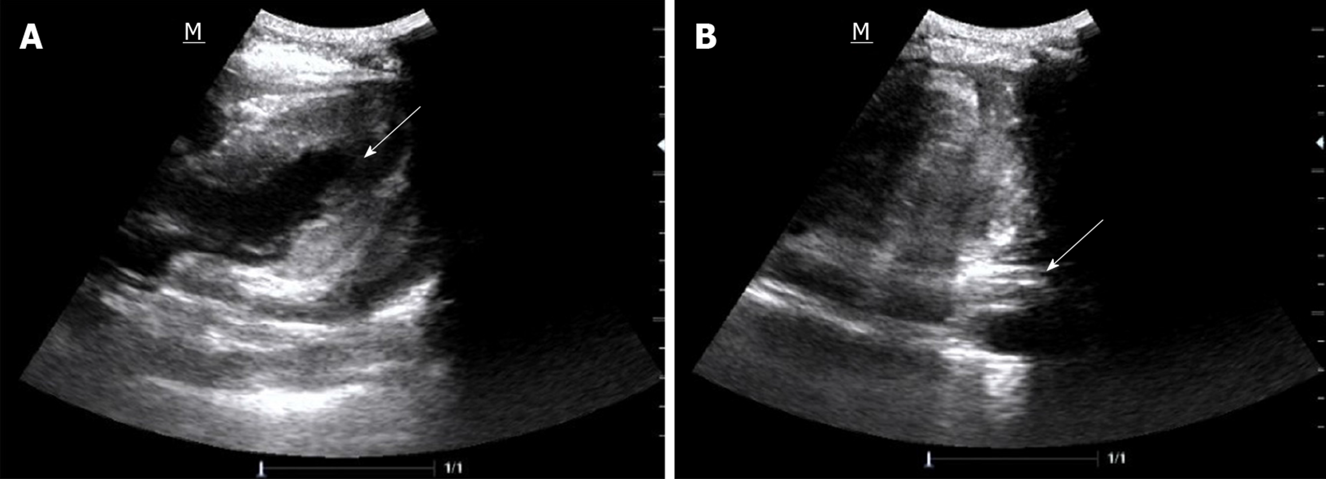
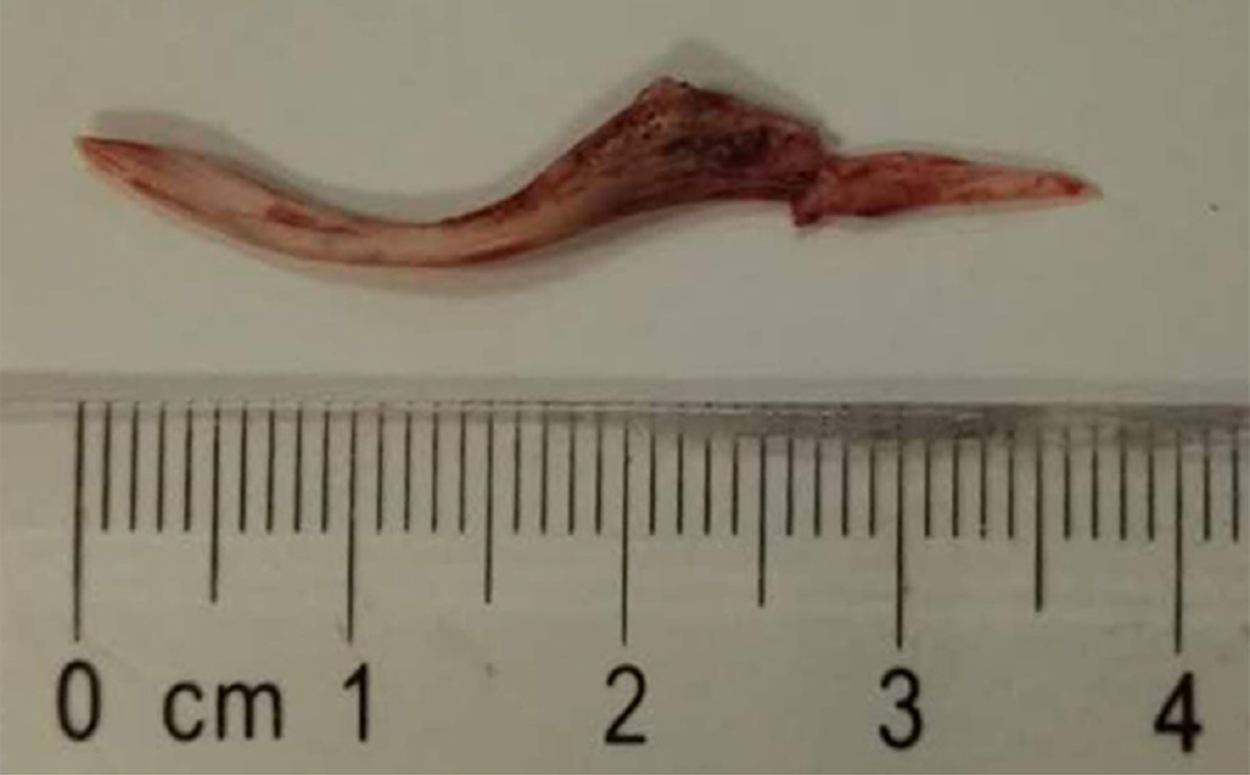
Considering that the lowest blood pressure was 74/40 mmHg and it was not possible to maintain normal blood pressure with the help of dopamine, the patient was transferred to intensive care unit (ICU) for further analysis. Subsequently, he required high doses of norepinephrine to maintain blood pressure. Follow-up bedside cardiac ultrasound showed pericardial effusion (Figure 1A). Pericardial tamponade was considered. The patients then underwent pericardium puncture drainage with some 220 mL pericardial effusion; consequently, the blood pressure increased to 110/75 mmHg, while the CTN-I increased to 2.29 ng/mL. At 18:24 (see Table 1), the symptom of chest pain worsened and the blood pressure was decreasing. Bedside cardiac ultrasound revealed a large number of pericardial blood clots prompting cardiac tamponade (Figure 1B). Our medical team including cardiology, ICU, and thoracic surgery, taking into consideration the unexplained hemopericardium, decided to preform emergency thoracotomy and pericardium exploratory surgery with the prior consent from patients’ relatives at 19:57. During the operation, the sharp foreign body, a long fish bone measuring about 3.7 cm (Figure 2), was found at the surface of the pericardium and penetrating the left ventricular myocardium to some blood vessels.
| Date | Time | Event |
| 10.24 | 19:00 | Acute chest pain |
| 10.24-10.25 | 19:00-04:00 | Local hospital |
| 10.25 | 04:00-06:00 | Emergency Department of our hospital |
| 06:00-07:17 | Emergency percutaneous coronary angiography | |
| 07:17-11:05 | Progressive circulation and respiration failure | |
| 11:05 | Transferred to Intensive Care Unit | |
| 12:00 | Pericardiocentesis | |
| 12:00-18:24 | Progressive cardiac tamponade | |
| 19:57-20:55 | Emergency thoracotomy and pericardium exploratory surgery | |
| 20:55 | Return to Intensive Care Unit | |
| 20:55-23:30 | Hemodynamic stabilization | |
| 10.28 | Extubated tracheal intubation | |
| 10.29 | Transferred to Thoracic Surgery Department | |
| 11.12 | Discharged |
Finally, the patient was diagnosed with fish bone-induced myocardial injury.
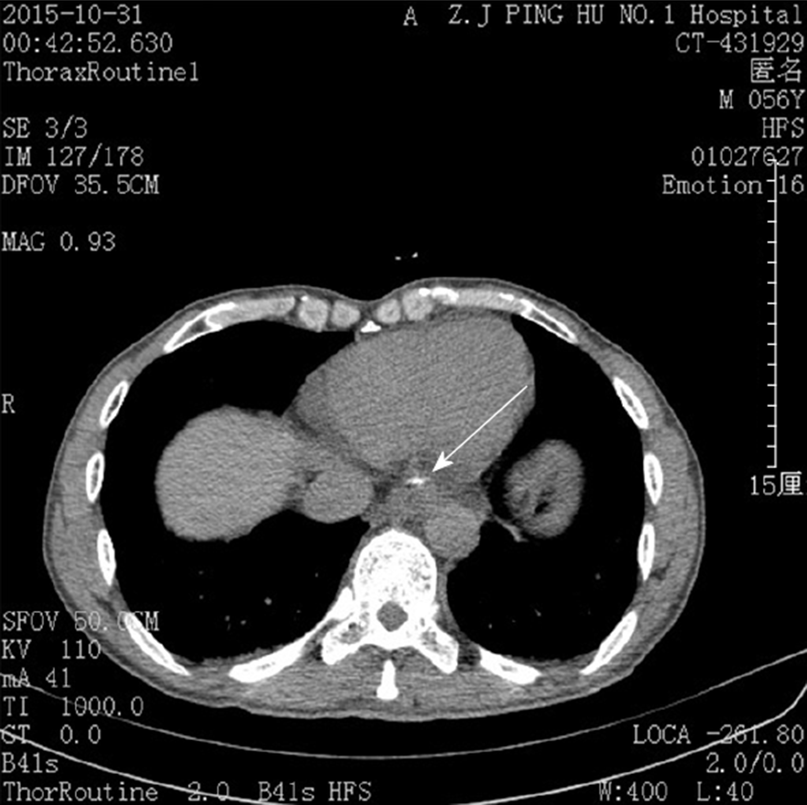
After removing the fish bone and mending the damaged blood vessels, the patient returned to ICU at 20:55. Reviewing the initial chest CT at local hospital (Figure 3), a strip-shaped high-density shadow at the bottom of the esophagus was found. After being repeatedly inquired of the medical history, the patient admitted to eating fish at dinner, but could not recall swallowing a fish bone.
The postoperative course was uneventful and CTN-I fell to 0.25 ng/mL. The patient was successfully extubated on day 3, transferred to thoracic surgery department next day, and discharged in good clinical condition after half a month.
Acute myocardial infarction or angina is the most common cause of ACP, which is mainly diagnosed by using ECG and blood biochemical test[2]. In our case, the patient was misdiagnosed with acute myocardial infarction. His emergency percutaneous coronary angiography was negative. Bedside cardiac ultrasound showed pericardial effusion and pericardium puncture drainage improved the circulation. Nevertheless, persistent pericardial bleeding, unclear bleeding causes, and clot formation led to poor drainage, eventually resulting in worsening of cardiac tamponade symptoms. Considering the possibility of coronary artery injury and lack of the capability of emergency coronary artery bypass at our hospital, our medical team was highly concerned that thoracotomy hemostasis would significantly increase the risk for the patient, while he was reaching the condition when it was no longer safe for him to be transferred to another hospital. We conferred with the patient’s family and obtained their consent for emergency exploratory thoracotomy. Finally, a diagnosis of fish bone leading to pericardial bleeding was confirmed.
Our patient was extremely lucky to survive this penetrating heart injury. Actually, in retrospect to the whole treatment, there are still many aspects worth reflecting on: First, the primary clinician failed to adequately and carefully examine the patient’s history. The existing literature shows that ACP caused by ingested fish bone resulting in esophageal or cardiac injury is not relatively uncommon[3-8]. Foreign body ingestion is an important diagnostic clue. Thus, the diagnostic challenge we faced with this patient was that he could not recall swallowing a fish bone, which partly contributed to the initial misdiagnosis. Second, chest CT examination is the preferred technique for suspected esophageal foreign body[9]. In this case, the patient underwent CT examination at a local hospital; however, the clinicians failed to observe a high-density shadow in the lower esophagus. Moreover, the primary clinician at our hospital did not personally read the chest CT, which further contributed to the misdiagnosis. Finally, we failed to timely recheck chest CT, cardiac ultrasound and so on after negative results of emergency coronary angiography, which led to further deterioration of the patient’s condition.
Therefore, for patients with chest pain, it is necessary to consider the possibility of foreign body in the esophagus or even in the heart. Careful history taking and the corresponding inspection can help to avoid unnecessary damage and safeguard patients from unnecessary pain.
Foreign body is a very rare cause of chest pain. Careful history taking and the corresponding inspection can help to avoid unnecessary damage and safeguard patients from unnecessary pain.
Manuscript source: Unsolicited manuscript
Specialty type: Medicine, Research and Experimental
Country of origin: China
Peer-review report classification
Grade A (Excellent): 0
Grade B (Very good): B
Grade C (Good): 0
Grade D (Fair): 0
Grade E (Poor): 0
P-Reviewer: Firstenberg MS S-Editor: Zhang L L-Editor: Wang TQ E-Editor: Liu JH
| 1. | Goodacre S, Cross E, Arnold J, Angelini K, Capewell S, Nicholl J. The health care burden of acute chest pain. Heart. 2005;91:229-230. [RCA] [PubMed] [DOI] [Full Text] [Cited by in Crossref: 292] [Cited by in RCA: 333] [Article Influence: 16.7] [Reference Citation Analysis (0)] |
| 2. | Fothergill NJ, Hunt MT, Touquet R. Audit of patients with chest pain presenting to an accident and emergency department over a 6-month period. Arch Emerg Med. 1993;10:155-160. [RCA] [PubMed] [DOI] [Full Text] [Cited by in Crossref: 27] [Cited by in RCA: 32] [Article Influence: 1.0] [Reference Citation Analysis (0)] |
| 3. | Hokama A, Uechi K, Takeshima E, Kobashigawa C, Iraha A, Kinjo T, Kishimoto K, Kinjo F, Fujita J. A fish bone perforation of the esophagus. Endoscopy. 2014;46 Suppl 1:E216-E217. [RCA] [PubMed] [DOI] [Full Text] [Cited by in Crossref: 4] [Cited by in RCA: 5] [Article Influence: 0.5] [Reference Citation Analysis (0)] |
| 4. | Kunishige H, Myojin K, Ishibashi Y, Ishii K, Kawasaki M, Oka J. Perforation of the esophagus by a fish bone leading to an infected pseudoaneurysm of the thoracic aorta. Gen Thorac Cardiovasc Surg. 2008;56:427-429. [RCA] [PubMed] [DOI] [Full Text] [Cited by in Crossref: 15] [Cited by in RCA: 16] [Article Influence: 0.9] [Reference Citation Analysis (0)] |
| 5. | Sharland MG, McCaughan BC. Perforation of the esophagus by a fish bone leading to cardiac tamponade. Ann Thorac Surg. 1993;56:969-971. [RCA] [PubMed] [DOI] [Full Text] [Cited by in Crossref: 47] [Cited by in RCA: 41] [Article Influence: 1.3] [Reference Citation Analysis (0)] |
| 6. | Kawakami M, Chigira H, Nitta S, Sato H, Takahashi H, Nakada T. Pyopneumothorax due to perforation of the esophagus with an ingested fish bone. A case report. Sci Rep Res Inst Tohoku Univ Med. 1978;25:1-4. [PubMed] |
| 7. | Jougon J, Minniti A, Moralès P, Laurent F, Velly JF. Retroesophageal hematoma caused by fish bone perforation of the esophagus. Asian Cardiovasc Thorac Ann. 2002;10:280-281. [RCA] [PubMed] [DOI] [Full Text] [Cited by in Crossref: 4] [Cited by in RCA: 5] [Article Influence: 0.2] [Reference Citation Analysis (0)] |
| 8. | Chen MF, Lou XJ, Yu XQ, Hu DL, Lu J, Jin HW, Ji ZZ, He HX. One case of a fish bone penetrating the esophageal wall into the heart. Zhonghua Xiaohua Neijing Zazhi. 2012;29:73-73. [DOI] [Full Text] |
| 9. | Birk M, Bauerfeind P, Deprez PH, Häfner M, Hartmann D, Hassan C, Hucl T, Lesur G, Aabakken L, Meining A. Removal of foreign bodies in the upper gastrointestinal tract in adults: European Society of Gastrointestinal Endoscopy (ESGE) Clinical Guideline. Endoscopy. 2016;48:489-496. [RCA] [PubMed] [DOI] [Full Text] [Cited by in Crossref: 274] [Cited by in RCA: 379] [Article Influence: 42.1] [Reference Citation Analysis (0)] |


