Published online Oct 26, 2019. doi: 10.12998/wjcc.v7.i20.3276
Peer-review started: April 17, 2019
First decision: June 10, 2019
Revised: August 13, 2019
Accepted: August 27, 2019
Article in press: August 27, 2019
Published online: October 26, 2019
Processing time: 192 Days and 1.9 Hours
Complications associated with upper gastrointestinal (UGI) endoscopy are uncommon, and rarely involve those of cardiovascular nature. We report herein a unique case of spontaneous superior mesenteric artery dissection (SMAD) after UGI pandenoscopy.
A 45-year-old man who had previously undergone UGI panendoscopy and colonoscopy during a voluntary health check-up at our facility was admitted to the emergency room (ER) at the same facility due to persistent epigastric pain with radiation to the back. At the ER, the patient did not present notable abnormalities upon physical, conscious, or laboratory examinations apart from mild tenderness in the epigastric abdomen. Acute abdominal aortic dissection was suspected, and abdominal contrast-enhanced computed tomography confirmed SMAD. He was then transferred to the cardiovascular ward and treated conservatively with fasting, prostaglandin E1, and aspirin. The patient recovered and returned home soon after, and was symptom-free 6 months after discharge from the facility.
SMAD after UGI panendoscopic procedure is a previously unreported complication. Awareness of this complication and associated sequela is warranted.
Core tip: Although upper gastrointestinal panendoscopy is relatively safe and complications are uncommon, we encountered herein a rare case of post-procedure spontaneous superior mesenteric artery dissection (SMAD) in a middle-aged man. After panendoscopy, the patient experienced abdominal pain extending to back immediately and persisting for two weeks. Upon examination, there were no obvious physical or laboratory signs of organ perforation and peritonitis. SMAD was suspected and confirmed by contrast-enhanced CT of the abdomen. While the patient recovered without sequelae after being treated conservatively in the cardiovascular ward, SMAD is a potentially lethal condition and awareness of its association with panendoscopy is warranted.
- Citation: Ou Yang CM, Yen YT, Chua CH, Wu CC, Chu KE, Hung TI. Spontaneous superior mesenteric artery dissection following upper gastrointestinal panendoscopy: A case report and literature review. World J Clin Cases 2019; 7(20): 3276-3281
- URL: https://www.wjgnet.com/2307-8960/full/v7/i20/3276.htm
- DOI: https://dx.doi.org/10.12998/wjcc.v7.i20.3276
Upper gastrointestinal (UGI) panendoscopy is a procedure that allows a clear view of the mucosal layer of the esophagus, stomach, and proximal duodenum. Patients with upper abdominal symptoms including pain, dyspepsia, regurgitation, or bleeding suggestive of reflux, ulcers, tumors, or vascular abnormalities in the UGI are indicated for diagnostic panendoscopy examinations[1]. Although panendoscopy performed in the UGI carries a low risk of complications, the adverse complication rates of UGI panendoscopy have been reported to be 1 in 200 to 1 in 10000 while the mortality rates between none to 1 in 2000 by several large-series studies[2-5]. Although major adverse complications related to diagnostic UGI panendoscopy are rare, they can include potentially life-threatening cardiopulmonary adverse events, infection, mechanical perforation, and bleeding[6]. An estimated cardiopulmonary event rate of 1 in 170 and a mortality rate of 1 in 10000 from among 140000 UGI endoscopic procedures were suggested by data collected from the Clinical Outcomes Research Initiative database[7]. Risk factors for complications associated with panendoscopy include pre-existing disease of cardiopulmonary or systemic nature, advanced age, and frailty[6]. Other than clinical signs suggesting of adverse events, patients can at times experience abdominal discomfort after gastrointestinal (GI) endoscopic procedures, and this is usually treated as a mild complication rather than an early symptom of more severe conditions. Spontaneous superior mesenteric artery dissection (SMAD) is an uncommon cause of abdominal pain and has never been described to associate with UGI endoscopy in the medical literature. We report herein a unique case presenting with acute abdominal symptoms after UGI endoscopy procedure who was later diagnosed with SMAD, possibly as a complication. The patient was managed promptly after diagnosis and recovered well. Isolated spontaneous SMAD is a rare disorder, and only 106 cases were detailed in the medical literature[8]. Despite the rarity, differential diagnosis of acute abdominal pain after UGI panendoscopy should include SMAD, which is a treatable condition that can lead to detrimental sequela when left unattended.
A 45-year-old man presented to the emergency department of our hospital with a chief complaint of epigastric pain with radiation to the back.
The patient experienced persistent abdominal pain for two weeks, starting from UGI panendoscopic and colonoscopic procedures performed at our facility. The pain had persisted for 2 wk before he returned to our hospital for further examination.
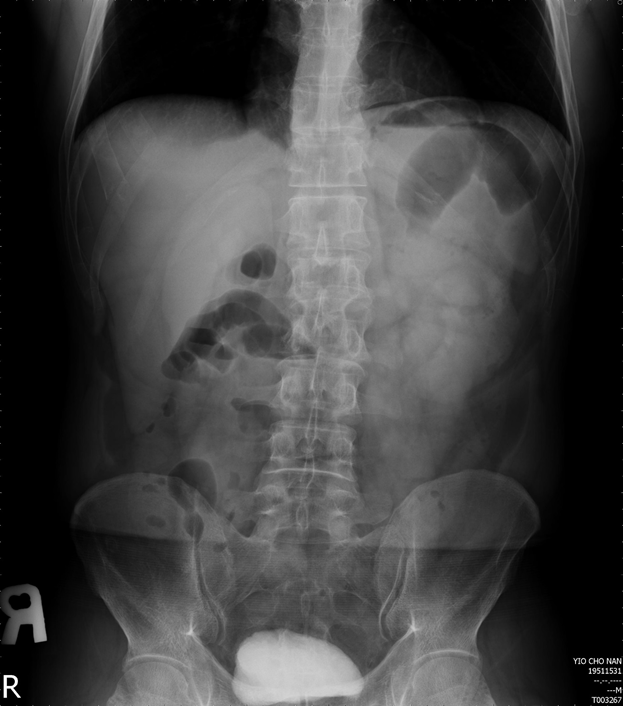
The patient had a history of untreated hypertension and hyperlipidemia. He experienced immediate abdominal pain after endoscopic procedures performed during a voluntary health checkup and brought this to our attention while resting in the recovery room. Four hours later, plain abdominal standing X-ray was taken and an apparently normal small bowel ileus pattern without subphrenic free air was found (Figure 1). Consequently, the patient was treated conservatively using menthol packing of his abdomen and allowed to go home in Tai Tung, which was in the eastern part of Taiwan, about 5 h drive from our facility. During the following 2 wk, he experienced persistent abdominal symptoms and sought help from several local clinics/hospitals, where he received conservative treatments mainly for gastritis or enteritis. As the discomfort did not improve, the patient contacted us for help 14 d after the initial endoscopic procedures and was transferred to our emergency room (ER) for further management.
At the ER, the patient complained of persistent epigastric pain radiating to the back with abdominal distension for the past 2 wk, but appeared otherwise easy-looking. There was no fever or chillness, and his vital signs were normal. Physical examination of the abdomen revealed mild epigastric tenderness without any signs suggestive of peritonitis. Soft and mild distended abdomen was found while no muscle guarding or board-like abdomen was observed. Since mechanical hollow organ perforation and peritonitis were both ruled out after thorough examination of the patient, acute abdominal aortic dissection was suspected.
Laboratory examination performed at the ER included complete blood count and biochemistry examinations. All data were within normal limits.
After thorough physical and laboratory examinations upon returning to ER, the patient was subjected to contrast-enhanced computed tomography (CT) of the abdomen. Isolated spontaneous SMAD was revealed (Figure 2). The dissection began from just below the orifice of the superior mesenteric artery with intra-mural hematoma and without stenosis. Although the intra-mural hematoma compressed the true lumen, there was no suggestive signs of bowel ischemia, bowel thickening, or ascites.
The patient was diagnosed with Type III SMAD as a complication of UGI panendoscopy.
Based on the diagnosis, the patient was admitted to our cardiovascular ward, where conservative treatments with blood pressure control, fasting, vasodilation (prostaglandin E1, 60 μg in 500 mL of 5% dextrose in 0.9% normal saline, 40 mL/h continuous infusion for 4 d) and antiplatelet agent (aspirin 100 mg q.d. for 4 d) were given.
The patient’s abdominal symptoms and back pain markedly improved on the subsequent day following admittance, and oral intake was initiated 2 d later. He was discharged from the hospital 4 d after admittance in stable condition. The patient was symptom-free 6 mo after discharge.
Up to date, UGI endoscopy related spontaneous SMAD relevant to the current case has never been reported in the medical literature. The clinical presentations of spontaneous SMAD are vast and wide, ranging from asymptomatic incidental finding to acute catastrophic bowel ischemia or aneurysmal spontaneous SMAD, and can only be correctly recognized upon CT examination. The initial differential diagnosis of acute abdominal discomfort after GI endoscopic procedures must first rule out hollow organ perforation. For the patient presented herein, subphrenic free air was not found upon standing plain abdominal X-ray taken 4 h after the examination, and thus received conservative menthol packing. However, due to the severity and persistence of abdominal pain in the face of minimal abdominal findings when the patient returned to our ER 2 wk later, conditions of vascular origin were suspected and examined. The pathophysiology of spontaneous SMAD is not yet clear, though risk factors include hypertension, connective tissue disorders, vasculitis, atherosclerosis, and trauma to the aorta[9]. High blood pressure (140-146/86-90) and hyperlipidemia were found in this patient during his stay in our health screening clinic, although he had no prior history of relevant medication. Furthermore, no spiking in blood pressure during the conscious sedation endoscopic procedures was found when we retrospectively reviewed his blood pressure records. The endoscopist who performed the procedure is well-experienced (> 10 years) and had performed about 1000 cases of UGI panendoscopy per year. Solis et al[10] hypothesized that dissections are likely caused by stress on the anterior wall of the artery. Thus, this hypothesis corresponds with the location of dissection in the currently presented case, where gaseous insufflation of the duodenum during panendoscopy may have provided the source of pressure on the anterior wall of the superior mesenteric artery. In any case, immediate presentation of the patient’s abdominal pain and direct association of spontaneous SMAD after the panendoscopy was not available as the diagnosis was confirmed two weeks later at the ER.
Accurate diagnosis of SMAD relies on abdominal CT, and treatment modalities are usually based on CT findings together with the clinical presentation of the disease. Sakamoto et al[11] categorised SMAD into four types according to the CT imaging features: Type I involves a patent false lumen with both entry and re-entry; Type II involves a "cul-de-sac"-shaped false lumen without re-entry; Type III involves a thrombotic false lumen with an ulcer-like projection; and Type IV involves a completely thrombotic false lumen with an ulcer-like projection. According to the type of SMAD, conservative treatment is recommended for Types I and IV while long-term follow-up should be given for Types II and III due to the possibility of bowel necrosis or rupture. Our patient presented a Type III SMAD and he was treated with fasting, blood pressure control, and antiplatelet therapy. In addition, prostaglandin E1 as a vasodilator was administered as suggested by Totsugawa et al[12]. Despite the full recovery of the patient presented in the current report, failure in medical management can occur in 10%-45% of patients. Patients who develop signs of worsening intestinal ischemia disregarding initial medical management may require aggressive abdominal exploration to evaluate the bowel and resect nonviable segments[13]. On the other hand, around 16%-34% of spontaneous SMAD cases appear asymptomatic and most of them were discovered incidentally[9,14,15]. Single-centered and systematic studies suggest that most asymptomatic spontaneous SMAD have a benign disease course and can be conservatively treated[9,14,15]. In the case reported herein, it is difficult to determine whether the patient experienced a direct procedural complication or an exacerbation of pre-existing asymptomatic SMAD secondary to panendoscopy. In any case, panendoscopy is useful for screening individuals at high risks for UGI cancers[1] and also an elective examination of adult voluntary health check-ups, but the presence of an appropriate indication and associated contraindication should always be thoroughly evaluated prior to procedure[16].
SMAD is a rare condition that requires specific treatment modalities depending on disease presentation. Failure to recognize the condition and treat correctly can lead to catastrophic consequences. We hope with the presentation of this unique case of UGI panendoscopy-associated SMAD, awareness of possible complications with vascular-origin will increase when treating patients with acute abdominal symptoms after endoscopic treatments.
Manuscript source: Unsolicited manuscript
Specialty type: Medicine, research and experimental
Country of origin: Taiwan
Peer-review report classification
Grade A (Excellent): 0
Grade B (Very good): 0
Grade C (Good): C, C
Grade D (Fair): 0
Grade E (Poor): 0
P-Reviewer: Lieto E, Cianci P S-Editor: Tang JZ L-Editor: Wang TQ E-Editor: Wu YXJ
| 1. | ASGE Standards of Practice Committee. Early DS, Ben-Menachem T, Decker GA, Evans JA, Fanelli RD, Fisher DA, Fukami N, Hwang JH, Jain R, Jue TL, Khan KM, Malpas PM, Maple JT, Sharaf RS, Dominitz JA, Cash BD. Appropriate use of GI endoscopy. Gastrointest Endosc. 2012;75:1127-1131. [RCA] [PubMed] [DOI] [Full Text] [Cited by in Crossref: 156] [Cited by in RCA: 179] [Article Influence: 13.8] [Reference Citation Analysis (0)] |
| 2. | Silvis SE, Nebel O, Rogers G, Sugawa C, Mandelstam P. Endoscopic complications. Results of the 1974 American Society for Gastrointestinal Endoscopy Survey. JAMA. 1976;235:928-930. [RCA] [PubMed] [DOI] [Full Text] [Cited by in Crossref: 89] [Cited by in RCA: 138] [Article Influence: 2.8] [Reference Citation Analysis (0)] |
| 3. | Quine MA, Bell GD, McCloy RF, Charlton JE, Devlin HB, Hopkins A. Prospective audit of upper gastrointestinal endoscopy in two regions of England: safety, staffing, and sedation methods. Gut. 1995;36:462-467. [RCA] [PubMed] [DOI] [Full Text] [Cited by in Crossref: 249] [Cited by in RCA: 239] [Article Influence: 8.0] [Reference Citation Analysis (0)] |
| 4. | Sieg A, Hachmoeller-Eisenbach U, Eisenbach T. Prospective evaluation of complications in outpatient GI endoscopy: a survey among German gastroenterologists. Gastrointest Endosc. 2001;53:620-627. [RCA] [PubMed] [DOI] [Full Text] [Cited by in Crossref: 210] [Cited by in RCA: 211] [Article Influence: 8.8] [Reference Citation Analysis (0)] |
| 5. | Heuss LT, Froehlich F, Beglinger C. Changing patterns of sedation and monitoring practice during endoscopy: results of a nationwide survey in Switzerland. Endoscopy. 2005;37:161-166. [RCA] [PubMed] [DOI] [Full Text] [Cited by in Crossref: 100] [Cited by in RCA: 99] [Article Influence: 5.0] [Reference Citation Analysis (0)] |
| 6. | ASGE Standards of Practice Committee. Ben-Menachem T, Decker GA, Early DS, Evans J, Fanelli RD, Fisher DA, Fisher L, Fukami N, Hwang JH, Ikenberry SO, Jain R, Jue TL, Khan KM, Krinsky ML, Malpas PM, Maple JT, Sharaf RN, Dominitz JA, Cash BD. Adverse events of upper GI endoscopy. Gastrointest Endosc. 2012;76:707-718. [RCA] [PubMed] [DOI] [Full Text] [Cited by in Crossref: 204] [Cited by in RCA: 249] [Article Influence: 19.2] [Reference Citation Analysis (2)] |
| 7. | Sharma VK, Nguyen CC, Crowell MD, Lieberman DA, de Garmo P, Fleischer DE. A national study of cardiopulmonary unplanned events after GI endoscopy. Gastrointest Endosc. 2007;66:27-34. [RCA] [PubMed] [DOI] [Full Text] [Cited by in Crossref: 291] [Cited by in RCA: 286] [Article Influence: 15.9] [Reference Citation Analysis (0)] |
| 8. | Nakamura K, Nozue M, Sakakibara Y, Kuramoto K, Satoh M, Kobayashi S, Kashimura H, Fukutomi H, Todoroki T, Fukao K. Natural history of a spontaneous dissecting aneurysm of the proximal superior mesenteric artery: report of a case. Surg Today. 1997;27:272-274. [RCA] [PubMed] [DOI] [Full Text] [Cited by in Crossref: 29] [Cited by in RCA: 28] [Article Influence: 1.0] [Reference Citation Analysis (0)] |
| 9. | Morgan CE, Mansukhani NA, Eskandari MK, Rodriguez HE. Ten-year review of isolated spontaneous mesenteric arterial dissections. J Vasc Surg. 2018;67:1134-1142. [RCA] [PubMed] [DOI] [Full Text] [Cited by in Crossref: 20] [Cited by in RCA: 35] [Article Influence: 4.4] [Reference Citation Analysis (0)] |
| 10. | Solis MM, Ranval TJ, McFarland DR, Eidt JF. Surgical treatment of superior mesenteric artery dissecting aneurysm and simultaneous celiac artery compression. Ann Vasc Surg. 1993;7:457-462. [RCA] [PubMed] [DOI] [Full Text] [Cited by in Crossref: 112] [Cited by in RCA: 93] [Article Influence: 2.9] [Reference Citation Analysis (0)] |
| 11. | Sakamoto I, Ogawa Y, Sueyoshi E, Fukui K, Murakami T, Uetani M. Imaging appearances and management of isolated spontaneous dissection of the superior mesenteric artery. Eur J Radiol. 2007;64:103-110. [RCA] [PubMed] [DOI] [Full Text] [Cited by in Crossref: 183] [Cited by in RCA: 177] [Article Influence: 9.8] [Reference Citation Analysis (0)] |
| 12. | Totsugawa T, Kuinose M, Ishida A, Tamaki T, Yoshitaka H, Tsushima Y. Spontaneous dissection of the superior mesenteric artery as a rare cause of acute abdomen: report of two cases. Acta Med Okayama. 2009;63:157-160. [RCA] [PubMed] [DOI] [Full Text] [Cited by in RCA: 3] [Reference Citation Analysis (0)] |
| 13. | Sparks SR, Vasquez JC, Bergan JJ, Owens EL. Failure of nonoperative management of isolated superior mesenteric artery dissection. Ann Vasc Surg. 2000;14:105-109. [RCA] [PubMed] [DOI] [Full Text] [Cited by in Crossref: 90] [Cited by in RCA: 93] [Article Influence: 3.7] [Reference Citation Analysis (0)] |
| 14. | Karaolanis G, Antonopoulos C, Tsilimigras DI, Moris D, Moulakakis K. Spontaneous isolated superior mesenteric artery dissection: Systematic review and meta-analysis. Vascular. 2019;27:324-337. [RCA] [PubMed] [DOI] [Full Text] [Cited by in Crossref: 29] [Cited by in RCA: 16] [Article Influence: 2.7] [Reference Citation Analysis (0)] |
| 15. | Wang J, He Y, Zhao J, Yuan D, Xu H, Ma Y, Huang B, Yang Y, Bian H, Wang Z. Systematic review and meta-analysis of current evidence in spontaneous isolated celiac and superior mesenteric artery dissection. J Vasc Surg. 2018;68:1228-1240.e9. [RCA] [PubMed] [DOI] [Full Text] [Cited by in Crossref: 75] [Cited by in RCA: 63] [Article Influence: 9.0] [Reference Citation Analysis (0)] |
| 16. | Kang SH, Hyun JJ. Preparation and patient evaluation for safe gastrointestinal endoscopy. Clin Endosc. 2013;46:212-218. [RCA] [PubMed] [DOI] [Full Text] [Full Text (PDF)] [Cited by in Crossref: 13] [Cited by in RCA: 13] [Article Influence: 1.1] [Reference Citation Analysis (0)] |


