Published online Oct 26, 2019. doi: 10.12998/wjcc.v7.i20.3208
Peer-review started: May 21, 2019
First decision: July 30, 2019
Revised: September 2, 2019
Accepted: September 11, 2019
Article in press: September 11, 2019
Published online: October 26, 2019
Processing time: 164 Days and 18.9 Hours
Cavovarus foot is a common form of foot deformity in children, which is clinically characterized by an abnormal increase of the longitudinal arch of the foot, and it can be simultaneously complicated with forefoot pronation and varus, rearfoot varus, Achilles tendon contracture, or cock-up toe deformity. Muscle force imbalance is the primary cause of such deformity. Many diseases can lead to muscle force imbalance, such as tethered cord syndrome, cerebral palsy, Charcot-Marie-Tooth disease, and trauma. At present, many surgical treatments are available for cavovarus foot. For older children, priority should be given to midfoot osteotomy and fusion. Since complications such as abnormal foot length, foot stiffness, and abnormal gait tend to develop postoperatively, it is important to preserve the joints and correct the deformity as much as possible. Adequate soft tissue release and muscle balance are the keys to correcting the deformity and avoiding its postoperative recurrence.
To assess the efficacy of soft tissue release combined with joint-sparing osteotomy in the treatment of cavovarus foot deformity in older children.
The clinical data of 21 older children with cavovarus foot deformity (28 feet) who were treated surgically at the Ninth Department of Orthopedics of Jizhong Energy Xingtai Mining Group General Hospital from November 2014 to July 2017 were retrospectively analyzed. The patients ranged in age from 10 to 14 years old, with an average age of 12.46 ± 1.20 years. Their main clinical manifestations were deformity, pain, and gait abnormality. The patients underwent magnetic resonance imaging of the lumbar spine, electromyographic examination, weight-bearing anteroposterior and lateral X-rays of the feet, and the Coleman block test. Surgical procedures including metatarsal fascia release, Achilles tendon or medial gastrocnemius lengthening, "V"-shaped osteotomy on the dorsal side of the metatarsal base, opening medial cuneiform wedge osteotomy, closing cuboid osteotomy, anterior transfer of the posterior tibial tendon, peroneus longus-to-brevis transfer, and calcaneal sliding osteotomy to correct hindfoot varus deformity were performed. After surgery, long leg plaster casts were applied, the plaster casts were removed 6 wk later, Kirschner wires were removed, and functional exercise was initiated. The patients began weight-bearing walk 3 mo after surgery. Therapeutic effects were evaluated using the Wicart grading system, and Meary’s angles and Hibbs’ angles were measured based on X-ray images obtained preoperatively and at last follow-up to assess their changes.
The patients were followed for 6 to 32 mo, with an average follow-up period of 17.68 ± 6.290 mo. Bone healing at the osteotomy site was achieved at 3 mo in all cases. According to the Wicart grading system, very good results were achieved in 18 feet, good in 7, and fair in 3, with a very good/good rate of 89.3%. At last follow-up, mean Meary’s angle was 6.36° ± 1.810°, and mean Hibbs’ angle was 160.21° ± 4.167°, both of which were significantly improved compared with preoperative values (24.11° ± 2.948° and 135.86° ± 5.345°, respectively; P < 0.001 for both). No complications such as infection, skin necrosis, or bone nonunion occurred.
Soft tissue release combined with joint-sparing osteotomy has appreciated efficacy in the treatment of cavovarus foot deformity in older children.
Core tip: The treatment of cavovarus foot in older children was based on the etiology and preoperative evaluations. Soft tissue release and muscle balancing as well as extraarticular osteotomy were performed according to the apex of deformity. The deformity was corrected, the flexibility of the foot was retained, the comfort of the foot was increased, and the good gait was obtained.
- Citation: Chen ZY, Wu ZY, An YH, Dong LF, He J, Chen R. Soft tissue release combined with joint-sparing osteotomy for treatment of cavovarus foot deformity in older children: Analysis of 21 cases. World J Clin Cases 2019; 7(20): 3208-3216
- URL: https://www.wjgnet.com/2307-8960/full/v7/i20/3208.htm
- DOI: https://dx.doi.org/10.12998/wjcc.v7.i20.3208
Cavovarus foot is a common form of foot deformity in children which is clinically characterized by an abnormal increase of the longitudinal arch of the foot, and it can be simultaneously complicated with forefoot pronation and varus, rearfoot varus, Achilles tendon contracture, or cock-up toe deformity. Muscle force imbalance is the primary cause of such deformity[1]. Traditionally, it is believed that cavovarus foot deformity is caused by anterior tibial muscle dysfunction[2,3], whereas telipes cavus caused by Charcot-Marie-Tooth (CMT) disease[4] is believed to be caused by plantar intrinsic muscle paralysis[5]. Currently, diverse treatments are available for telipes cavus, but there is no standard treatment. Improper treatment will lead to residual deformity or recurrence. Therefore, personalized surgical treatment strategies should be formulated based on preoperative assessment of the deformity and muscle force of the affected foot[6]. At present, the research of cavovarus foot deformity is focused on how to fully correct the deformity, reduce its recurrence, and avoid postoperative foot stiffness. Taking these factors into full consideration, this study retrospectively analyzed the clinical data of 21 older children with cavovarus foot deformity (28 feet) who underwent surgical treatment at our hospital from November 2014 to July 2017, which is reported as follows.
The clinical data of 21 older children with cavovarus foot deformity (28 feet) who were treated surgically at the Ninth Department of Orthopedics of Jizhong Energy Xingtai Mining Group General Hospital from November 2014 to July 2017 were retrospectively analyzed. There were 11 boys and 10 girls. They ranged in age from 10 to 14 years old, with an average age of 12.46 ± 1.20 years. Bilateral involvement was observed in 7 cases and unilateral involvement in 14 cases (6 on the left and 8 on the right). There were 13 feet in 12 patients with idiopathic disease, 6 feet in 3 patients with CMT disease, 6 feet in 3 patients with lumbar meningiocele, and 3 feet in 3 patients with cerebral palsy. All the children had no obvious history of trauma, with the deformity appearing gradually. The main reasons for seeking medical care were gait abnormality, altered foot appearance, and lateral plantar callus formation accompanied by pain. Main clinical manifestations were cavus with cock-up toe deformity, bony protrusion on the dorsal side of the midfoot, forefoot pronation and varus, rearfoot varus, lateral plantar cuticle thickening or callus formation, and contracture of the Achilles tendon or gastrocnemius muscle.
The Coleman block test was used to evaluate the presence of fixed deformity in the rearfoot or not[7,8].
Muscle force examination: The ankle joint of the affected foot was mostly located in the plantarflexion position, and it was difficult to reach the neutral position with active dorsal extension. The ankle joint was slightly improved with passive dorsal extension when the knee was in the knee extension position, and a more significant improvement in the knee flexion position indicated the contracture of the gastrocneum, for which medial gastrocnemius lengthening was performed, while the absence of an obvious improvement suggested the presence of Achilles tendon contracture, for which the Achilles tendon would be lengthened. The muscle forces for dorsal extension, plantar flexion, adduction, and eversion were classified as grades IV, V, V, and III-IV, respectively. Electromyographic examination findings supported the physical examination results. Restoring muscle balance is the key to treating the deformity and preventing recurrence.
Imaging evaluation: Preoperative weight-bearing anteroposterior and lateral X-rays of the feet (including the lower 1/3 of the tibia and fibula) were performed. The Meary’s angles for the 18 feet in this group of patients (the talar-first metatarsal angle measured on the lateral X-ray; normal value, 0°) were all greater than 15°, with an average angle of 24.11°; the Hibbs’ angles (the calcaneus-first metatarsal angle; normal range, 150°-175°) were all less than 150°, with an average value of 135.86°. Lateral and Coleman block view X-rays of the feet were performed to evaluate the apex of cavus deformity.
Magnetic resonance imaging (MRI) examination: All patients underwent MRI examination of the lumbar spine to determine whether tethered vertebral bodies or spinal cord lesions were present or not.
The patient was put in the supine position, with the hip of the affected side raised with a pad. After a tourniquet was applied, a medial incision was made at the end of the metatarsal fascia of the heel. The metatarsal fascia was tightened under the dorsal extension of the foot, and tissue scissors were used to cut off its attachment to the heel bone, resulting in the disappearance of plantar cord sign. According to preoperative physical examination results, if the foot cannot reach the neutral position with passive dorsal extension in both the knee extension and flexion positions, which suggests contracture of the Achilles tendon, the "Z"-lengthening of the Achilles tendon was performed; if the foot can reach the neutral position with passive dorsal extension in the knee flexion position, which suggests contracture of the gastrocneum, medial gastrocnemius lengthening was performed. A medial longitudinal incision was made at the tubercle of the scaphoid bone to reveal the posterior tibial tendon. Then, the posterior tibial tendon was cut off at the tubercle of the scaphoid bone, transferred via the interosseous membrane to the dorsalis pedis, and reconstructed at the lateral or medial cuneiform (chosen according to the preoperative assessment of forefoot varus deformity and eversion force; in case of mild varus deformity and fair eversion force, the posterior tibial tendon was reconstructed at the medial cuneiform, otherwise it was reconstructed at the lateral cuneiform). If lateral X-ray showed that the apex of the deformity was located at the first metatarsophalangeal joint, a "V"-shaped osteotomy was performed on the dorsal side of the base of the metatarsal, with the first metatarsal elevated and fixed with Kirschner wires. If the apex of the deformity was located at the tarsometatarsal joint, a longitudinal incision was first made at the cuboid bone to fully expose the cuboid bone and the short and long peroneal tendons. A closing "V"-shaped osteotomy was performed on the lateral side of the base to take out a sphenoid bone piece for further use. Then, an osteotomy parallel to the tarsometatarsal joint was performed at the medial cuneiform bone. Following "V"-shaped dilation, forefoot adduction was corrected, the sphenoid bone piece was implanted, and temporary fixation with Kirschner wires was performed. Then, peroneus longus-to-brevis transfer was performed. After conventional flushing of the incision with iodophor solution, a horizontal mattress suture of the skin was performed. At the sites with high tension, a sharp surgical blade was used to puncture the skin in parallel to the incision to reduce the tension. Long leg plaster casts were applied in the stretch knee position. All cases of cock-up toe deformity were not managed in the same stage. The patient was given passive exercise after surgery.
The plaster casts were removed 6 wk later, and X-ray was performed to observe the healing situation at the osteotomy site. The Kirschner wires were removed and functional exercise was initiated. The patient began to walk 3 mo after the surgery, with weight added gradually, and was reexamined every 3 mo thereafter.
The evaluations at last follow-up were performed using the Wicart grading system (Table 1)[5].
| Grade | Symptoms | Heel axis and Meary’s angle (M°) |
| Very good | None | Valgus, 0° ≤ M < 15° or neutral/mild varus, 0° ≤ M < 5° |
| Good | None | Valgus, 15° ≤ M < 20° or neutral, 5° ≤ M < 20° or varus, 5° ≤ M < 15° or -15° ≤ M < 0° (mild overcorrection) |
| Fair | None | Valgus/neutral, M > 20° or varus, M > 15° or M < -15° (overcorrection) |
| Poor | Pain/sprains | Recurrence or need for triple arthrodesis |
All statistical analyses were performed using SPSS13.0 software. Meary’s angles and Hibbs’ angles were measured based on X-ray images obtained preoperatively and at last follow-up, and the values between before and after surgery were compared using Student's t-test, with P values < 0.05 considered statistically significant.
All the 28 feet underwent plantar fascia release and peroneus longus-to-brevis transfer. The posterior posterior tibial tendon was transferred to the lateral cuneiform tendon in 18 feet, and to the medial cuneiform tendon in 10 feet. Twenty-five feet underwent "Z"-lengthening of the Achilles tendon, 3 underwent medial gastrocnemius lengthening, 10 underwent "V"-shaped osteotomy at the first metatarsal base, 18 underwent "V"-shaped osteotomy at both the medial cuneiform bone and cuboid bone, and 18 underwent calcaneal osteotomy. Cock-up and claw toe deformities were not treated.
The patients were followed for 6 to 32 mo, with an average follow-up period of 17.68 ± 6.290 mo. No complications such as infection, skin necrosis, or bone nonunion occurred. Bone healing at the osteotomy site was achieved at 3 mo in all cases. At last follow-up, mean Meary’s angle was 6.36° ± 1.810°, and mean Hibbs’ angle was 160.21° ± 4.167°, both of which were significantly improved compared with preoperative values (24.11° ± 2.948° and 135.86° ± 5.345°; t = 27.151 and 19.017, P < 0.001 for both; Table 2). According to the Wicart criteria, very good results were achieved in 18 feet, good in 7, and fair in 3, with a very good/good rate of 89.3%. Both cock-up and claw toe deformities were improved after passive functional exercise and weigh-bearing walking.
| Time | Meary’s angle (°) | Hibbs’ angle (°) |
| Before surgery | 24.11 ± 2.948 | 135.86 ± 5.345 |
| Last follow-up | 6.36 ± 1.810 | 160.21 ± 4.167 |
| t | 27.151 | 19.017 |
| P value | < 0.001 | < 0.001 |
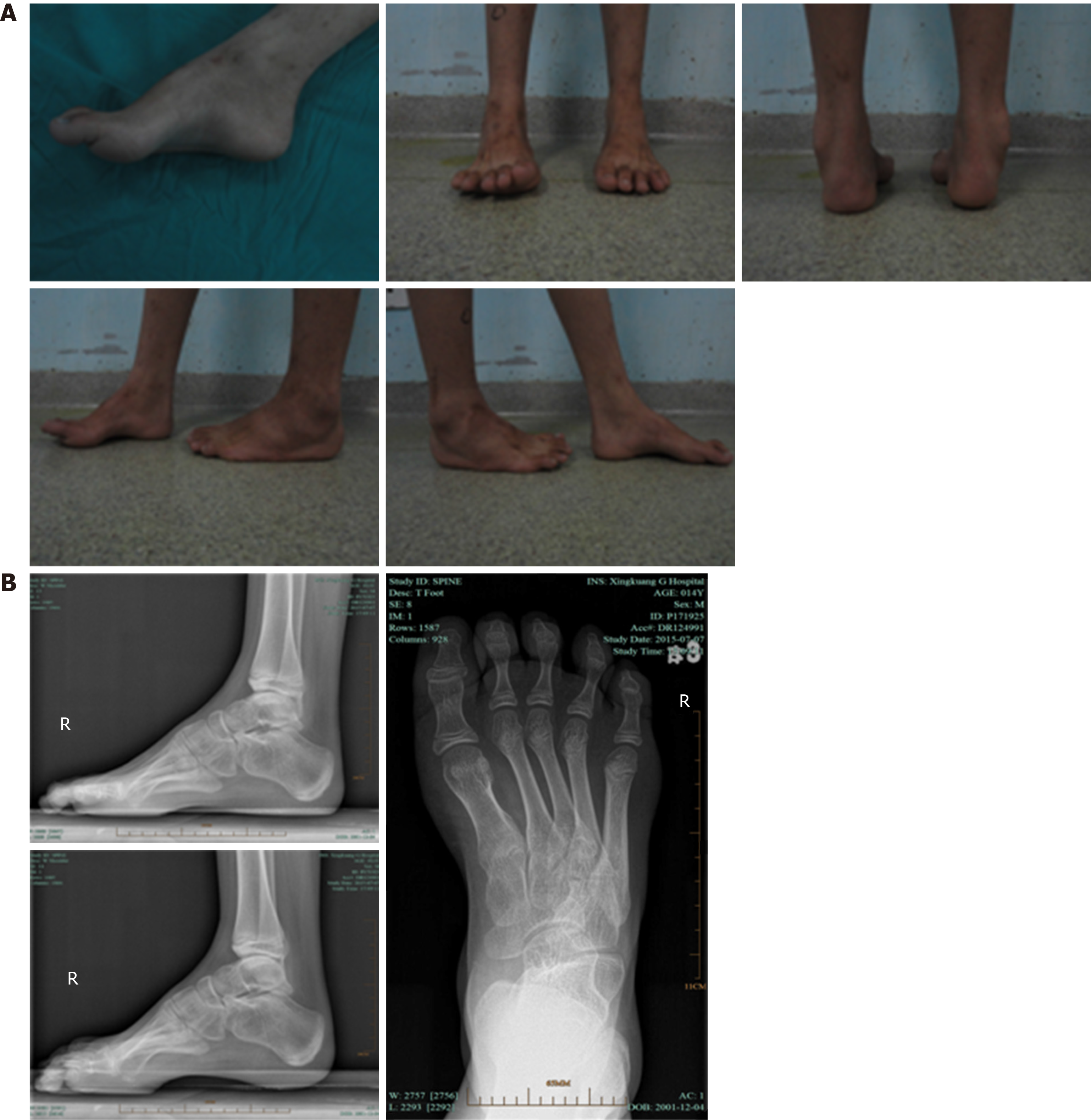
A 13-year-old boy had idiopathic bilateral cavovarus foot deformities in both weight-bearing and non-weight-bearing positions, cock-up toe deformity in the right foot, elevation of the medial longitudinal arch, forefoot pronation, and calcaneal varus (Figure 1).
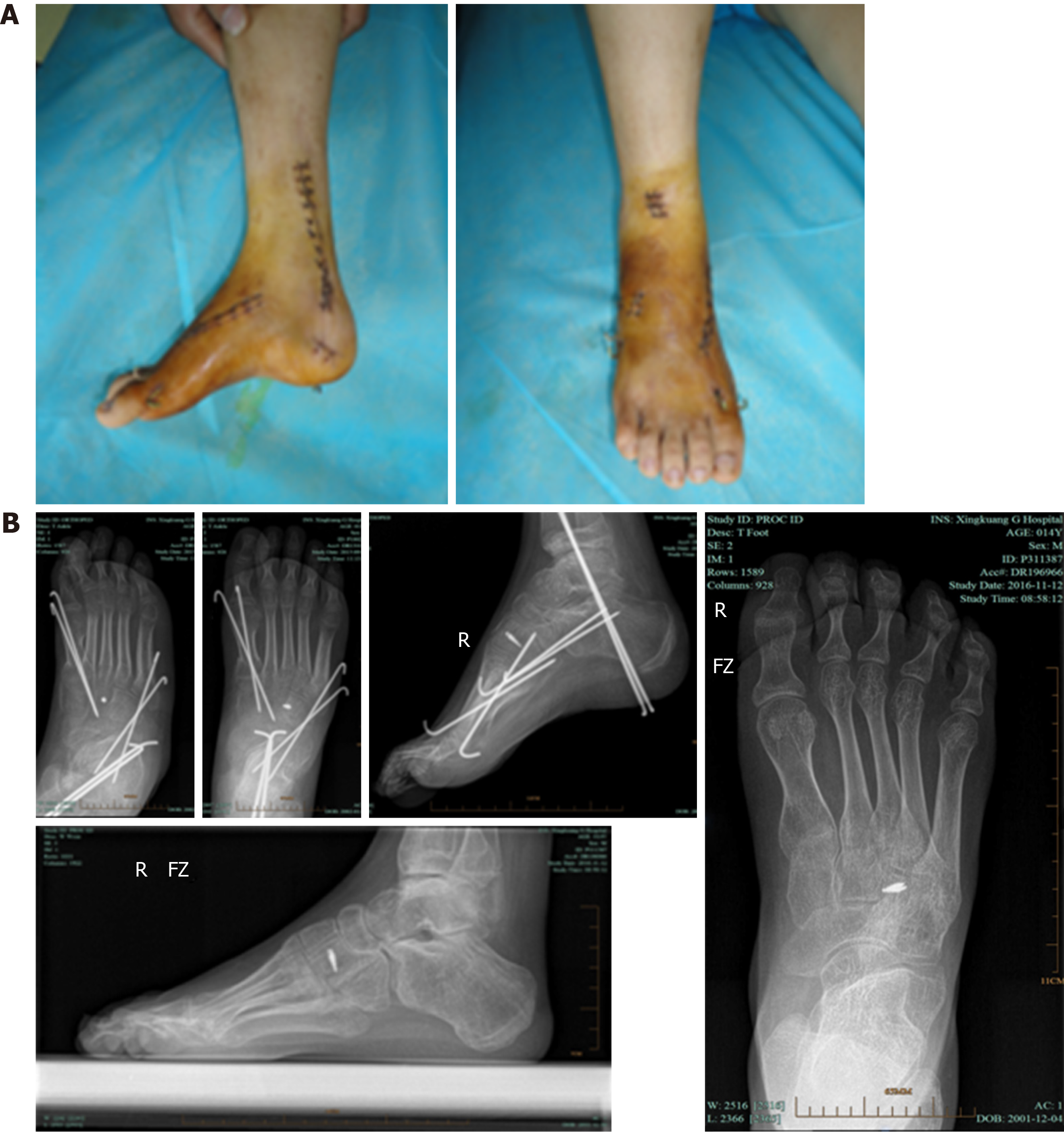
He underwent metatarsal fascia release, Achilles tendon lengthening, "V"-shaped osteotomy on the dorsal side of the first metatarsal base, opening medial cuneiform osteotomy, closing cuboid osteotomy, and transfer of the posterior tibial tendon to the the lateral cuneiform bone. Gross and X-ray examinations performed 6 wk after operation showed that he had a good recovery. Weight-bearing anteroposterior and lateral X-rays of the feet performed 18 mo after surgery also showed good results (Figure 2).
The shape of the normal foot is a tripod-like structure supported by the head of the first metatarsal, the head of the fifth metatarsal, and the calcaneus. The tripod effect directly leads to the widening of the lateral plantar margin of cavus foot, callus formation, and calcaneal varus[5]. Studies have proved that the main cause of cavovarus foot is the imbalance of medial and lateral muscles of the foot, and most patients have neuromuscular system lesions. Patients with cavovarus foot often have no obvious early symptoms, which is not easy to be found. With the increase in age and the further progression of muscle imbalance, the deformity gradually aggravates. Conservative treatment is not effective in improving such deformity. After the deformity aggravates, it cannot be fully corrected only by soft tissue release, because it has become a fixed deformity that can only be corrected by osteotomy.
Based on the etiology of cavus foot, when children with such disease seek medical care, the medical history should be asked, the relevant nervous system examinations (lumbar MRI and electromyography of lower limbs) should be performed, the deformity should be classified, and the basic diseases should be evaluated to determine whether treatment is needed. Since cavus foot is a compound deformity, the forefoot, midfoot, and rearfoot need to be evaluated separately and corrected one by one to obtain satisfactory therapeutic effects[9,10]. Adequate soft tissue release and intraoperative muscle balance play an important role in the correction of deformity and prevention of postoperative recurrence. Early-stage cavus foot is mostly a malleable deformity, which can be corrected by massage. With the increase in age and bone mass, the deformity further aggravates and cannot be corrected by manual massage alone. At this stage, the deformity becomes stiff, causing foot discomfort in children, including fatigue and local pain in the forefoot, and even gait instability[11-13]. The joint capsule, ligament, and tendon all need to be fully released to correct the deformity, and the tendon that leads to varus and cavus deformities needs to be transferred. Since the force of the transferred tendon is reduced, the balance of medial and lateral muscles should be ensured to reduce the incidence of postoperative recurrence.
All the subjects in this study were older children with cavus foot deformity. Considering their well-developed feet and sufficient bone mass as well as the feasibility of bone surgery, joint-sparing osteotomy should be performed to preserve the joint. Soft tissue balance was required to correct the deformity and prevent its recurrence. According to the severity of deformity, the procedure was designed preoperatively as two steps: soft tissue surgery and bone surgery[14]. In soft tissue surgery, medial gastrocnemius lengthening and "Z"-lengthening of the Achilles tendon were first performed according to preoperative evaluations to correct the equinovarus deformity. After the affected foot was dorsiflexed and the head of the first metatarsal was elevated, metatarsal fasciotomy was performed at the calcaneal insertion point. Strengthening the short peroneal tendon with the long peroneal tendon can not only strengthen the force of foot abduction, but also eliminate the factors causing cavus deformity at the first metatarsal. Anterior transfer of the posterior tibial tendon increased the forces of dorsal extension and valgus. In bone surgery, osteotomy was performed in different ways depending on the apex of the deformity. The principle of osteotomy was to preserve the joint as much as possible and perform a joint-sparing osteotomy. For cavus forefoot, a closing V-shaped osteotomy was performed on the dorsal side of the first metatarsal base, and the head of the first metatarsal was raised to correct the cavus deformity at the first metatarsal. When the apex of cavus deformity was located in the medial cuneiform, the osteotomy site was chosen at the medial cuneiform and the cuboid, and the medial cuneiform was opened to correct forefoot adduction and reduce high arch. For forefoot pronation deformity, which often leads to increased weight-bearing at the lateral margin of the foot while walking with load, and callus formation at the fifth metatarsal base, cuboid osteotomy was performed to raise the base of the fifth metatarsal and makes the forefoot abduct. In cases with concomitant fixed calcaneal varus, calcaneal valgus osteotomy was performed to correct the varus[15].
Wu et al[16] used the procedure reported by Japas[17], in which primary release of plantar soft tissue was combined with tarsal “V”-shaped tarsal osteotomy, to treat seven cases of idiopathic cavus foot deformity in children over 6 years old, achieving a very good/good rate of 83. 3%. Yan et al[18] utilized different treatment methods in 27 children (41 feet) with different types of cavus foot deformity. In cases with first ray cavus foot deformity, wedge osteotomy was performed on the dorsal side of the first metatarsal base; for medial column cavus foot deformity, closing wedge osteotomy on the dorsal side of the first metatarsal base combined with opening wedge osteotomy on the plantar side of the first, second, and third cuneiforms was performed; for cavus forefoot deformity, closing wedge osteotomy on the dorsal side of the cuneonavicular joint was performed. The patients were followed for an average period of 28 mo. According to the Wicart et al[19] criteria, very good therapeutic effects were achieved in 34 feet, and good achieved in 7. This finding suggested that accurate assessment of deformity and selection of proper treatment are the keys to achieving good therapeutic effects. Zhang et al[20] performed personalized treatment of cavus foot deformity, and achieved good therapeutic effects. Qin et al[21-23] proposed that the general treatment strategy for ankle-foot deformity associated with spina bifida can be summarized as four basic principles: Correction of deformity, balancing of muscle forces, stabilization of joints, and preservation of foot elasticity. They considered that muscle balance can be recovered by soft tissue release in children under 12 years old. For children over 12 years old and adults, orthopedic surgery for bone deformity was performed after sufficient soft tissue release and muscle balancing, and all patients underwent fixation with an external bone fixator postoperatively. Patients with satisfactory intraoperative deformity correction were all fixed with combined external fixation device. For those who did not achieve satisfactory results, the Ilizarov external fixator was applied and slowly adjusted after the operation to gradually correct the deformity. Finally, a good therapeutic effect was achieved. Sanpera et al[24] assessed dorsal hemiepiphyseal arrest of the first metatarsal, which enabled the metatarsal growth of the first metatarsal to be faster than the dorsal growth and reduced the apex of the cavus foot deformity by asymmetrically interfering with bone growth. At the same time, a plantar release of the metatarsal fascia decreased the tension on the metatarsal side of the foot and reduced the "windlass" effect, thus reducing the apex of the deformity. This method is less invasive and allows for early weight-bearing walking postoperatively. However, this method requires the growth potential of the proximal epiphysis of the first metatarsal in children. A total of 15 patients with 26 feet were included in that study. The average age at surgery was 10 years (range, 7-13 years) for boys and 11 years (range, 9-12 years) for girls. Twenty-four feet of the 13 patients were followed, with an average follow-up period of 28 mo. Compared with preoperative values, Meary’s angle and talo-calcaneal angle at the final follow-up were significantly improved. Complications including breakage of the plate and proximal screw misplacement occurred in three patients. Mubarak et al[1] addressed cavus deformities in children using stepwise osteotomies. First, a closing wedge osteotomy of the first metatarsal was performed to elevate the first metatarsal and reduce the apex of the high arch. Second, an opening plantar wedge osteotomy of the medial cuneiform and a closing cuboid wedge osteotomy were performed to correct the varus deformity of the forefoot and increased its flexibility. Second and third metatarsal osteotomies were performed if needed. Finally, calcaneal sliding osteotomies were performed to correct hindfoot varus deformity. Plantar fasciotomy and peroneus longus-to-brevis transfer were performed for soft tissue release. A total of 13 children with 20 feet were treated. The changes of Meary’s angle and Hibbs’ angle showed good results, and no obvious complications occurred.
In conclusion, for older children with cavovarus foot, the deformity should be fully evaluated and a good surgical plan should be designed in order to obtain good therapeutic effects. Bone deformity should be corrected and the joint should be preserved as much as possible to avoid postoperative stiffness. Adequate soft tissue release and muscle balance are the keys to correcting the deformity and avoiding its postoperative recurrence.
Cavovarus foot is a common form of foot deformity in children. It can be simultaneously complicated with forefoot pronation and varus, rearfoot varus, Achilles tendon contracture, or cock-up toe deformity. Many diseases can lead to muscle force imbalance. At present, many surgical treatments are available for cavovarus foot. For older children, priority should be given to midfoot osteotomy and fusion. It is important to preserve the joints and correct the deformity as much as possible.
Currently, diverse treatments are available for cavovarus foot, but there is no standard treatment. Improper treatment will lead to residual deformity or recurrence. Therefore, personalized surgical treatment strategies should be formulated based on preoperative assessment of the deformity and muscle force of the affected foot.
This study aimed to assess the efficacy of soft tissue release combined with joint-sparing osteotomy in the treatment of cavovarus foot deformity in older children.
Clinical data of 21 older children with cavovarus foot deformity were retrospectively analyzed. The patients underwent magnetic resonance imaging of the lumbar spine, electromyographic examination, weight-bearing anteroposterior and lateral X-rays of the feet, and the Coleman block test. Surgical procedures were performed. Therapeutic effects were evaluated. Meary’s angles and Hibbs’ angles were measured based on X-ray images.
Very good results were achieved in 18 feet, good in 7, and fair in 3, with a very good/good rate of 89.3%. At last follow-up, mean Meary’s angle was 6.36° ± 1.810°, and mean Hibbs’ angle was 160.21° ± 4.167°, both of which were significantly improved compared with preoperative values. No complications such as infection, skin necrosis, or bone nonunion occurred.
Soft tissue release combined with joint-sparing osteotomy has appreciated efficacy in the treatment of cavovarus foot deformity in older children, and it can correct the deformity, reduce postoperative recurrence, and preserve the flexibility of the foot as much as possible.
Although satisfactory efficacy can be achieved by soft tissue release combined with joint-sparing osteotomy in children with cavovarus foot deformity, long-tern follow-up data are needed to confirm our conclusion. Future search should utilize big data analysis to formulate accurate treatment plan to minimize complications while guaranteeing the best therapeutic effect.
Manuscript source: Unsolicited manuscript
Specialty type: Medicine, research and experimental
Country of origin: China
Peer-review report classification
Grade A (Excellent): 0
Grade B (Very good): B, B
Grade C (Good): 0
Grade D (Fair): 0
Grade E (Poor): 0
P-Reviewer: Andersen V, Kruis W S-Editor: Wang JL L-Editor: Wang TQ E-Editor: Liu JH
| 1. | Mubarak SJ, Van Valin SE. Osteotomies of the foot for cavus deformities in children. J Pediatr Orthop. 2009;29:294-299. [RCA] [PubMed] [DOI] [Full Text] [Cited by in Crossref: 41] [Cited by in RCA: 32] [Article Influence: 2.0] [Reference Citation Analysis (0)] |
| 2. | Mosca VS. The cavus foot. J Pediatr Orthop. 2001;21:423-424. [PubMed] |
| 3. | Schwend RM, Drennan JC. Cavus foot deformity in children. J Am Acad Orthop Surg. 2003;11:201-211. [PubMed] |
| 4. | Karakis I, Gregas M, Darras BT, Kang PB, Jones HR. Clinical correlates of Charcot-Marie-Tooth disease in patients with pes cavus deformities. Muscle Nerve. 2013;47:488-492. [RCA] [PubMed] [DOI] [Full Text] [Cited by in Crossref: 19] [Cited by in RCA: 21] [Article Influence: 1.8] [Reference Citation Analysis (0)] |
| 5. | Wicart P. Cavus foot, from neonates to adolescents. Orthop Traumatol Surg Res. 2012;98:813-828. [RCA] [PubMed] [DOI] [Full Text] [Cited by in Crossref: 45] [Cited by in RCA: 38] [Article Influence: 2.9] [Reference Citation Analysis (0)] |
| 6. | Dragoni M, Farsetti P, Vena G, Bellini D, Maglione P, Ippolito E. Ponseti Treatment of Rigid Residual Deformity in Congenital Clubfoot After Walking Age. J Bone Joint Surg Am. 2016;98:1706-1712. [RCA] [PubMed] [DOI] [Full Text] [Cited by in Crossref: 9] [Cited by in RCA: 8] [Article Influence: 0.9] [Reference Citation Analysis (0)] |
| 7. | Klaue K. Hindfoot issues in the treatment of the cavovarus foot. Foot Ankle Clin. 2008;13:221-227, vi. [RCA] [PubMed] [DOI] [Full Text] [Cited by in Crossref: 12] [Cited by in RCA: 9] [Article Influence: 0.5] [Reference Citation Analysis (0)] |
| 8. | Alexander IJ, Johnson KA. Assessment and management of pes cavus in Charcot-Marie-tooth disease. Clin Orthop Relat Res. 1989;273-281. [PubMed] |
| 9. | Nagai MK, Chan G, Guille JT, Kumar SJ, Scavina M, Mackenzie WG. Prevalence of Charcot-Marie-Tooth disease in patients who have bilateral cavovarus feet. J Pediatr Orthop. 2006;26:438-443. [RCA] [PubMed] [DOI] [Full Text] [Cited by in Crossref: 51] [Cited by in RCA: 38] [Article Influence: 2.0] [Reference Citation Analysis (0)] |
| 10. | Reeves CL, Shane AM, Zappasodi F, Payne T. Surgical Correction of Rigid Equinovarus Contracture Utilizing Extensive Soft Tissue Release. Clin Podiatr Med Surg. 2016;33:139-152. [RCA] [PubMed] [DOI] [Full Text] [Cited by in Crossref: 6] [Cited by in RCA: 7] [Article Influence: 0.8] [Reference Citation Analysis (0)] |
| 11. | Fernández-Seguín LM, Diaz Mancha JA, Sánchez Rodríguez R, Escamilla Martínez E, Gómez Martín B, Ramos Ortega J. Comparison of plantar pressures and contact area between normal and cavus foot. Gait Posture. 2014;39:789-792. [RCA] [PubMed] [DOI] [Full Text] [Cited by in Crossref: 56] [Cited by in RCA: 63] [Article Influence: 5.7] [Reference Citation Analysis (0)] |
| 12. | Najafi B, Wrobel JS, Burns J. Mechanism of orthotic therapy for the painful cavus foot deformity. J Foot Ankle Res. 2014;7:2. [RCA] [PubMed] [DOI] [Full Text] [Full Text (PDF)] [Cited by in Crossref: 21] [Cited by in RCA: 19] [Article Influence: 1.7] [Reference Citation Analysis (0)] |
| 13. | Eleswarapu AS, Yamini B, Bielski RJ. Evaluating the Cavus Foot. Pediatr Ann. 2016;45:e218-e222. [RCA] [PubMed] [DOI] [Full Text] [Cited by in Crossref: 13] [Cited by in RCA: 10] [Article Influence: 1.1] [Reference Citation Analysis (0)] |
| 14. | Kadakia AR. The cavus foot. Foot Ankle Clin. 2013;18:xiii-xxiv. [RCA] [PubMed] [DOI] [Full Text] [Cited by in Crossref: 6] [Cited by in RCA: 4] [Article Influence: 0.3] [Reference Citation Analysis (0)] |
| 15. | Sammarco GJ, Taylor R. Cavovarus foot treated with combined calcaneus and metatarsal osteotomies. Foot Ankle Int. 2001;22:19-30. [RCA] [PubMed] [DOI] [Full Text] [Cited by in Crossref: 76] [Cited by in RCA: 60] [Article Influence: 2.5] [Reference Citation Analysis (0)] |
| 16. | Wu JY, Mei HB, Liu K, He RG, Tang J, Hu X, Ye WH. The clinical research for idiopathic pes cavus in children with Japas operation. Linchuang Xiaoer Waike Zazhi. 2008;29:11-14. |
| 17. | Japas LM. Surgical treatment of pes cavus by tarsal V-osteotomy. Preliminary report. J Bone Joint Surg Am. 1968;50:927-944. [PubMed] |
| 18. | Yan GS, Yang Z, Lu M, Zhu ZH, Zhang JL, Guo Y. Cavovarus foot in children: evaluation of deformity and choice of treatment. Linchuang Xiaoer Waike Zazhi. 2015;36:496-500. |
| 19. | Wicart P, Seringe R. Plantar opening-wedge osteotomy of cuneiform bones combined with selective plantar release and dwyer osteotomy for pes cavovarus in children. J Pediatr Orthop. 2006;26:100-108. [RCA] [PubMed] [DOI] [Full Text] [Cited by in Crossref: 47] [Cited by in RCA: 38] [Article Influence: 2.0] [Reference Citation Analysis (0)] |
| 20. | Zhang WJ, Zhang Y, Xu SQ, Li P. Analysis of efficacy of individualized treatment of varus clubfoot. Zhongguo Gu Yu Guanjie Sunshang Zazhi. 2017;32:1045-1047. |
| 21. | Qin SH, Ge JZ, Guo BF. [Clinical analysis of 107 patients with foot and ankle deformities caused by spinal bifida]. Zhonghua Wai Ke Za Zhi. 2010;48:900-903. [PubMed] |
| 22. | Qin SH, Guo BF, Zheng XJ, Jiao SF, Xia HT, Peng AM, Pan Q, Zang JC, Wang ZJ. [Domestic external fixator application in the treatment of limb deformities: 7 289 cases application report]. Zhonghua Wai Ke Za Zhi. 2017;55:678-683. [RCA] [PubMed] [DOI] [Full Text] [Cited by in RCA: 4] [Reference Citation Analysis (0)] |
| 23. | Jiao SF, Qin SH, Ren LX, Ge JZ, Wu HF, Wang ZJ, Zheng XJ. [Combined procedure for the treatment of ankle and foot deformities secondary to spina bifida]. Zhongguo Gu Shang. 2012;25:237-240. [PubMed] |
| 24. | Sanpera I, Frontera-Juan G, Sanpera-Iglesias J, Corominas-Frances L. Innovative treatment for pes cavovarus: a pilot study of 13 children. Acta Orthop. 2018;89:668-673. [RCA] [PubMed] [DOI] [Full Text] [Full Text (PDF)] [Cited by in Crossref: 4] [Cited by in RCA: 4] [Article Influence: 0.6] [Reference Citation Analysis (0)] |


