Copyright
©The Author(s) 2019.
World J Clin Cases. Oct 6, 2019; 7(19): 3033-3038
Published online Oct 6, 2019. doi: 10.12998/wjcc.v7.i19.3033
Published online Oct 6, 2019. doi: 10.12998/wjcc.v7.i19.3033
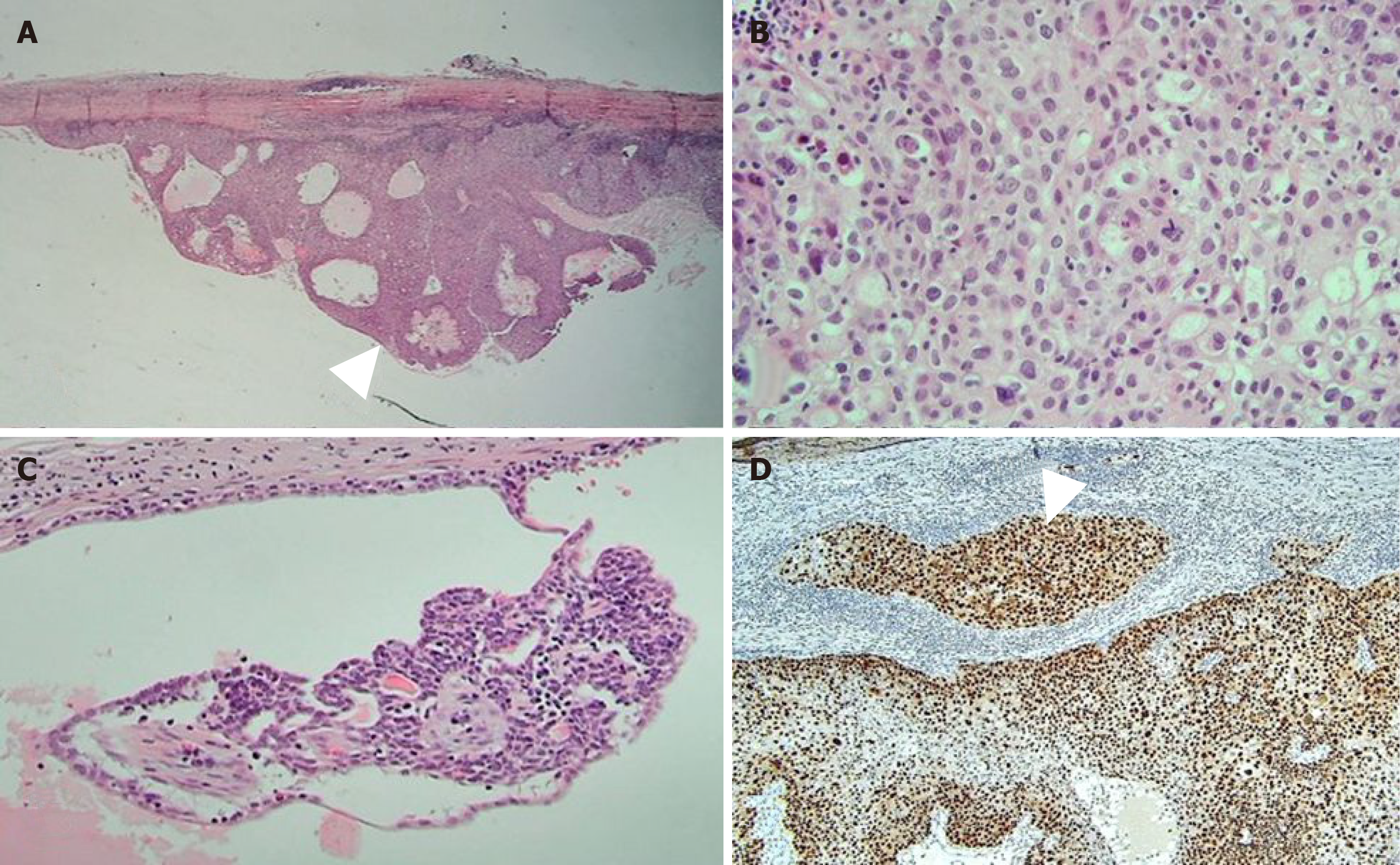
Figure 3 Photomicrographs of the cystic mass.
A: Photomicrograph demonstrating predominantly cystic mass with solid portion of carcinoma (arrowhead) [Hematoxylin and Eosin (HE) staining, × 20 magnification]; B: The carcinoma was composed of epithelial cells with clear or eosinophilic cytoplasm, prominent nucleoli, and frequent mitosis (HE staining, × 400 magnification); C: Residual benign tissue was smaller than 0.1 cm (HE staining, × 200 magnification); D: Microinvasion to fibrous cystic wall was noted (arrowhead) (P53 staining, × 100 magnification).
- Citation: An JK, Woo JJ, Hong YO. Malignant sweat gland tumor of breast arising in pre-existing benign tumor: A case report. World J Clin Cases 2019; 7(19): 3033-3038
- URL: https://www.wjgnet.com/2307-8960/full/v7/i19/3033.htm
- DOI: https://dx.doi.org/10.12998/wjcc.v7.i19.3033


