Copyright
©The Author(s) 2019.
World J Clin Cases. Sep 26, 2019; 7(18): 2871-2878
Published online Sep 26, 2019. doi: 10.12998/wjcc.v7.i18.2871
Published online Sep 26, 2019. doi: 10.12998/wjcc.v7.i18.2871
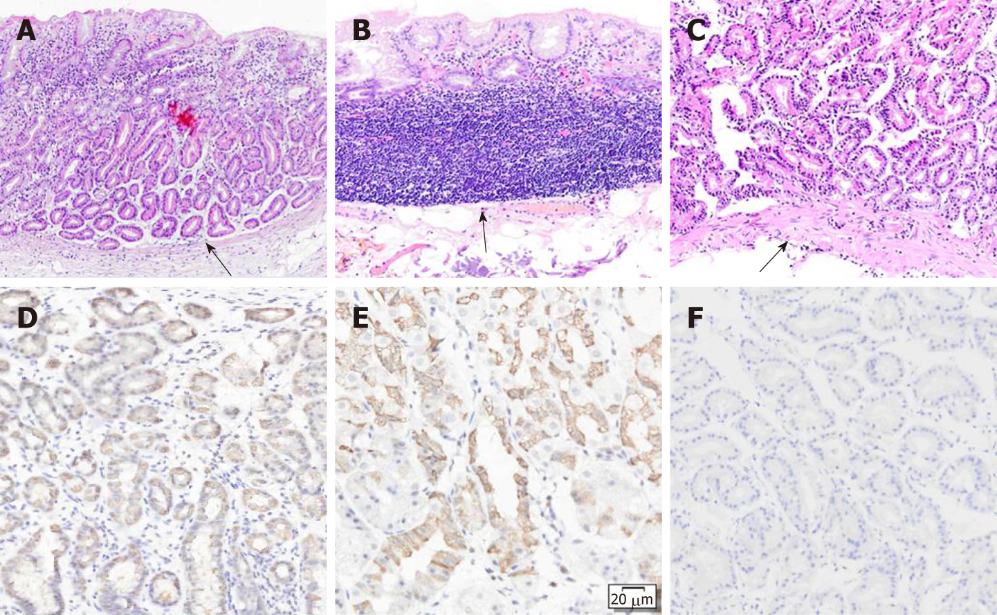
Figure 4 Hematoxylin and eosin staining.
A: Hematoxylin and eosin staining showed that the gastric corpus lesion was located in the deep layer of the lamina propria and did not invade into the muscularis mucosa. The lesion did not extend to the superficial epithelium; B: Hematoxylin and eosin staining showed no detectable lesion in the gastric fundus but only the biopsy scar. The mucularis mucosa of the ESD-resected specimen was intact; C: Hematoxylin and eosin staining of the fundic biopsy showing that the mucularis mucosa was intact; D-F: Immunostaining showed that both lesions were positive for pepsinogen I (D) and MUC6 (E) and partially positive for H+/K+-ATPase (F).
- Citation: Chen O, Shao ZY, Qiu X, Zhang GP. Multiple gastric adenocarcinoma of fundic gland type: A case report. World J Clin Cases 2019; 7(18): 2871-2878
- URL: https://www.wjgnet.com/2307-8960/full/v7/i18/2871.htm
- DOI: https://dx.doi.org/10.12998/wjcc.v7.i18.2871


