Published online Sep 6, 2019. doi: 10.12998/wjcc.v7.i17.2562
Peer-review started: April 4, 2019
First decision: June 19, 2019
Revised: June 27, 2019
Accepted: July 20, 2019
Article in press: July 20, 2019
Published online: September 6, 2019
Processing time: 161 Days and 18.5 Hours
Injuries to multiple fingertips pose a significant treatment dilemma. Numerous reconstructive options exist, all with the ultimate goal of restoring function and sensibility to the injured fingertips.
A 24-year-old male suffered injury to multiple fingertips of the right hand, resulting in exposed distal phalanges of the middle, ring, and small fingers. The amputated distal stumps were not possible for replantation. Free flap coverage was selected in order to achieve better functional outcome. The fingertip defects were covered by performing a right second toe split tibial flap using local anesthesia at the harvest site and brachial plexus nerve block for the right upper extremity. At 6-month follow-up, all three of the reconstructed fingertips had some preserved nail growth, Semmes-Weinstein Monofilaments testing was equal to the contralateral side and the Static Two-Point Discrimination were comparable to the contralateral side.
This report provides a novel reconstructive option for the management of multiple fingertip injuries and demonstrates the utility of supermicrosurgery in management of these injuries.
Core tip: Injuries to multiple fingertips pose a significant treatment dilemma. Numerous reconstructive options exist, all with the ultimate goal of restoring function and sensibility to the injured fingertips. We present a case of a split tibial flap of the second toe utilized to treat multiple fingertip injuries, resulting in satisfactory restoration of function and sensation. This report provides a novel reconstructive option for the management of multiple fingertip injuries and demonstrates the utility of supermicrosurgery in management of these injuries.
- Citation: Wang KL, Zhang ZQ, Buckwalter JA, Yang Y. Supermicrosurgery in fingertip defects-split tibial flap of the second toe to reconstruct multiple fingertip defects: A case report. World J Clin Cases 2019; 7(17): 2562-2566
- URL: https://www.wjgnet.com/2307-8960/full/v7/i17/2562.htm
- DOI: https://dx.doi.org/10.12998/wjcc.v7.i17.2562
The fingertips are the most frequently injured part of the hand[1,2]. The underlying finger pulps play a vital role in functions of daily life such as sensation, fine touch, and grip perception. It is imperative to manage fingertip injuries with the goal of obtaining a painless, functional finger with protective sensation[3].
Given the complexity of fingertip injuries, successful replantation of amputated finger pulp is not always possible. Therefore, numerous surgical options have been reported for finger pulp reconstruction with good functional and sensory outcomes, such as V-Y advancement, pedicle flaps, free flaps, and toe pulp transfer[1,2,4-15].
The purpose of this report is to describe a novel reconstructive method to restore function and sensation after injury to three finger pulps by using the split second toe tibial flap.
A 24-year-old male presented to our hospital, with a shearing, crush-type injury to multiple fingertips of the right hand, resulting in exposed distal phalanges of the middle, ring, and small fingers. The amputated distal stumps were retrieved, but due to the nature of the injury, replantation was not possible.
The patient was a healthy, non-smoker, without signs of tinea pedis or unguium. A variety of reconstruction strategies were presented to the patient and his family, including stump revision amputation, V-Y flap advancement, and pedicle or free flap coverage.
After consideration of the options, the patient elected for free flap coverage in order to potentially achieve better functional recovery.
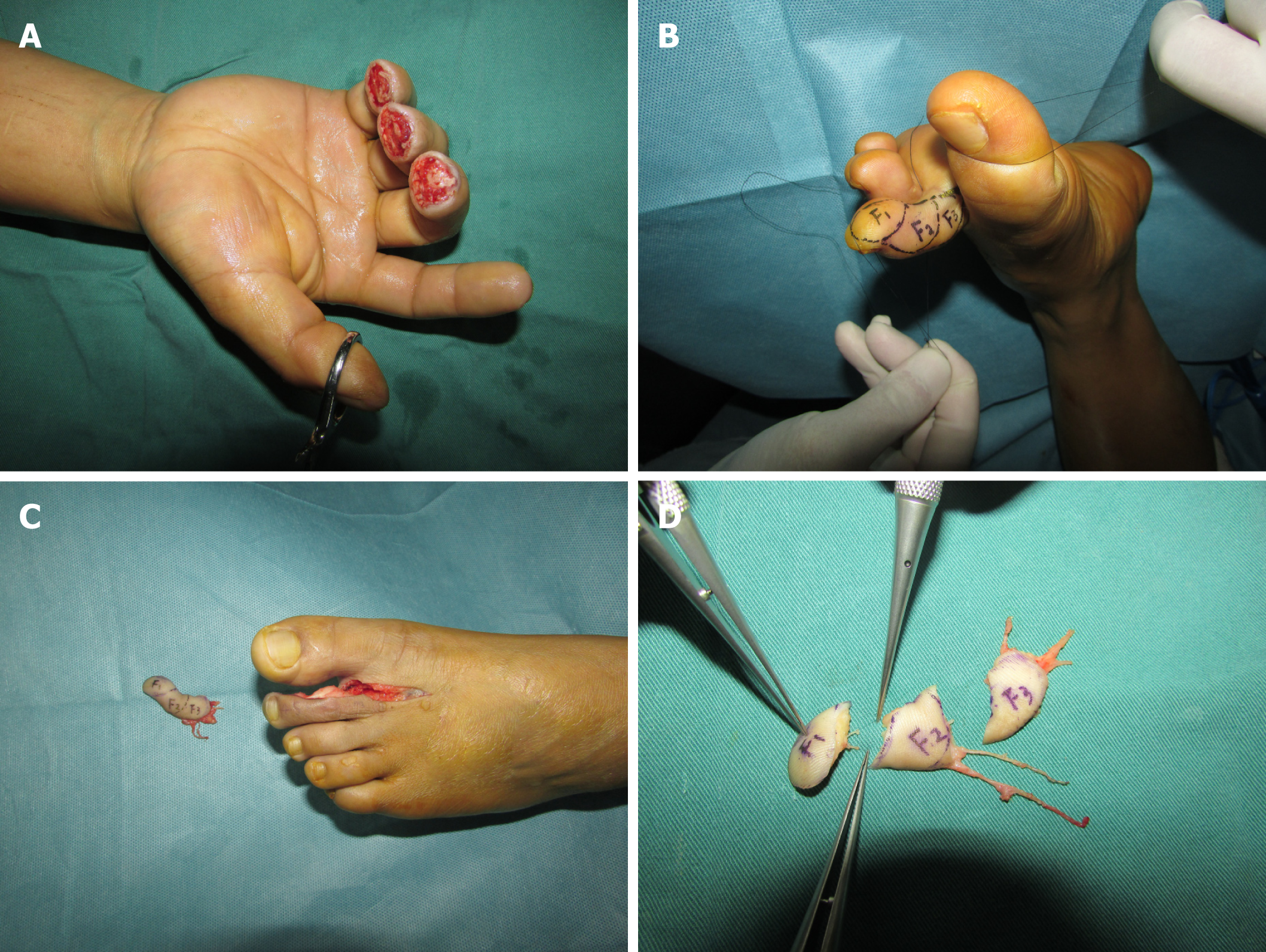
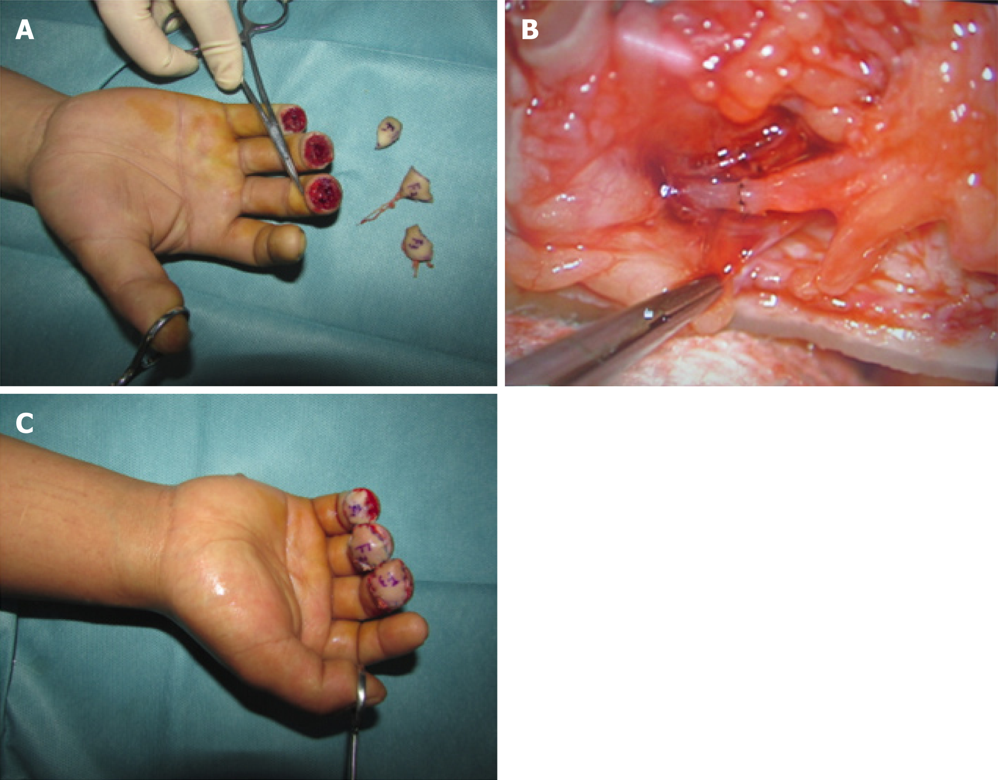
Initially, the patient underwent irrigation and debridement of the injured fingertips with vacuum-assisted closure (VAC) to cover the wounds. Three days later, we covered the fingertip defects by performing a right second toe split tibial flap using local anesthesia at the harvest site and brachial plexus nerve block for the right upper extremity. The defects of the right middle, ring, and small finger measured 1.8 cm2 × 1.5 cm2, 1.5 cm2 × 1.2 cm2, and 1.2 cm2 × 1.0 cm2, respectively (Figure 1A). A 1.6 cm2 × 4.5 cm2 tibial second toe flap was then harvested and accordingly split into three small flaps as F1, F2, and F3 under the microscope with respective flap areas of 1.3 cm2 × 1.0 cm2, 1.6 cm2 × 1.2 cm2, and 1.8 cm2 × 1.5 cm2 (Figure 1B-D). The F3, F2, and F1 were transferred to middle, ring, and little fingertips with the one artery (the diameter for F3, F2, F1 was 0.8, 0.5, 0.4 mm, respectively), one vein (the diameter for F3, F2, F1 was 0.9, 0.8, 0.5 mm, respectively), and one nerve (the diameter for F3, F2, F1 was 1.0, 0.8, 0.6 mm, respectively) (Figure 2A-C). The anastomoses were completed to the recipient sites by using 11-0 Prolene suture under the microscope, and the donor site was closed with full thickness skin graft.
Post-operatively, Cefuroxime were administered for three days; Papaverine hydro-chloride was administered intramuscularly (30 mg, q6h→qd, day1→day 7) for seven days; and Heparin was continually administered intravenously (12500 IU every 24 h) for seven days. Blood coagulation function was monitored every two days to ensure the activated partial thromboplastin time exceeded no more than 1.5 times of the normal level. The patient remained non-weight bearing for both the right upper extremity and right lower extremity, and was discharged from the hospital without any noted complications.
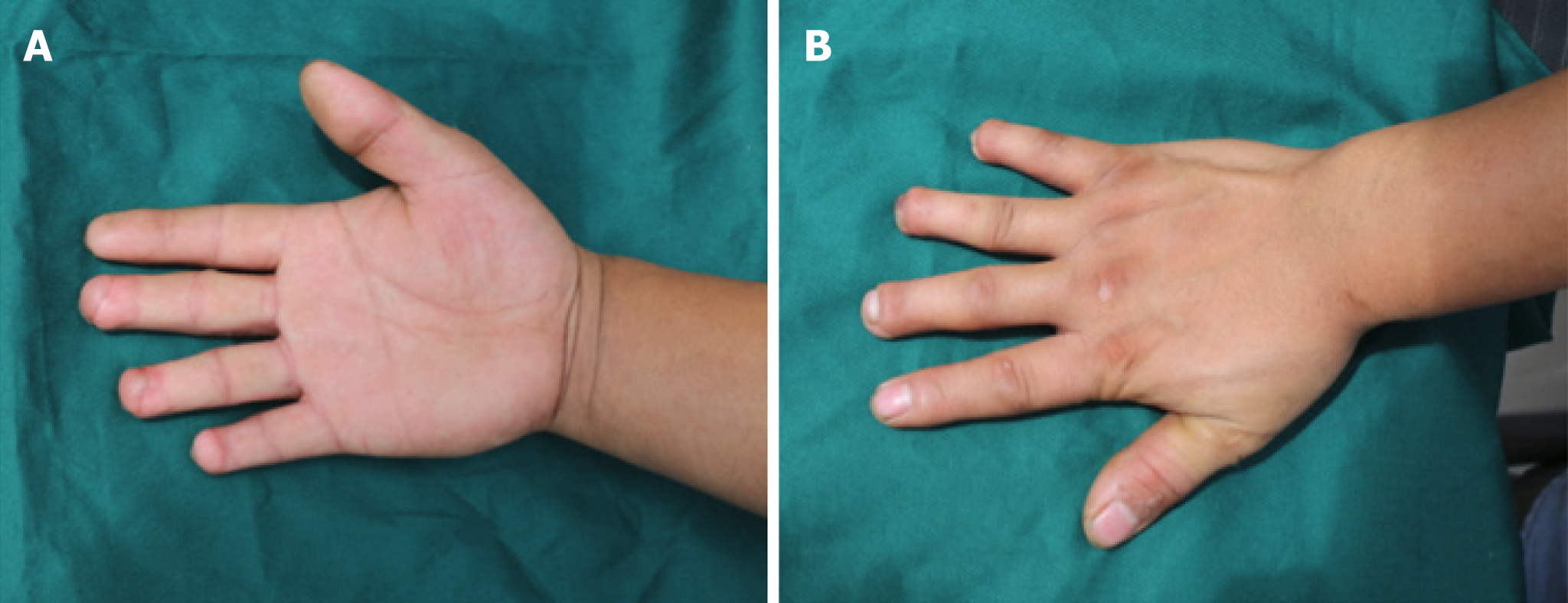
At 6-mo follow-up, the patient had no donor site or harvest site complications (Figure 3A and B), and reported no residual pain or cold intolerance. All three of the reconstructed fingertips had some preserved nail growth, measuring 17 mm, 13 mm, and 10 mm of the middle, ring, and small fingers, respectively. Semmes-Weinstein Monofilament testing was equal to the contralateral side with the 2.83 of the right middle, ring, and small fingertips. The Static Two-Point Discrimination (s2PD) measurements were 8 mm, 9 mm, and 8 mm in the right middle, ring, and small fingertips, while in the contralateral side the measurements were 6 mm, 5 mm and 4 mm, respectively.
The fingertips perform unique and significant functions of daily life such as sensation, fine handling, and gripping[1,2], and they are the most frequently injured part of the hand. For these reasons, it is of vital importance to treat fingertip injuries with careful strategy to achieve pain-free, quality functional and sensory outcomes. In cases where replantation of an amputated fingertip is not feasible, numerous reconstructive options exist[2-16]. When considering the appropriate surgical technique and reconstruction options, a surgeon should deliberate on the characteristics of the injury, the advantages and disadvantages of the reconstructive method, and the potential for recovering of function, especially for cases involving multiple fingertips injuries.
This case report offers a novel reconstructive option for the management of multiple fingertip injuries. In this case, the patient had no surgical or post-operative complications, and reported no pain, cold intolerance, or functional deficits. Although the patient reported no perceived functional or sensory deficits, the s2PD measurements were decreased compared to contralateral side. These s2PD measurements were all less than 10mm and Semmes-Weinstein Monofilaments testing was equal to the contralateral side, indicating a satisfactory sensory outcome.
The concept of supermicrosurgery has found increasing clinical applications in recent years[16]. With supermicrosurgery, the dissection and anastomosis of very small caliber structures with minimal donor-site morbidity had become a reasonable option. The split tibial second toe flap utilized for this patient is an advanced application of supermicrosurgery, in which the multiple defects were covered with one single split flap. This report is noteworthy for its originality and application of super-microsurgery. Supermicrosurgery should continue to be explored and considered as a feasible option for fingertip defect reconstruction and other diseases treatment such as lymphedema, nerve repair, organ transplantation, and free flaps in the future.
Manuscript source: Unsolicited Manuscript
Specialty type: Medicine, research and experimental
Country of origin: United States
Peer-review report classification
Grade A (Excellent): 0
Grade B (Very good): B
Grade C (Good): 0
Grade D (Fair): 0
Grade E (Poor): 0
P-Reviewer: Ünver B S-Editor: Cui LJ L-Editor: A E-Editor: Xing YX
| 1. | Panattoni JB, De Ona IR, Ahmed MM. Reconstruction of fingertip injuries: surgical tips and avoiding complications. J Hand Surg Am. 2015;40:1016-1024. [RCA] [PubMed] [DOI] [Full Text] [Cited by in Crossref: 24] [Cited by in RCA: 34] [Article Influence: 3.4] [Reference Citation Analysis (0)] |
| 2. | Lemmon JA, Janis JE, Rohrich RJ. Soft-tissue injuries of the fingertip: methods of evaluation and treatment. An algorithmic approach. Plast Reconstr Surg. 2008;122:105e-117e. [RCA] [PubMed] [DOI] [Full Text] [Cited by in Crossref: 56] [Cited by in RCA: 77] [Article Influence: 4.5] [Reference Citation Analysis (0)] |
| 3. | Lee DH, Mignemi ME, Crosby SN. Fingertip injuries: an update on management. J Am Acad Orthop Surg. 2013;21:756-766. [RCA] [PubMed] [DOI] [Full Text] [Cited by in Crossref: 14] [Cited by in RCA: 14] [Article Influence: 1.2] [Reference Citation Analysis (0)] |
| 4. | Song D, Pafitanis G, Yang P, Narushima M, Li Z, Liu L, Wang Z. Innervated dorsoradial perforator free flap: A reliable supermicrosurgery fingertip reconstruction technique. J Plast Reconstr Aesthet Surg. 2017;70:1001-1008. [RCA] [PubMed] [DOI] [Full Text] [Cited by in Crossref: 7] [Cited by in RCA: 9] [Article Influence: 1.1] [Reference Citation Analysis (0)] |
| 5. | Shao X, Chen C, Zhang X, Yu Y, Ren D, Lu L. Coverage of fingertip defect using a dorsal island pedicle flap including both dorsal digital nerves. J Hand Surg Am. 2009;34:1474-1481. [RCA] [PubMed] [DOI] [Full Text] [Cited by in Crossref: 26] [Cited by in RCA: 29] [Article Influence: 1.8] [Reference Citation Analysis (0)] |
| 6. | Ni F, Appleton SE, Chen B, Wang B. Aesthetic and functional reconstruction of fingertip and pulp defects with pivot flaps. J Hand Surg Am. 2012;37:1806-1811. [RCA] [PubMed] [DOI] [Full Text] [Cited by in Crossref: 17] [Cited by in RCA: 17] [Article Influence: 1.3] [Reference Citation Analysis (0)] |
| 7. | Lim GJ, Yam AK, Lee JY, Lam-Chuan T. The spiral flap for fingertip resurfacing: short-term and long-term results. J Hand Surg Am. 2008;33:340-347. [RCA] [PubMed] [DOI] [Full Text] [Cited by in Crossref: 22] [Cited by in RCA: 22] [Article Influence: 1.3] [Reference Citation Analysis (0)] |
| 8. | Huang YC, Liu Y, Chen TH. Use of homodigital reverse island flaps for distal digital reconstruction. J Trauma. 2010;68:429-433. [RCA] [PubMed] [DOI] [Full Text] [Cited by in Crossref: 13] [Cited by in RCA: 16] [Article Influence: 1.1] [Reference Citation Analysis (0)] |
| 9. | Xianyu M, Lei C, Laijin L, Zhigang L. Reconstruction of finger-pulp defect with a homodigital laterodorsal fasciocutaneous flap distally based on the dorsal branches of the proper palmar digital artery. Injury. 2009;40:1346-1350. [RCA] [PubMed] [DOI] [Full Text] [Cited by in Crossref: 15] [Cited by in RCA: 18] [Article Influence: 1.1] [Reference Citation Analysis (0)] |
| 10. | Regmi S, Gu JX, Zhang NC, Liu HJ. A Systematic Review of Outcomes and Complications of Primary Fingertip Reconstruction Using Reverse-Flow Homodigital Island Flaps. Aesthetic Plast Surg. 2016;40:277-283. [RCA] [PubMed] [DOI] [Full Text] [Cited by in Crossref: 30] [Cited by in RCA: 27] [Article Influence: 3.0] [Reference Citation Analysis (0)] |
| 11. | Acar MA, Güzel Y, Güleç A, Türkmen F, Erkoçak ÖF, Yılmaz G. Reconstruction of multiple fingertip injuries with reverse flow homodigital flap. Injury. 2014;45:1569-1573. [RCA] [PubMed] [DOI] [Full Text] [Cited by in Crossref: 16] [Cited by in RCA: 15] [Article Influence: 1.4] [Reference Citation Analysis (0)] |
| 12. | Karamese M, Akatekin A, Abac M, Koplay TG, Tosun Z. Fingertip Reconstruction With Reverse Adipofascial Homodigital Flap. Ann Plast Surg. 2015;75:158-162. [RCA] [PubMed] [DOI] [Full Text] [Cited by in Crossref: 10] [Cited by in RCA: 11] [Article Influence: 1.2] [Reference Citation Analysis (0)] |
| 13. | Kim KS, Yoo SI, Kim DY, Lee SY, Cho BH. Fingertip reconstruction using a volar flap based on the transverse palmar branch of the digital artery. Ann Plast Surg. 2001;47:263-268. [RCA] [PubMed] [DOI] [Full Text] [Cited by in Crossref: 27] [Cited by in RCA: 29] [Article Influence: 1.2] [Reference Citation Analysis (0)] |
| 14. | Borman H, Maral T, Tancer M. Fingertip reconstruction using two variations of direct-flow homodigital neurovascular island flaps. Ann Plast Surg. 2000;45:24-30. [RCA] [PubMed] [DOI] [Full Text] [Cited by in Crossref: 22] [Cited by in RCA: 22] [Article Influence: 0.9] [Reference Citation Analysis (0)] |
| 15. | Varitimidis SE, Dailiana ZH, Zibis AH, Hantes M, Bargiotas K, Malizos KN. Restoration of function and sensitivity utilizing a homodigital neurovascular island flap after amputation injuries of the fingertip. J Hand Surg Br. 2005;30:338-342. [RCA] [PubMed] [DOI] [Full Text] [Cited by in Crossref: 39] [Cited by in RCA: 34] [Article Influence: 1.7] [Reference Citation Analysis (0)] |
| 16. | Masia J, Olivares L, Koshima I, Teo TC, Suominen S, Van Landuyt K, Demirtas Y, Becker C, Pons G, Garusi C, Mitsunaga N. Barcelona consensus on supermicrosurgery. J Reconstr Microsurg. 2014;30:53-58. [RCA] [PubMed] [DOI] [Full Text] [Cited by in Crossref: 57] [Cited by in RCA: 79] [Article Influence: 6.6] [Reference Citation Analysis (0)] |


