Copyright
©The Author(s) 2019.
World J Clin Cases. Aug 6, 2019; 7(15): 2120-2127
Published online Aug 6, 2019. doi: 10.12998/wjcc.v7.i15.2120
Published online Aug 6, 2019. doi: 10.12998/wjcc.v7.i15.2120
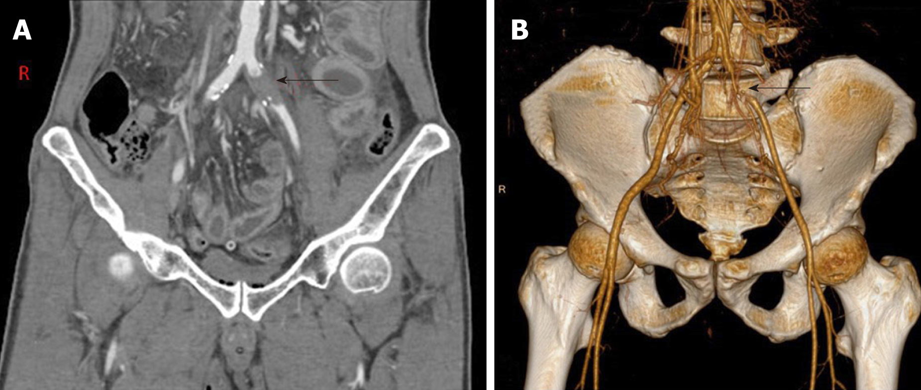
Figure 1 Abdominal computed tomography findings of a common iliac artery occlusion.
A: Abdominal computed tomography angiography confirmed a 2.5 cm occlusion of the left common iliac artery, with preservation of the common iliac bifurcation and distal arterial tree. There was no evidence of active arterial bleeding or pseudoaneurysm formation (arrow); B: Three-dimensional computed tomography angiogram reconstruction showed obvious occlusion of the left common iliac artery (arrow).
- Citation: Zhou YX, Ji Y, Chen J, Yang X, Zhou Q, Lv J. Common iliac artery occlusion with small intestinal transection caused by blunt abdominal trauma: A case report and review of the literature. World J Clin Cases 2019; 7(15): 2120-2127
- URL: https://www.wjgnet.com/2307-8960/full/v7/i15/2120.htm
- DOI: https://dx.doi.org/10.12998/wjcc.v7.i15.2120


