Published online Aug 6, 2019. doi: 10.12998/wjcc.v7.i15.2003
Peer-review started: March 28, 2019
First decision: May 31, 2019
Revised: June 12, 2019
Accepted: July 3, 2019
Article in press: July 4, 2019
Published online: August 6, 2019
Processing time: 133 Days and 1.2 Hours
Heart defects are the most common congenital malformations in fetuses. Fetal cardiac structure and function abnormalities lead to changes in ventricular volume. As ventricular volume is an important index for evaluating fetal cardiovascular development, an effective and reliable method for measuring fetal ventricular volume and cardiac function is necessary for accurate ultrasonic diagnosis and effective clinical treatment. The new intelligent spatiotemporal image correlation (iSTIC) technology acquires high-resolution volumetric images. In this study, the iSTIC technique was used to measure right ventricular volume and to evaluate right ventricular systolic function to provide a more accurate and convenient evaluation of fetal heart function.
To investigate the value of iSTIC in evaluating right ventricular volume and systolic function in normal fetuses.
Between October 2014 and September 2015, a total of 123 pregnant women received prenatal ultrasound examinations in our hospital. iSTIC technology was used to acquire the entire fetal cardiac volume with off-line analysis using QLAB software. Cardiac systolic and diastolic phases were defined by opening of the atrioventricular valve and the subsequent closure of the atrioventricular valve. The volumetric data of the two phases were measured by manual tracking and summation of multiple slices and recording of the right ventricular end-systolic volume and the right ventricular end-diastolic volume. The data were used to calculate the right stroke volume, the right cardiac output, and the right ejection fraction. The correlations of changes between the above-mentioned indices and gestational age were analyzed. The right ventricular volumes of 30 randomly selected cases were measured twice by the same sonographer, and the intra-observer agreement measurements were calculated.
Among the 123 normal fetuses, the mean right ventricular end-diastolic volume increased from 0.99 ± 0.34 mL at 22 wk gestation to 3.69 ± 0.36 mL at 35+6 wk gestation. The mean right ventricular end-systolic volume increased from 0.43 ± 0.18 mL at 22 wk gestation to 1.36 ± 0.22 mL at 35+6 wk gestation. The mean right stroke volume increased from 0.62 ± 0.29 mL at 22 wk gestation to 2.33 ± 0.18 mL at 35+6 wk gestation. The mean right cardiac output increased from 92.23 ± 40.67 mL/min at 22 wk gestation to 335.83 ± 32.75 mL/min at 35+6 wk gestation. Right ventricular end-diastolic volume, right ventricular end-systolic volume, right stroke volume, and right cardiac output all increased with gestational age and the correlations were linear (P < 0.01). Right ejection fraction had no apparent correlation with gestational age (P > 0.05).
Fetal right ventricular volume can be quantitatively measured using iSTIC technology with relative ease and high repeatability. iSTIC technology is expected to provide a new method for clinical evaluation of fetal cardiac function.
Core tip: Heart defects are the most common congenital malformations in fetuses. Fetal cardiac structure and function abnormalities often lead to changes in ventricular volume. Numerous studies have focused on fetal heart functions, which have not been widely accepted. Between October 2014 and September 2015, the intelligent spatiotemporal image correlation technique was used to measure right ventricular volume in 123 normal fetuses and to evaluate right ventricular systolic function to provide a new method for more accurate and convenient evaluation of fetal heart function.
- Citation: Sun JX, Cai AL, Xie LM. Evaluation of right ventricular volume and systolic function in normal fetuses using intelligent spatiotemporal image correlation. World J Clin Cases 2019; 7(15): 2003-2012
- URL: https://www.wjgnet.com/2307-8960/full/v7/i15/2003.htm
- DOI: https://dx.doi.org/10.12998/wjcc.v7.i15.2003
Heart defects are the most common congenital malformations in fetuses with an incidence six times greater than chromosome abnormalities and four times greater than neural tube defects[1,2]. The diagnosis of congenital heart disease is a challenge using prenatal ultrasound and is the focus of perinatal medical research. Congenital heart disease can be divided into structural and functional abnormalities. Indeed, fetal cardiac structure and function abnormalities often lead to changes in ventricular volume. As ventricular volume is an important index for evaluating fetal cardiovascular development, an effective and reliable method for measuring fetal ventricular volume and cardiac function is necessary for accurate ultrasonic diagnosis and effective clinical treatment of congenital heart disease.
Numerous studies have focused on fetal heart function in an effort to evaluate cardiac function in the healthy fetus and in those in states of cardiac decompen-sation[3]. The accuracy of traditional two-dimensional (2D) ultrasound in the quantitative evaluation of fetal heart function has not been widely accepted[4-15]. Spatiotemporal image correlation (STIC) technology overcomes many of the shortcomings of conventional 2D ultrasound in the measurement of fetal ventricular volume and evaluation of fetal cardiac function. Recently, a number of studies have used STIC technology in combination with organ computer-aided analysis software to measure fetal ventricular volume and evaluate heart function with proven accuracy and feasibility[4-7,16-27]. However, STIC still has some limitations and the imaging principle determines that STIC is not a real-time three-dimensional (3D) imaging technology. One-way scanning using the sensors during the scanning process execution is slow, which leads to relatively long image acquisition time. Therefore, STIC is vulnerable to the effects of fetal and maternal respiration, resulting in degradation of image quality[21,23].
The new intelligent STIC (iSTIC) technology adopts an electronic matrix type of probe, which is composed of thousands of vibrating bits, thus generating real-time 3D images with acquired data. The new iSTIC technology acquires high-resolution volumetric images of one cardiac cycle in only 2 s, thus reducing the effects of fetal movement on the image. iSTIC realizes real-time 3D visualization of the fetal heart, which has been shown to have many advantages[24,25,27-29]. In the present study, the iSTIC technique was used to measure right ventricular volume in normal fetuses and to evaluate right ventricular systolic function to provide a new method for more accurate and convenient evaluation of fetal heart function.
One hundred twenty-three healthy gravidas with singleton gestations who visited our hospital for prenatal ultrasonography between October 2014 and September 2015 were included in the study. The maternal age range was 22–36 years (average, 26.60 ± 3.54 years). The gestational age range was 22–35+6 wk (mean, 28 ± 3.79 wk), including 20 cases at 22–23+6 wk, 24 cases at 24–25+6 wk, 20 cases at 26–27+6 wk, 16 cases at 28–29+6 wk, 18 cases at 30–31+6 wk, 15 cases at 32–33+6 wk, and 10 cases at 34–35+6 wk. The inclusion criteria were as follows: (1) singleton pregnancy; (2) fetal biological measurement indicators consistent with the corresponding gestational age; (3) no obvious fetal anatomic abnormalities shown on 2D ultrasound examination; (4) pregnant women were informed of the purpose of the evaluation, and informed consent was obtained before image acquisition; and (5) fetal heart image acquired using real-time 3D iSTIC technology was free of artifacts.
An IU22 3D color Doppler ultrasound diagnostic instrument from Philips Company (Bothell, WA, USA) was used with a 2-4 MHz electronic matrix probe. QLAB software was used for image analysis and post-processing.
The information of the last menstrual period and the gestational age in the pregnant women was obtained and recorded. The women were asked to lie flat, and a fetal echo program was begun with transabdominal scanning using an X6-1 probe. They were advised to hold their breath during the acquisition process to avoid interference of motion artifacts. Using a 3D/4D iSTIC technology model for a 4-chamber view of the fetal heart, the 3D/4D sampling frame location was adjusted and the sampling range assured 3D/4D volume data acquisition. The sampling frame included the entire fetal heart. The fetal heart rate was measured and recorded after completion of data acquisition. All images were stored in a built-in hard disk in the machine for later offline analysis.
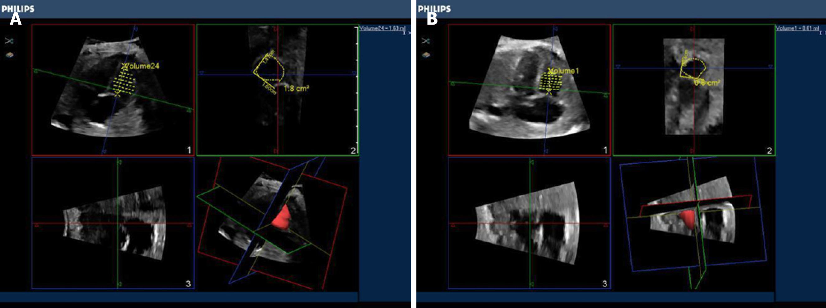
(1) Post-processing of 3D image. The image stored in the built-in hard disk in the machine was extracted for analysis using QLAB software. The atrioventricular valve opening and closing framing at end systole and diastole in the entire cardiac cycle were observed to measure the fetal right ventricular volume. End systole was defined as the moment before atrioventricular valve opening, and end diastole was defined as the moment immediately after the atrioventricular valve closed. The center point of the central position of the right ventricle was adjusted using stacked contours to draw a line along the endocardium of the right ventricular apex to the edge of the tricuspid valve, the software was then automatically stratified followed by manual outlining of the contour of each layer of the lining of the heart at the short axis view of the heart including the trabecular muscle and regulating bundle. Computer software automatically calculated the volume of the right ventricle (Figure 1). The right ventricular end-diastolic volume (REDV) and the right ventricular end-systolic volume (RESV) were measured by the same observer three times to obtain the average value.
(2) The heart function index was calculated as follows: The right stroke volume (RSV) = REDV- RESV; the right cardiac output (RCO) = RSV × fetal heart rate; the right ejection fraction = RSV/REDV.
(3) The paired Student's t test was performed by randomly retrieved images from 30 cases with the fetal right ventricular volume independently measured by two observers (twice for each observer or twice by the same observer) to evaluate the consistency of the measurement. The average value by each observer was compared.
Data input and processing were carried out using SPSS 23.0 statistical software. The quantitative data conforming to normal distribution were presented as x ± s. Spearman rank correlation analysis was used to determine the relationship between fetal right ventricular volume and cardiac function parameter changes with gestational age. In addition, the paired Student's t test was used to evaluate the consistency of the measurement by the same observer and between different observers. Differences with P < 0.05 were considered statistically significant.
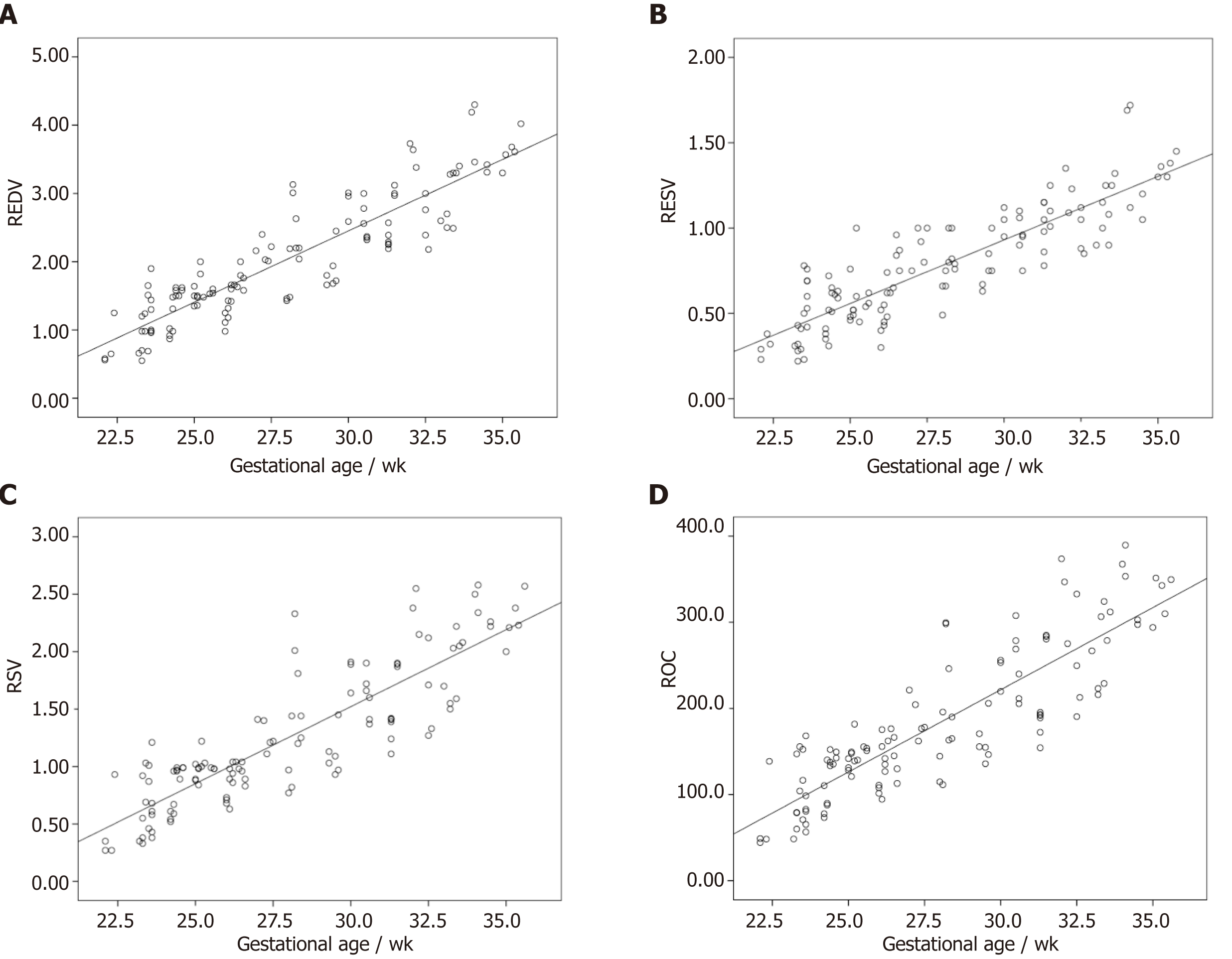
The changes in normal fetal REDV, RESV, RSV, and RCO with gestational age showed significant linear correlations, and the correlation coefficients were 0.903, 0.874, 0.866 and 0.865, respectively (P < 0.01, Figure 2, Table 1). Right ejection fraction did not change as gestational age increased, was relatively constant throughout the entire pregnancy, and was not significantly correlated with gestational age (P > 0.05, Table 1). Measurement of fetal right ventricular volume and cardiac function was carried out using the iSTIC technique (Table 2).
| Gestational age | REDV | RESV | RSV | RCO | REF |
| r | 0.903 | 0.874 | 0.866 | 0.865 | 0.120 |
| P | < 0.001 | < 0.001 | < 0.001 | < 0.001 | 0.187 |
| Gestational age, wk | Case | REDV, mL | RESV, mL | RSV, mL | RCO, mL/min | REF |
| 22–23+6 | 20 | 0.99 ± 0.34 | 0.43 ± 0.18 | 0.62 ± 0.29 | 92.23 ± 40.67 | 0.58 ± 0.12 |
| 24–25+6 | 24 | 1.43 ± 0.28 | 0.55 ± 0.15 | 0.90 ± 0.18 | 132.10 ± 26.61 | 0.62 ± 0.58 |
| 26–27+6 | 20 | 1.66 ± 0.39 | 0.69 ± 0.20 | 0.98 ± 0.22 | 149.25 ± 34.68 | 0.59 ± 0.06 |
| 28–29+6 | 16 | 2.06 ± 0.53 | 0.77 ± 0.15 | 1.29 ± 0.44 | 181.12 ± 57.03 | 0.61 ± 0.06 |
| 30–31+6 | 18 | 2.61 ± 0.33 | 1.00 ± 0.14 | 1.60 ± 0.26 | 232.58 ± 45.60 | 0.61 ± 0.05 |
| 32–33+6 | 15 | 2.98 ± 0.49 | 1.10 ± 0.17 | 1.88 ± 0.39 | 275.86 ± 55.43 | 0.63 ± 0.04 |
| 34–35+6 | 10 | 3.69 ± 0.36 | 1.36 ± 0.22 | 2.33 ± 0.18 | 335.83 ± 32.75 | 0.63 ± 0.03 |
The intraclass correlation coefficients (ICC) of REDV measured by the same observer was 0.989 with a 95% confidence interval (CI) of 0.978–0.995. The ICC of RESV was 0.978 with a 95%CI of 0.955–0.989. The ICC of REDV measured by different observers was 0.988 with a 95%CI of 0.975–0.944. The ICC of RESV was 0.988 with a 95%CI of 0.900–0.977. There was no statistical difference between the same observer and different observers when applying the iSTIC technique to measure RESV and REDV using the consistency test, and the correlation coefficients were 0.989, 0.951, 0.990, and 0.980, respectively. The results showed that there was particularly good consistency between the same observer and different observers (P < 0.001, Table 3).
| n | Observer A | Observer B | t | P | r | P | ICC | 95%CI | |
| REDV | 30 | 1.54 ± 0.82 | 1.51 ± 0.79 | 1.417 | 0.167 | 0.989 | < 0.001 | 0.988 | 0.975-0.994 |
| RESV | 30 | 0.62 ± 0.36 | 0.61 ± 0.37 | 0.384 | 0.704 | 0.951 | < 0.001 | 0.951 | 0.900-0.977 |
| First measurement | Second measurement | ||||||||
| REDV | 30 | 1.54 ± 0.81 | 1.54 ± 0.84 | 0.196 | 0.846 | 0.990 | < 0.001 | 0.989 | 0.978-0.995 |
| RESV | 30 | 0.63 ± 0.36 | 0.61 ± 0.37 | 1.731 | 0.094 | 0.980 | < 0.001 | 0.978 | 0.955-0.989 |
Fetal cardiac function can be affected by a variety of conditions and prenatal interventions. Accurate assessment of fetal cardiac function could help to understand the process of fetal heart disease, improve the accuracy of diagnosis, and determine prenatal interventions[2,5,6,16,19,20,22,23,26,30-39]. Two-dimensional ultrasound is still the traditional gold standard for diagnosis of fetal congenital heart diseases[17,20,21] but has a number of limitations in the quantitative evaluation of fetal ventricular volume and cardiac function. When abnormal fetal heart structure causes significant cardiac geometric morphology changes, accurate quantitative assessment of fetal ventricular volume and cardiac function by 2D ultrasound is difficult[3,34].
Application of the STIC imaging technique in fetal heart disorders has been studied extensively, which adds the time factor into the process of 3D data acquisition with continuous unidirectional scanning of the target area by the sensor to obtain the 3D volume data composed of each 2D slice. After completion of volume data acquisition, the software can automatically group 2D sections at the same time to reorganize the image of the entire fetal cardiac cycle, with end-systolic and end-diastolic phases of the cardiac cycle image defined according to opening and closing of the atrioven-tricular valve in post-processing[3,16,17,20-23,29,36,38-42]. Such technology overcomes the imaginary heart geometry by 2D ultrasound along with a relatively simple image acquisition process and fewer requirements for operator experience. The imaging principle determines that STIC is not a real-time imaging technology but a delayed signal. Therefore, when an abnormality of the fetal heart leads to ventricular desynchrony, the maximum and minimum volume of the fetal ventricle cannot be accurately estimated using this method.
In contrast, iSTIC can generate real-time 3D images with an electronic matrix probe used vertically, and a horizontally-arranged 2D probe to conduct matrix volume imaging. The acoustic beam can automatically rotate for large-range scanning, which can significantly reduce the acquisition time of 3D image data and improve work efficiency. The acquisition time of one image by STIC is usually 8–12 s, but iSTIC only requires 2 s. With the imaging time significantly shortened, iSTIC has solved the contradiction between slow STIC imaging and a rapid fetal heart rate and reduced the effects of fetal and maternal respiration and other factors on the quality of the image. The image resolution is thus increased[29,43,44]. In theory, accuracy of the iSTIC technique in the measurement of fetal ventricular volume is higher than STIC, which also reduces the exposure time of the fetus to ultrasound.
In this study, iSTIC technology was used to evaluate the heart function of 123 normal fetuses. The results showed that the fetal right ventricular volume, right ventricular stroke output, and RCO increased as gestational age increased, thus showing a good correlation with gestational age. Right ejection fraction did not change as the gestational age increased, which was relatively constant throughout the pregnancy and consistent with a previous report[23]. During the entire process of fetal growth and development, the circulatory system plays a critical role. Unlike adults, the fetal right ventricle has the main circulating function. As the right ventricular volume continues to increase, the increased RCO with gestational age can meet the oxygen and nutrient demands for fetal development. In contrast, due to the anatomic and structural characteristics of the right ventricle, which has an irregular shape with the regulation bundle at the apex as well as a rougher endocardial surface compared with the left ventricle, it is particularly important to find an accurate and reliable method to measure fetal right ventricular volume and cardiac function indices.
This study had some shortcomings, such as small sample size and the manual delineation of the lining of the heart in post-processing, which requires clear endocardial imaging and multiple measurements to obtain an average value and reduce measurement errors as far as possible. Due to the rough endocardium surface of the right ventricle, measurement error is inevitable. In addition, there is currently no gold standard for the assessment of fetal heart function to compare the data obtained by iSTIC technology.
In summary, iSTIC technology can be used for the quantitative measurement of fetal right ventricular volume and the evaluation of right ventricular systolic function, which has shown a number of advantages, and the application of iSTIC technology in prenatal diagnosis is worthy of further study and discussion.
Heart defects are the most common congenital malformations in fetuses. Fetal cardiac structure and function abnormalities lead to changes in ventricular volume. As ventricular volume is an important index for evaluating fetal cardiovascular development, an effective and reliable method for measuring fetal ventricular volume and cardiac function is necessary for accurate ultrasonic diagnosis and effective clinical treatment. The new intelligent spatiotemporal image correlation (iSTIC) technology acquires high-resolution volumetric images. Numerous studies have focused on fetal heart function which have not been widely accepted. The iSTIC technique was used to measure right ventricular volume in 123 normal fetuses, and to evaluate right ventricular systolic function to provide a new method for more accurate and convenient evaluation of fetal heart function.
The iSTIC technique was used to provide a new method for more accurate and convenient evaluation of fetal heart function.
One hundred twenty-three healthy gravidas with singleton gestations who visited our hospital for prenatal ultrasonography between October 2014 and September 2015 were included in the study.
The women were asked to lie flat, and a fetal echo program was begun with transabdominal scanning using an X6-1 probe. Using a 3D/4D iSTIC technology model for a 4-chamber view of the fetal heart, the 3D/4D sampling frame location was adjusted and the sampling range assured 3D/4D volume data acquisition and the sampling frame included the entire fetal heart. The fetal heart rate was measured and recorded after completion of data acquisition.
The changes in normal fetal right ventricular end-diastolic volume, right ventricular end-systolic volume, right stroke volume, and right cardiac output with gestational age showed significant linear correlations. Right ejection fraction did not change as gestational age increased, was relatively constant throughout the entire pregnancy, and was not significantly correlated with gestational age.
iSTIC technology can be used for the quantitative measurement of fetal right ventricular volume and the evaluation of right ventricular systolic function.
iSTIC can generate real-time 3D images, only requires 2 s, and is the best method now. The direction of the future research is to improve the scanning time.
Manuscript source: Unsolicited manuscript
Specialty type: Medicine, research and experimental
Country of origin: China
Peer-review report classification
Grade A (Excellent): 0
Grade B (Very good): B
Grade C (Good): C
Grade D (Fair): 0
Grade E (Poor): 0
P-Reviewer: Dave M, Lowenberg M S-Editor: Wang JL L-Editor: Filipodia E-Editor: Xing YX
| 1. | Carvalho JS, Mavrides E, Shinebourne EA, Campbell S, Thilaganathan B. Improving the effectiveness of routine prenatal screening for major congenital heart defects. Heart. 2002;88:387-391. [RCA] [PubMed] [DOI] [Full Text] [Cited by in Crossref: 244] [Cited by in RCA: 219] [Article Influence: 9.5] [Reference Citation Analysis (0)] |
| 2. | Tedesco GD, de Souza Bezerra M, Barros FS, Martins WP, Nardozza LM, Carrilho MC, Moron AF, Carvalho FH, Rolo LC, Araujo Júnior E. Reference Ranges of Fetal Cardiac Biometric Parameters Using Three-Dimensional Ultrasound with Spatiotemporal Image Correlation M Mode and Their Applicability in Congenital Heart Diseases. Pediatr Cardiol. 2017;38:271-279. [RCA] [PubMed] [DOI] [Full Text] [Cited by in Crossref: 10] [Cited by in RCA: 9] [Article Influence: 1.1] [Reference Citation Analysis (0)] |
| 3. | Godfrey ME, Messing B, Cohen SM, Valsky DV, Yagel S. Functional assessment of the fetal heart: a review. Ultrasound Obstet Gynecol. 2012;39:131-144. [RCA] [PubMed] [DOI] [Full Text] [Cited by in Crossref: 68] [Cited by in RCA: 69] [Article Influence: 5.3] [Reference Citation Analysis (0)] |
| 4. | Simioni C, Nardozza LM, Araujo Júnior E, Rolo LC, Terasaka OA, Zamith MM, Moron AF. Fetal cardiac function assessed by spatio-temporal image correlation. Arch Gynecol Obstet. 2011;284:253-260. [RCA] [PubMed] [DOI] [Full Text] [Cited by in Crossref: 8] [Cited by in RCA: 9] [Article Influence: 0.6] [Reference Citation Analysis (0)] |
| 5. | Simioni C, Araujo Júnior E, Martins WP, Rolo LC, Rocha LA, Nardozza LM, Moron AF. Fetal cardiac output and ejection fraction by spatio-temporal image correlation (STIC): comparison between male and female fetuses. Rev Bras Cir Cardiovasc. 2012;27:275-282. [RCA] [PubMed] [DOI] [Full Text] [Cited by in Crossref: 1] [Cited by in RCA: 9] [Article Influence: 0.8] [Reference Citation Analysis (0)] |
| 6. | Simioni C, Nardozza LM, Araujo Júnior E, Rolo LC, Zamith M, Caetano AC, Moron AF. Heart stroke volume, cardiac output, and ejection fraction in 265 normal fetus in the second half of gestation assessed by 4D ultrasound using spatio-temporal image correlation. J Matern Fetal Neonatal Med. 2011;24:1159-1167. [RCA] [PubMed] [DOI] [Full Text] [Cited by in Crossref: 27] [Cited by in RCA: 34] [Article Influence: 2.4] [Reference Citation Analysis (0)] |
| 7. | Rolo LC, Marcondes Machado Nardozza L, Araujo Júnior E, Simioni C, Maccagnano Zamith M, Fernandes Moron A. Reference curve of the fetal ventricular septum area by the STIC method: preliminary study. Arq Bras Cardiol. 2011;96:386-392. [RCA] [PubMed] [DOI] [Full Text] [Cited by in Crossref: 8] [Cited by in RCA: 9] [Article Influence: 0.6] [Reference Citation Analysis (0)] |
| 8. | Araujo Júnior E, Rolo LC, Rocha LA, Nardozza LM, Moron AF. The value of 3D and 4D assessments of the fetal heart. Int J Womens Health. 2014;6:501-507. [RCA] [PubMed] [DOI] [Full Text] [Full Text (PDF)] [Cited by in Crossref: 16] [Cited by in RCA: 19] [Article Influence: 1.7] [Reference Citation Analysis (0)] |
| 9. | Adler DG, Gabr M, Taylor LJ, Witt B, Pleskow D. Initial report of transesophageal EUS-guided intraparenchymal lung mass core biopsy: Findings and outcomes in two cases. Endosc Ultrasound. 2018;7:413-417. [RCA] [PubMed] [DOI] [Full Text] [Full Text (PDF)] [Cited by in Crossref: 6] [Cited by in RCA: 7] [Article Influence: 1.0] [Reference Citation Analysis (0)] |
| 10. | Sawada H, Chen JZ, Wright BC, Sheppard MB, Lu HS, Daugherty A. Heterogeneity of Aortic Smooth Muscle Cells: A Determinant for Regional Characteristics of Thoracic Aortic Aneurysms? J Transl Int Med. 2018;6:93-96. [RCA] [PubMed] [DOI] [Full Text] [Full Text (PDF)] [Cited by in Crossref: 15] [Cited by in RCA: 21] [Article Influence: 3.0] [Reference Citation Analysis (0)] |
| 11. | Castro-Pocas FM, Araújo TP, Ferreira ML, Saraiva MM. The role of endoscopic ultrasound in a case of lung cancer with jaundice. Endosc Ultrasound. 2018;7:279-281. [RCA] [PubMed] [DOI] [Full Text] [Full Text (PDF)] [Cited by in Crossref: 3] [Cited by in RCA: 5] [Article Influence: 0.7] [Reference Citation Analysis (0)] |
| 12. | Das UN. Is Aortic Aneurysm Preventable? J Transl Int Med. 2017;5:72-78. [RCA] [PubMed] [DOI] [Full Text] [Cited by in Crossref: 6] [Cited by in RCA: 6] [Article Influence: 0.8] [Reference Citation Analysis (0)] |
| 13. | Qin C, Wei B, Ma Z. Endobronchial ultrasound: Echoing in the field of pediatrics. Endosc Ultrasound. 2018;7:371-375. [RCA] [PubMed] [DOI] [Full Text] [Full Text (PDF)] [Cited by in Crossref: 3] [Cited by in RCA: 4] [Article Influence: 0.6] [Reference Citation Analysis (0)] |
| 14. | Wang J, Ouyang N, Qu L, Lin T, Zhang X, Yu Y, Jiang C, Xie L, Wang L, Wang Z, Ren S, Chen S, Huang J, Liu F, Huang W, Qin X. Effect of MTHFR A1298C and MTRR A66G Genetic Mutations on Homocysteine Levels in the Chinese Population: A Systematic Review and Meta-analysis. J Transl Int Med. 2017;5:220-229. [RCA] [PubMed] [DOI] [Full Text] [Cited by in Crossref: 7] [Cited by in RCA: 7] [Article Influence: 0.9] [Reference Citation Analysis (0)] |
| 15. | Sahai AV. EUS is trending! Endosc Ultrasound. 2018;7:353-355. [RCA] [PubMed] [DOI] [Full Text] [Full Text (PDF)] [Cited by in Crossref: 8] [Cited by in RCA: 8] [Article Influence: 1.1] [Reference Citation Analysis (0)] |
| 16. | Gómez O, Soveral I, Bennasar M, Crispi F, Masoller N, Marimon E, Bartrons J, Gratacós E, Martinez JM. Accuracy of Fetal Echocardiography in the Differential Diagnosis between Truncus Arteriosus and Pulmonary Atresia with Ventricular Septal Defect. Fetal Diagn Ther. 2016;39:90-99. [RCA] [PubMed] [DOI] [Full Text] [Cited by in Crossref: 19] [Cited by in RCA: 21] [Article Influence: 2.1] [Reference Citation Analysis (0)] |
| 17. | Bravo-Valenzuela NJ, Peixoto AB, Nardozza LM, Souza AS, Araujo Júnior E. Applicability and technical aspects of two-dimensional ultrasonography for assessment of fetal heart function. Med Ultrason. 2017;19:94-101. [RCA] [PubMed] [DOI] [Full Text] [Cited by in Crossref: 13] [Cited by in RCA: 17] [Article Influence: 2.1] [Reference Citation Analysis (0)] |
| 18. | He Y, Wang J, Gu X, Zhang Y, Han J, Liu X, Li Z. Application of spatio-temporal image correlation technology in the diagnosis of fetal cardiac abnormalities. Exp Ther Med. 2013;5:1637-1642. [RCA] [PubMed] [DOI] [Full Text] [Full Text (PDF)] [Cited by in Crossref: 11] [Cited by in RCA: 11] [Article Influence: 0.9] [Reference Citation Analysis (0)] |
| 19. | DeVore GR. Assessing fetal cardiac ventricular function. Semin Fetal Neonatal Med. 2005;10:515-541. [RCA] [PubMed] [DOI] [Full Text] [Cited by in Crossref: 68] [Cited by in RCA: 69] [Article Influence: 3.5] [Reference Citation Analysis (0)] |
| 20. | Araujo Júnior E, Tonni G, Bravo-Valenzuela NJ, Da Silva Costa F, Meagher S. Assessment of Fetal Congenital Heart Diseases by 4-Dimensional Ultrasound Using Spatiotemporal Image Correlation: Pictorial Review. Ultrasound Q. 2018;34:11-17. [RCA] [PubMed] [DOI] [Full Text] [Cited by in Crossref: 8] [Cited by in RCA: 9] [Article Influence: 1.3] [Reference Citation Analysis (0)] |
| 21. | Rocha LA, Rolo LC, Barros FS, Nardozza LM, Moron AF, Araujo Júnior E. Assessment of Quality of Fetal Heart Views by 3D/4D Ultrasonography Using Spatio-Temporal Image Correlation in the Second and Third Trimesters of Pregnancy. Echocardiography. 2015;32:1015-1021. [RCA] [PubMed] [DOI] [Full Text] [Cited by in Crossref: 14] [Cited by in RCA: 14] [Article Influence: 1.3] [Reference Citation Analysis (0)] |
| 22. | Rolo LC, Nardozza LM, Araujo Júnior E, Simioni C, Zamith MM, Moron AF. [Assessment of the fetal mitral and tricuspid valves areas development by three-dimensional ultrasonography]. Rev Bras Ginecol Obstet. 2010;32:426-432. [RCA] [PubMed] [DOI] [Full Text] [Cited by in Crossref: 7] [Cited by in RCA: 7] [Article Influence: 0.5] [Reference Citation Analysis (0)] |
| 23. | Zhang J, Zhou Q, Peng Q, Zhao Y, Gong Z. [Assessment of the right ventricle function of fetus by spatio-temporal image correlation]. Zhong Nan Da Xue Xue Bao Yi Xue Ban. 2015;40:486-494. [RCA] [PubMed] [DOI] [Full Text] [Cited by in RCA: 1] [Reference Citation Analysis (0)] |
| 24. | Xiong Y, Liu T, Wu Y, Xu JF, Ting YH, Yeung Leung T, Lau TK. Comparison of real-time three-dimensional echocardiography and spatiotemporal image correlation in assessment of fetal interventricular septum. J Matern Fetal Neonatal Med. 2012;25:2333-2338. [RCA] [PubMed] [DOI] [Full Text] [Cited by in Crossref: 14] [Cited by in RCA: 15] [Article Influence: 1.2] [Reference Citation Analysis (0)] |
| 25. | Xiong Y, Liu T, Gan HJ, Wu Y, Xu JF, Ting YH, Leung TY, Lau TK. Detection of the fetal conotruncal anomalies using real-time three-dimensional echocardiography with live xPlane imaging of the fetal ductal arch view. Prenat Diagn. 2013;33:462-466. [RCA] [PubMed] [DOI] [Full Text] [Cited by in Crossref: 7] [Cited by in RCA: 8] [Article Influence: 0.7] [Reference Citation Analysis (0)] |
| 26. | Zhang J, Zhou Q, Zhao Y, Peng Q, Gong Z, Long X. Evaluation of right ventricular function in fetal hypoplastic left heart syndrome using spatio-temporal image correlation (STIC). Cardiovasc Ultrasound. 2016;14:12. [RCA] [PubMed] [DOI] [Full Text] [Full Text (PDF)] [Cited by in Crossref: 4] [Cited by in RCA: 5] [Article Influence: 0.6] [Reference Citation Analysis (0)] |
| 27. | Xiong Y, Chen M, Chan LW, Ting YH, Fung TY, Leung TY, Lau TK. Scan the fetal heart by real-time three-dimensional echocardiography with live xPlane imaging. J Matern Fetal Neonatal Med. 2012;25:324-328. [RCA] [PubMed] [DOI] [Full Text] [Cited by in Crossref: 7] [Cited by in RCA: 8] [Article Influence: 0.6] [Reference Citation Analysis (0)] |
| 28. | Lu Y, Yang T, Luo H, Deng F, Cai Q, Sun W, Song H. Visualization and quantitation of fetal movements by real-time three-dimensional ultrasound with live xPlane imaging in the first trimester of pregnancy. Croat Med J. 2016;57:474-481. [RCA] [PubMed] [DOI] [Full Text] [Full Text (PDF)] [Cited by in Crossref: 3] [Cited by in RCA: 5] [Article Influence: 0.6] [Reference Citation Analysis (0)] |
| 29. | Zhu M, Ashraf M, Zhang Z, Streiff C, Shimada E, Kimura S, Schaller T, Song X, Sahn DJ. Real Time Three-Dimensional Echocardiographic Evaluations of Fetal Left Ventricular Stroke Volume, Mass, and Myocardial Strain: In Vitro and In Vivo Experimental Study. Echocardiography. 2015;32:1697-1706. [RCA] [PubMed] [DOI] [Full Text] [Cited by in Crossref: 5] [Cited by in RCA: 5] [Article Influence: 0.5] [Reference Citation Analysis (0)] |
| 30. | Meyer-Wittkopf M, Cole A, Cooper SG, Schmidt S, Sholler GF. Three-dimensional quantitative echocardiographic assessment of ventricular volume in healthy human fetuses and in fetuses with congenital heart disease. J Ultrasound Med. 2001;20:317-327. [PubMed] [DOI] [Full Text] |
| 31. | Blessberger H, Binder T. NON-invasive imaging: Two dimensional speckle tracking echocardiography: basic principles. Heart. 2010;96:716-722. [RCA] [PubMed] [DOI] [Full Text] [Cited by in Crossref: 226] [Cited by in RCA: 255] [Article Influence: 17.0] [Reference Citation Analysis (0)] |
| 32. | Blessberger H, Binder T. Two dimensional speckle tracking echocardiography: clinical applications. Heart. 2010;96:2032-2040. [RCA] [PubMed] [DOI] [Full Text] [Cited by in Crossref: 80] [Cited by in RCA: 84] [Article Influence: 6.0] [Reference Citation Analysis (0)] |
| 33. | Zhao L, Wu Y, Chen S, Ren Y, Chen P, Niu J, Li C, Sun K. Feasibility Study on Prenatal Cardiac Screening Using Four-Dimensional Ultrasound with Spatiotemporal Image Correlation: A Multicenter Study. PLoS One. 2016;11:e0157477. [RCA] [PubMed] [DOI] [Full Text] [Full Text (PDF)] [Cited by in Crossref: 10] [Cited by in RCA: 10] [Article Influence: 1.1] [Reference Citation Analysis (0)] |
| 34. | Rolo LC, Santana EF, da Silva PH, Costa Fda S, Nardozza LM, Tonni G, Moron AF, Araujo Júnior E. Fetal cardiac interventricular septum: volume assessment by 3D/4D ultrasound using spatio-temporal image correlation (STIC) and virtual organ computer-aided analysis (VOCAL). J Matern Fetal Neonatal Med. 2015;28:1388-1393. [RCA] [PubMed] [DOI] [Full Text] [Cited by in Crossref: 14] [Cited by in RCA: 16] [Article Influence: 1.5] [Reference Citation Analysis (0)] |
| 35. | Hamill N, Yeo L, Romero R, Hassan SS, Myers SA, Mittal P, Kusanovic JP, Balasubramaniam M, Chaiworapongsa T, Vaisbuch E, Espinoza J, Gotsch F, Goncalves LF, Lee W. Fetal cardiac ventricular volume, cardiac output, and ejection fraction determined with 4-dimensional ultrasound using spatiotemporal image correlation and virtual organ computer-aided analysis. Am J Obstet Gynecol. 2011;205:76.e1-76.10. [RCA] [PubMed] [DOI] [Full Text] [Full Text (PDF)] [Cited by in Crossref: 57] [Cited by in RCA: 55] [Article Influence: 3.9] [Reference Citation Analysis (0)] |
| 36. | Messing B, Cohen SM, Valsky DV, Shen O, Rosenak D, Lipschuetz M, Yagel S. Fetal heart ventricular mass obtained by STIC acquisition combined with inversion mode and VOCAL. Ultrasound Obstet Gynecol. 2011;38:191-197. [RCA] [PubMed] [DOI] [Full Text] [Cited by in Crossref: 25] [Cited by in RCA: 25] [Article Influence: 1.8] [Reference Citation Analysis (0)] |
| 37. | Barros FS, Moron AF, Rolo LC, Rocha LA, Martins WP, Tonni G, Nardozza LM, Araujo Júnior E. Fetal myocardial wall area: constructing a reference range by means of spatiotemporal image correlation in the rendering mode. Fetal Diagn Ther. 2015;37:44-50. [RCA] [PubMed] [DOI] [Full Text] [Cited by in Crossref: 9] [Cited by in RCA: 14] [Article Influence: 1.3] [Reference Citation Analysis (0)] |
| 38. | Turan S, Turan OM, Desai A, Harman CR, Baschat AA. First-trimester fetal cardiac examination using spatiotemporal image correlation, tomographic ultrasound and color Doppler imaging for the diagnosis of complex congenital heart disease in high-risk patients. Ultrasound Obstet Gynecol. 2014;44:562-567. [RCA] [PubMed] [DOI] [Full Text] [Cited by in Crossref: 34] [Cited by in RCA: 22] [Article Influence: 2.0] [Reference Citation Analysis (0)] |
| 39. | Tanis JC, Mohammed N, Bennasar M, Martinez JM, Bijnens B, Crispi F, Gratacos E. Online versus offline spatiotemporal image correlation (STIC) M-mode for the evaluation of cardiac longitudinal annular displacement in fetal growth restriction. J Matern Fetal Neonatal Med. 2018;31:1845-1850. [RCA] [PubMed] [DOI] [Full Text] [Cited by in Crossref: 4] [Cited by in RCA: 6] [Article Influence: 0.8] [Reference Citation Analysis (0)] |
| 40. | Yagel S, Cohen SM, Shapiro I, Valsky DV. 3D and 4D ultrasound in fetal cardiac scanning: a new look at the fetal heart. Ultrasound Obstet Gynecol. 2007;29:81-95. [RCA] [PubMed] [DOI] [Full Text] [Cited by in Crossref: 120] [Cited by in RCA: 105] [Article Influence: 5.8] [Reference Citation Analysis (0)] |
| 41. | Tedesco GD, de Souza Bezerra M, Barros FSB, Martins WP, Nardozza LMM, Mattar R, Moron AF, Rolo LC, Araujo Júnior E. Fetal Heart Function by Tricuspid Annular Plane Systolic Excursion and Ventricular Shortening Fraction Using STIC M-Mode: Reference Ranges and Validation. Am J Perinatol. 2017;34:1354-1361. [RCA] [PubMed] [DOI] [Full Text] [Cited by in Crossref: 5] [Cited by in RCA: 6] [Article Influence: 0.8] [Reference Citation Analysis (0)] |
| 42. | Messing B, Gilboa Y, Lipschuetz M, Valsky DV, Cohen SM, Yagel S. Fetal tricuspid annular plane systolic excursion (f-TAPSE): evaluation of fetal right heart systolic function with conventional M-mode ultrasound and spatiotemporal image correlation (STIC) M-mode. Ultrasound Obstet Gynecol. 2013;42:182-188. [RCA] [PubMed] [DOI] [Full Text] [Cited by in Crossref: 42] [Cited by in RCA: 52] [Article Influence: 4.3] [Reference Citation Analysis (0)] |
| 43. | Acar P, Battle L, Dulac Y, Peyre M, Dubourdieu H, Hascoet S, Groussolles M, Vayssière C. Real-time three-dimensional foetal echocardiography using a new transabdominal xMATRIX array transducer. Arch Cardiovasc Dis. 2014;107:4-9. [RCA] [PubMed] [DOI] [Full Text] [Cited by in Crossref: 8] [Cited by in RCA: 9] [Article Influence: 0.8] [Reference Citation Analysis (0)] |
| 44. | Liu J, Wang Y, Zhao H, Liu W. Spatio-temporal image correlation rendering mode visualizes the specific location and surrounding structure of ventricular septal defect. Clin Anat. 2019;32:408-420. [RCA] [PubMed] [DOI] [Full Text] [Cited by in Crossref: 3] [Cited by in RCA: 3] [Article Influence: 0.5] [Reference Citation Analysis (0)] |


