Published online Sep 16, 2016. doi: 10.12998/wjcc.v4.i9.290
Peer-review started: April 6, 2016
First decision: May 17, 2016
Revised: June 8, 2016
Accepted: July 11, 2016
Article in press: July 13, 2016
Published online: September 16, 2016
Processing time: 158 Days and 3 Hours
This case report describes the surgical removal of cementoblastoma associated with apicoectomy and endodontic therapy. The patient, an 18-year-old man, presented pain in the region of the mandibular body on the right side. On clinical exam, bone expansion was observed in the region at the bottom of the vestibular sulcus, pain on palpation, slight extrusion of tooth 46 with presence of pulp vitality. Radiographic exams demonstrated the presence of a radiopaque area and discrete radiolucent halo associated with the root of tooth 46, suggesting the diagnosis of cementoblastoma. Endodontic treatment of tooth 46 was performed and exeresis of the lesion by apicoectomy. Twelve months after the first surgery, recurrence of the lesion was observed, and a new apicoectomy was necessary, this time up to the middle third of the root. Clinical radiographic control 12 mo after the second surgical intervention demonstrated absence of signs and symptoms, radiographic repair, with tooth 46 shown to be fully functional.
Core tip: The present clinical case demonstrated that surgical treatment associated with endodontic treatment was effective for the treatment of cementoblastoma. However, the patient must be followed-up due to the possibility of recurrence of this lesion. The importance of these findings demonstrated that the treatment of cementoblastoma may be conservative with maintenance of the affected teeth.
- Citation: Costa BC, de Oliveira GJPL, Chaves MDGAM, da Costa RR, Gabrielli MFR, Guerreiro-Tanomaru JM, Tanomaru-Filho M. Surgical treatment of cementoblastoma associated with apicoectomy and endodontic therapy: Case report. World J Clinical Cases 2016; 4(9): 290-295
- URL: https://www.wjgnet.com/2307-8960/full/v4/i9/290.htm
- DOI: https://dx.doi.org/10.12998/wjcc.v4.i9.290
The cementoblastoma is a benign odontogenic tumor of ectomesenchymal origin that preferentially affects the roots of mandibular molars or premolars, in patients in the age range of 20-30 years, with slight prevalence in the male sex[1-3]. Its low prevalence (less than 6% of all odontogenic tumors)[4,5], generates difficulties with documentation about the standard treatment for this tumor[1-3,6].
Due to the high rates of recurrence (approximately 31.7%) associated with incomplete removal of the lesion[1], the treatment of cementoblastoma most indicated in the literature, is removal of the tooth together with the tumor[6,7]. However, endodontic treatment of the affected tooth associated with apicoectomy during removal of the lesion is cited as an alternative[1], allowing the tooth to be maintained in function[1].
Conservative treatment of cementoblastoma lesions allows the tooth to be maintained. However, this has rarely been documented in the literature. Therefore, the aim of this case report was to demonstrate the treatment of cementoblastoma by means of endodontic treatment and removal of the lesion associated with apicoectomy and maintenance of the affected tooth after two surgical interventions. Follow-up of one year after the second surgical intervention showed clinical and radiographic success.
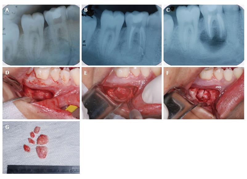
The patient, an 18-year-old man, presented to the service of the Endodontic Clinic at the Dental School of Araraquara (FOAr-Unesp) with complaints of pain in the mandibular body on the right side for the past 3 mo. On clinical exam, the patient presented with discrete facial asymmetry, bone expansion in the region at the bottom of the vestibular sulcus, pain on palpation, slight extrusion of tooth 46, and positive response to the pulp vitality test. In the imaging exams, a radiopaque mass was observed, measuring approximately 1.5 mm in diameter, with discrete radiolucent halo associated with the root apex of tooth 45 (Figure 1A).
Considering the clinical and imaging exams, the treatment initially proposed was worn of the occlusal surface of the tooth involved, to alleviate the painful symptoms. After 30 d of follow-up, the patient returned because of the pain. In view of the new situation, and considering the diagnostic hypothesis of cementoblastoma, the option taken was to perform endodontic treatment of tooth 46 and exeresis of the lesion by apicoectomy and peripheral osteotomy (Figure 1B-G).
Endodontic treatment (pulpectomy) was performed in two sessions with the use of a calcium hydroxide-based intracanal dressing. Biomechanical preparation was performed by using crown-down technique, and manual instruments (K files 10 and 15) associated with rotary instrumentation, by using an electric motor and ProDesign S files of the Easy® System, in accordance with the protocol of the manufacturer (Easy Equipamentos Odontológicos®, Belo Horizonte - MG, Brazil). Surgical intervention was performed to remove the lesion in the apical third portion of the root associated with the lesion.
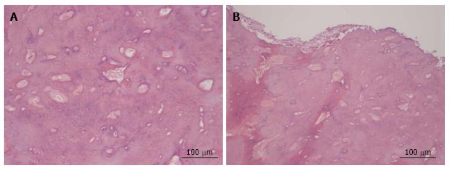
The lesion removed had the aspect of a rounded mass of hardened consistency, measuring 1.3 mm in diameter. The tissue was sent for histopathological exam that provided description of the material as being calcified, presenting superimposed lamellae and presence of dentin united to cementoid material. In the central portion of the lesion, a cementoid structure with blood vessels was verified, presenting superimposed lamellae and basophilic material, while the peripheral portion of the lesion presented irregular fibrous tissue, with an aspect of cementoid tissue and presence of blood vessels (Figure 2A and B). According to the histopathological report, the diagnosis presented was that of cementoblastoma.
In the cone beam computed tomography exam performed during follow-up of the case after one year, recurrence of the lesion was observed (Figure 3). In view of the new condition, the option taken was to perform a second paraendodontic surgery. In this procedure, a more aggressive root section was performed up to the middle level of the roots affected by the lesion (Figure 4A). At present the patient is undergoing post-operative follow-up of 12 mo, without painful symptoms and showing complete remission of the lesion (Figure 4B). The Table 1 shows the different types of treatment and recurrence rates of the cementoblastoma demonstrated in case series previously reported in the literature.
| Ref. | No. of cases | Type of treatment | Recurrence rates |
| Abrams et al[9],1974 | 7 | Extraction of the affected tooth | No recurrences after 6-10 yr |
| Ulmansky et al[6], 1994 | 5 | Extraction of the affected tooth in two cases Association between the surgical enucleation of the lesion associated and the treatment in three cases | No recurrences after two years |
| Brannon et al[1], 2004 | 44 | En bloc resection in 5 cases Extraction of the involved tooth with concurrent tumor removal in 26 cases Root amputation with tumor removal in 2 cases Curettage of the lesion without tooth extraction in 6 cases Extraction of the involved tooth with no attempt to remove the tumor in four cases | There were recurrences in 13 cases (37.1%) between 4-24 mo after the treatment |
| Prakash et al[2], 2013 | 3 | Extraction of the affected tooth | No recurrences |
The cementoblastoma is a rare benign odontogenic lesion, and reports of cases documented represent a large part of the information with regard to therapeutic conduct[2,3,8]. Diagnosis of this lesion must be made by association of clinical, radiographic and histopathological methods[9,10]. It is important to perform differential diagnosis with other lesions that present characteristics to those of the cementoblastoma, such as cement-bone dysplasia, ossifying fibroma, hypercementosis and osteoblastoma[9,11].
In the case reported, all the methods cited were used to identify the lesion. Clinical exam demonstrated that the lesion promoted painful symptoms, increase in volume associated with the vestibule of tooth 46, and presence of pulp vitality. Radiographic exam detected the presence of a radiopaque lesion with radiolucent halo associated with the root of tooth 46. The histopathological exam demonstrated that the lesion presented a dense central region, with birefringent material similar to that of bone, with the presence of lines of reversion, while the peripheral portion presented foci of vascularization and connective tissue. All of these signs have been reported in the diagnostic reports of cementoblastoma[1-3,6,8,9,12].
The treatment applied in the case was removal of the lesion associated with a portion of the root surface after endodontic treatment. In spite of the presence of pulp vitality, in cases of cementoblastoma, the surgical act of removing the lesion and part of the tooth root must be performed after endodontic treatment[6]. This treatment has been applied in other studies with good clinical results and absence[13,14] of recurrence or rate of recurrence similar to that of treatment by tooth extraction[1]. However, removal of the affected tooth is still the option most indicated in case reports and previous studies[2,7,15].
One of the reasons proposed for applying removal of the tooth as treatment for cementoblastoma is the high rate of recurrence of these lesions, associated with its incomplete removal[1]. A series of cases has described that cases of cementoblastoma treated with a protocol similar to that performed in the present report may present recurrence of the lesion[6], as verified in this clinical case after one year of follow-up. Considering that the cementoblastoma arises from the uncontrolled proliferation of the cementoid matrix by cementoblasts[16] and that these cells are not present in the middle and cervical portions of the root[17], in the second surgical approach, removal of the root was performed up to the middle third, thereby eliminating all the cellular cement that could have led to the origin of the second lesion. However, this procedure may not eliminate the possibility of the lesion recurrence since other ethological factors as the uncontrolled induction of the cementoblasts differentiation by the epithelial rests of Malassez cells can be the trigger of the cementoblastoma lesions[18]. After one year of follow-up of this surgical procedure, repair with bone neoformation was verified in the region of the lesion.
Therefore, the authors concluded that the surgical treatment associated with endodontic treatment was effective for the treatment of cementoblastoma. However, follow-up must be performed due to the possibility of recurrence of this lesion. Moreover, apicoectomy must be performed at the level of the middle third of the root to prevent the remaining cementoblasts from inducing recurrence of the lesion.
The patient, an 18-year-old man with complaints of pain in the mandibular body on the right side for the past 3 mo.
Cementoblastoma.
Cement-bone dysplasia, ossifying fibroma, hypercementosis and osteoblastoma.
A radiopaque mass was observed, measuring approximately 1.5 mm in diameter, with discrete radiolucent halo associated with the root apex of tooth 45.
Cementoblastoma.
Endodontic treatment and surgical removal of the lesion.
The treatment of this condition normally is the tooth extraction. In this case report we propose a more conservative therapy. The association of the endodontic treatment and surgical removal of the lesion permits the maintenance of the tooth.
Apicoectomy must be performed at the level of the middle third of the root to prevent the remaining cementoblasts from inducing recurrence of the lesion.
The authors report on a surgical treatment of a cementoblastoma associated with apicoectomy and endodontic therapy. The case report is well written and Brannon’s series is reported as well as satisfactory literature review.
Manuscript source: Invited manuscript
Specialty type: Medicine
Country of origin: Brasil
Peer-review report classification
Grade A (Excellent): 0
Grade B (Very good): B, B
Grade C (Good): 0
Grade D (Fair): D
Grade E (Poor): 0
P- Reviewer: Razek AAKA, Yura S, Zhang ZM S- Editor: Ji FF L- Editor: A E- Editor: Lu YJ
| 1. | Brannon RB, Fowler CB, Carpenter WM, Corio RL. Cementoblastoma: an innocuous neoplasm? A clinicopathologic study of 44 cases and review of the literature with special emphasis on recurrence. Oral Surg Oral Med Oral Pathol Oral Radiol Endod. 2002;93:311-320. [RCA] [PubMed] [DOI] [Full Text] [Cited by in Crossref: 93] [Cited by in RCA: 70] [Article Influence: 3.0] [Reference Citation Analysis (0)] |
| 2. | Prakash R, Gill N, Goel S, Verma S. Cementoblastoma. A report of three cases. N Y State Dent J. 2013;79:41-43. [PubMed] |
| 3. | Jeyaraj CP. Clinicopathological study of a case of cementoblastoma and an update on review of literature. J Oral Maxillofac Surg Med Pathol. 2014;26:415-420. [RCA] [DOI] [Full Text] [Cited by in Crossref: 2] [Cited by in RCA: 2] [Article Influence: 0.2] [Reference Citation Analysis (0)] |
| 4. | Ohki K, Kumamoto H, Nitta Y, Nagasaka H, Kawamura H, Ooya K. Benign cementoblastoma involving multiple maxillary teeth: report of a case with a review of the literature. Oral Surg Oral Med Oral Pathol Oral Radiol Endod. 2004;97:53-58. [RCA] [PubMed] [DOI] [Full Text] [Cited by in Crossref: 37] [Cited by in RCA: 30] [Article Influence: 1.4] [Reference Citation Analysis (0)] |
| 5. | Avelar RL, Antunes AA, Santos Tde S, Andrade ES, Dourado E. Odontogenic tumors: clinical and pathology study of 238 cases. Braz J Otorhinolaryngol. 2008;74:668-673. [RCA] [PubMed] [DOI] [Full Text] [Full Text (PDF)] [Cited by in Crossref: 51] [Cited by in RCA: 56] [Article Influence: 3.3] [Reference Citation Analysis (0)] |
| 6. | Ulmansky M, Hjørting-Hansen E, Praetorius F, Haque MF. Benign cementoblastoma. A review and five new cases. Oral Surg Oral Med Oral Pathol. 1994;77:48-55. [RCA] [PubMed] [DOI] [Full Text] [Cited by in Crossref: 48] [Cited by in RCA: 35] [Article Influence: 1.1] [Reference Citation Analysis (0)] |
| 7. | Iannaci G, Luise R, Iezzi G, Piattelli A, Salierno A. Multiple cementoblastoma: a rare case report. Case Rep Dent. 2013;2013:828373. [RCA] [PubMed] [DOI] [Full Text] [Full Text (PDF)] [Cited by in Crossref: 2] [Cited by in RCA: 4] [Article Influence: 0.3] [Reference Citation Analysis (0)] |
| 8. | Sharma N. Benign cementoblastoma: A rare case report with review of literature. Contemp Clin Dent. 2014;5:92-94. [PubMed] |
| 9. | Abrams AM, Kirby JW, Melrose RJ. Cementoblastoma. A clinical-pathologic study of seven new cases. Oral Surg Oral Med Oral Pathol. 1974;38:394-403. [PubMed] |
| 10. | de Noronha Santos Netto J, Machado Cerri J, Miranda AM, Pires FR. Benign fibro-osseous lesions: clinicopathologic features from 143 cases diagnosed in an oral diagnosis setting. Oral Surg Oral Med Oral Pathol Oral Radiol. 2013;115:e56-e65. [RCA] [PubMed] [DOI] [Full Text] [Cited by in Crossref: 48] [Cited by in RCA: 49] [Article Influence: 3.8] [Reference Citation Analysis (0)] |
| 11. | Rao GS, Kamalapur MG, Acharya S. Focal cemento-osseous dysplasia masquerading as benign cementoblastoma: A diagnostic dilemma. J Oral Maxillofac Pathol. 2014;18:150. [RCA] [PubMed] [DOI] [Full Text] [Full Text (PDF)] [Cited by in Crossref: 7] [Cited by in RCA: 6] [Article Influence: 0.5] [Reference Citation Analysis (0)] |
| 12. | Mortazavi H, Baharvand M, Rahmani S, Jafari S, Parvaei P. Radiolucent rim as a possible diagnostic aid for differentiating jaw lesions. Imaging Sci Dent. 2015;45:253-261. [RCA] [PubMed] [DOI] [Full Text] [Full Text (PDF)] [Cited by in Crossref: 17] [Cited by in RCA: 15] [Article Influence: 1.5] [Reference Citation Analysis (0)] |
| 13. | Biggs JT, Benenati FW. Surgically treating a benign cementoblastoma while retaining the involved tooth. J Am Dent Assoc. 1995;126:1288-1290. [PubMed] |
| 14. | Gulses A, Bayar GR, Aydin C, Sencimen M. A case of a benign cementoblastoma treated by enucleation and apicoectomy. Gen Dent. 2012;60:e380-e382. [PubMed] |
| 15. | Harada H, Omura K, Mogi S, Okada N. Cementoblastoma arising in the maxilla of an 8-year-old boy: a case report. Int J Dent. 2011;2011:384578. [RCA] [PubMed] [DOI] [Full Text] [Full Text (PDF)] [Cited by in Crossref: 6] [Cited by in RCA: 7] [Article Influence: 0.5] [Reference Citation Analysis (0)] |
| 16. | Dadhich AS, Nilesh K. Cementoblastoma of posterior maxilla involving the maxillary sinus. Ann Maxillofac Surg. 2015;5:127-129. [RCA] [PubMed] [DOI] [Full Text] [Full Text (PDF)] [Cited by in Crossref: 7] [Cited by in RCA: 8] [Article Influence: 0.8] [Reference Citation Analysis (0)] |
| 17. | Nanci A, Bosshardt DD. Structure of periodontal tissues in health and disease. Periodontol 2000. 2006;40:11-28. [RCA] [PubMed] [DOI] [Full Text] [Cited by in Crossref: 320] [Cited by in RCA: 401] [Article Influence: 21.1] [Reference Citation Analysis (0)] |
| 18. | Farea M, Husein A, Halim AS, Berahim Z, Nurul AA, Mokhtar KI, Mokhtar K. Cementoblastic lineage formation in the cross-talk between stem cells of human exfoliated deciduous teeth and epithelial rests of Malassez cells. Clin Oral Investig. 2016;20:1181-1191. [RCA] [PubMed] [DOI] [Full Text] [Cited by in RCA: 1] [Reference Citation Analysis (0)] |


