Published online Aug 16, 2015. doi: 10.12998/wjcc.v3.i8.732
Peer-review started: March 17, 2015
First decision: April 13, 2015
Revised: May 16, 2015
Accepted: July 3, 2015
Article in press: July 9, 2015
Published online: August 16, 2015
Processing time: 157 Days and 7.9 Hours
Meckel’s diverticula incarcerated in a hernia were first described anecdotally by Littré, a French surgeon, in 1700. Meckel, a German anatomist and surgeon, explained the pathophysiology of this disease 100 years later. In addition, a congenital paraduodenal mesocolic hernia, known as a Treitz hernia, is a rare cause of small bowel obstruction. These hernias are caused by an abnormal rotation of the primitive midgut, resulting in a right or left paraduodenal hernia. We treated a patient presenting with pain and diagnosed extraluminal air in the abdomen after a computed tomography examination. We performed a laparotomy and found a combination of these two seldomly occurring congenital diseases, incarceration and perforation of Meckel’s diverticulum in a left paraduodenal hernia. We performed a thorough review of the literature, and this report is the first to describe a patient with a combination of these two rare conditions. We considered the case regarding the variety of terminology as well as the treatment options of these conditions.
Core tip: Meckel’s diverticula incarcerated in a hernia were first described anecdotally by Littré, a French surgeon, in 1700. Meckel, a German anatomist and surgeon, explained the pathophysiology of this disease 100 years later. In addition, a congenital paraduodenal mesocolic hernia, known as a Treitz hernia, is a rare cause of small bowel obstruction. We performed a thorough review of the literature, and this report is the first to describe a patient with a combination of these two seldomly occurring congenital diseases, incarceration and perforation of Meckel’s diverticulum in a left paraduodenal hernia.
- Citation: Gerdes C, Akkermann O, Krüger V, Gerdes A, Gerdes B. Incarceration of Meckel's diverticulum in a left paraduodenal Treitz' hernia. World J Clin Cases 2015; 3(8): 732-735
- URL: https://www.wjgnet.com/2307-8960/full/v3/i8/732.htm
- DOI: https://dx.doi.org/10.12998/wjcc.v3.i8.732
Internal hernias are the protrusion of viscus through an opening in the peritoneal or mesenteric fold[1]; they account for 0.2% to 0.9% of small bowel obstructions[2] and are classified as acquired or congenital. Littré’s hernia is a very rare type of hernia. In 1700, a French surgeon, Alexandre de Littré, first described a type of inguinal hernia that was different from known hernias[3]. An antimesenteric sacculation, not the whole bowel’s circumference, protruded through a defect in the abdominal wall, which explains why Littré’s patient did not die from a bowel obstruction. Littré could not discover a reason for the bowel eversion; however, 100 years later (1809), Meckel, an anatomist and surgeon, theorized that the persistence of the omphalomesenteric duct could be causative[3]. The omphalomesenteric duct, also known as the Ductus vitellinus, joins the midgut to the yolk sac. If the duct does not obliterate, a Meckel’s diverticulum remains, which is the most common congenital anomaly of the gastrointestinal tract. Only 4% to 16% of the patients with this true diverticulum experience complications[2] such as bleeding from ectopic gastric mucosa or obstruction caused by intussusceptions or adhesion bands[4].
For several decades, studies have described cases presenting with incarceration of Meckel’s diverticulum in different types of hernias, including inguinal, femoral or umbilical hernias[5], as well as in incarcerations in incisional hernias[6].
Another type of hernia, congenital paraduodenal mesocolic hernias, represents 53% of internal hernias[7]. Paraduodenal hernias, known as Treitz hernias, had been described by others before being defined for the first time by Treitz in 1857[1]. The two types of paraduodenal hernias are left and right, and left paraduodenal hernias occur more frequently than right ones[7]. Left paraduodenal hernias are caused by an abnormal rotation of the primitive midgut[8] when the small bowel invaginates into an avascular segment of the left mesocolon[9]. The bowel prolapses through the Landzert’s fossa behind the fourth segment, the ascending duodenum. It locates behind the inferior mesenteric vein and left colic artery[10] and becomes trapped between the mesocolon and the posterior abdominal wall in a hernial sac within the leaflets of the left colon mesentery[7]. Bowel loops are present between the stomach and pancreas. Recent studies have reported laparoscopic management of this condition[11].
We performed surgery on a patient with an incarceration of Meckel’s diverticulum in a left paraduodenal internal hernia.
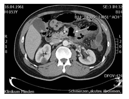
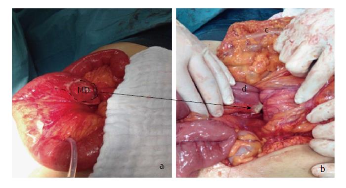
A 53-year-old man reported having unspecific moderate pressure for two weeks before presenting with sudden acute and severe pain in his upper abdomen. The patient consulted the emergency department of our hospital. During the clinical examination, we found moderate abdominal epigastric tenderness. The white blood cell count (16.9 g/L) and C-reactive protein were elevated (203.5 mg/L), and the patient underwent a computed tomography examination of the abdomen (Figure 1). Extraluminal air was detected beside the duodenojejunal flexure. Therefore, we performed surgery. During the laparotomy, we found that the small bowel was fixed adjacent to the duodenojejunal flexure, with the abdomen appearing to be “empty”. Small bowel was herniating through a tight, well-formed, fibrous ring. After having mobilized the non-ischemic bowel, we identified a Meckel’s diverticulum showing a fibrous ring at the base and perforation at the tip (Figure 2). The small bowel, including a 5-cm Meckel’s diverticulum that was located 60 cm oral of the ileocecal valve, was slipped through the hernial orifice of a left paraduodenal hernia. We repositioned the bowel and resected the diverticulum. Histologically, we found venous congestion of the diverticulum with a perforation at the tip and signs of peritonitis. After having opened the bursa omentalis by dividing the gastrocolic ligament, we opened the hernial sac and lavaged and drained the bursa. In the hernial sac, we found local fibrinous peritonitis. We closed the hernial orifice with an omental flap. The postoperative course of the patient was uneventful despite infection with clostridium difficile, which was treated with metronidazole.
The terminology in this medical field is highly variable. In a scientific paper in 1785, Gottlieb Richter, a professor of medicine in Göttingen, described the diversity of hernias. He counted intestinal wall hernias and Littré’s hernias among the small hernias that include all the hernias in which only one side of the bowel is incarcerated[3]. His descriptions are probably the reason that the clinical terminology of these hernias is confusing. All intestinal wall hernias are frequently referred to as Richter-Littré hernias or Littré’s hernias, particularly by German speakers. In 1888, Frederic Treves differentiated Littré’s hernias from Richter’s hernias[3]. A Littré’s hernia is present if the content of the hernia exists in a Meckel’s diverticulum whereas Richter’s hernia applies to all intestinal wall hernias.
Paraduodenal hernias as well as Meckel’s diverticula are rare, and their discovery is frequently delayed because they induce diffuse symptoms. The symptoms of Meckel’s diverticula could be similar to those of appendicitis. The diagnosis of paraduodenal hernias is challenging because of the lack of explicit symptoms. Paraduodenal hernias are rare causes of bowel obstruction, and if strangulation is present, the mortality rate could approach 50%[12]. There is a discussion of left paraduodenal hernias being identified as congenital mesocolic hernias[13].
To treat our patient, we considered the basic principles of hernia surgery by reduction and assessment of the hernial contents and correction of the defect. First, we repositioned the sac contents, which frequently consist of the majority of the small bowel, so that following the laparotomy, the surgeon accomplishes a lack of small bowel in the abdominal cavity[14], as in our case. After the reduction, we unexpectedly found the perforated Meckel’s diverticulum with a fibrous ring at its base. We suspected that the Meckel’s diverticulum was incarcerated in an additional pouch of the large hernial sac and that the fibrous ring had to be interpreted as an incarceration ring. This finding was confirmed histologically. We resected the Meckel’s diverticulum with a stapler. To prevent a postoperative abscess in the preformed hernial sac caused by contamination resulting from perforation of the Meckel’s diverticulum, we opened the bursa omentalis by transection of the gastrocolic ligament, resected the hernial sac and lavaged it. We performed a tension-free closure of the paraduodenal hernial orifice with the use of an omental flap to prevent recurrence of the hernia. To achieve that surgical goal, we used an absorbable running suture, taking care to avoid injury to the nearby mesenteric vessels. Our finding was an incarcerated and perforated Meckel’s diverticulum in a Treitz hernia.
In a thorough search of the literature, we could not find a similar case. Developing a hernia induced by a combination of these two seldomly seen congenital defects is a rare coincidence. There were several case reports of patients with a Littré’s hernia during the 20th century, as well as other rare conditions including Meckel’s diverticulum in an obturator hernia[5]. A recently published case described a patient with a congenital defect of the mesocolon in which an ileal loop with a Meckel’s diverticulum was prolapsed[4]. In that case, there was a defect in the right transverse mesocolon; however, the patient presented without a hernial sac, and thus there was no true internal hernia. To the best of our knowledge, this paper is the first report of an incarcerated and perforated Meckel’s diverticulum in a confirmed internal paraduodenal hernia.
Acute abdominal pain.
Temperate abdominal epigastric tenderness.
Diseases causing hollow organ perforation in the upper abdomen.
White blood cell count: 16.9 g/L; C-reactive protein: 203.5 mg/L.
Extraluminal air was found in the computed tomography-scan near the ascending duodenum.
Perforated Meckel’s diverticulum with venous congestion and peritonitis in a left paraduodenal hernia.
Reduction of the bowel, resection of the Meckel’s diverticulum and the hernia sac and closure of the hernial orifice with an omental flap.
This case report presents the clinical characteristics of a perforated Meckel’s diverticulum in a Treitz’ hernia. This new clinical situation has to be considered in differential diagnosis of acute abdomen.
The authors have described an interesting case.
P- Reviewer: Guan YS, Ivanov KD, Mirbagheri N S- Editor: Wang JL L- Editor: A E- Editor: Liu SQ
| 1. | Mehra R, Pujahari AK. Right paraduodenal hernia: report of two cases and review of literature. Gastroenterol Rep (Oxf). 2014;Epub ahead of print. [PubMed] |
| 2. | Sagar J, Kumar V, Shah DK. Meckel’s diverticulum: a systematic review. J R Soc Med. 2006;99:501-505. [RCA] [PubMed] [DOI] [Full Text] [Cited by in Crossref: 374] [Cited by in RCA: 196] [Article Influence: 10.3] [Reference Citation Analysis (0)] |
| 3. | Lauschke H, Kaminski M, Stratmann H, Hirner A. [Littré’s hernia--clinical aspects and review of the history]. Chirurg. 1999;70:953-956. [RCA] [PubMed] [DOI] [Full Text] [Cited by in Crossref: 6] [Cited by in RCA: 8] [Article Influence: 0.3] [Reference Citation Analysis (0)] |
| 4. | Wu SY, Ho MH, Hsu SD. Meckel’s diverticulum incarcerated in a transmesocolic internal hernia. World J Gastroenterol. 2014;20:13615-13619. [RCA] [PubMed] [DOI] [Full Text] [Full Text (PDF)] [Cited by in CrossRef: 8] [Cited by in RCA: 7] [Article Influence: 0.6] [Reference Citation Analysis (0)] |
| 5. | Jacob TJ, Gaikwad P, Tirkey AJ, Rajinikanth J, Raj JP, Muthusami JC. Perforated obturator Littre hernia. Can J Surg. 2009;52:E77-E78. [PubMed] |
| 6. | Citgez B, Yetkin G, Uludag M, Karakoc S, Akgun I, Ozsahin H. Littre’s hernia, an incarcerated ventral incisional hernia containing a strangulated meckel diverticulum: report of a case. Surg Today. 2011;41:576-578. [RCA] [PubMed] [DOI] [Full Text] [Cited by in Crossref: 14] [Cited by in RCA: 16] [Article Influence: 1.1] [Reference Citation Analysis (0)] |
| 7. | Shi Y, Felsted AE, Masand PM, Mothner BA, Nuchtern JG, Rodriguez JR, Vasudevan SA. Congenital left paraduodenal hernia causing chronic abdominal pain and abdominal catastrophe. Pediatrics. 2015;135:e1067-e1071. [RCA] [PubMed] [DOI] [Full Text] [Cited by in Crossref: 9] [Cited by in RCA: 9] [Article Influence: 0.9] [Reference Citation Analysis (0)] |
| 8. | Licciardello A, Rapisarda C, Conti P, Trombatore G. Small bowel obstruction caused by an unusual variant of paraduodenal hernia. The “middle congenital mesocolic hernia”: case report. J Gastrointest Surg. 2014;18:1514-1517. [RCA] [PubMed] [DOI] [Full Text] [Cited by in Crossref: 9] [Cited by in RCA: 9] [Article Influence: 0.8] [Reference Citation Analysis (0)] |
| 9. | Falk GA, Yurcisin BJ, Sell HS. Left paraduodenal hernia: case report and review of the literature. BMJ Case Rep. 2010;2010. [RCA] [PubMed] [DOI] [Full Text] [Cited by in Crossref: 14] [Cited by in RCA: 19] [Article Influence: 1.3] [Reference Citation Analysis (0)] |
| 10. | Meyers MA. Paraduodenal hernias. Radiologic and arteriographic diagnosis. Radiology. 1970;95:29-37. [RCA] [PubMed] [DOI] [Full Text] [Cited by in Crossref: 83] [Cited by in RCA: 60] [Article Influence: 1.1] [Reference Citation Analysis (0)] |
| 11. | Tomino T, Itoh S, Yoshida D, Nishida T, Kawanaka H, Ikeda T, Kohnoe S, Shirabe K, Maehara Y. Right paraduodenal hernia successfully treated with laparoscopic surgery. Asian J Endosc Surg. 2015;8:87-90. [RCA] [PubMed] [DOI] [Full Text] [Cited by in Crossref: 10] [Cited by in RCA: 14] [Article Influence: 1.4] [Reference Citation Analysis (0)] |
| 12. | Akyildiz H, Artis T, Sozuer E, Akcan A, Kucuk C, Sensoy E, Karahan I. Internal hernia: complex diagnostic and therapeutic problem. Int J Surg. 2009;7:334-337. [RCA] [PubMed] [DOI] [Full Text] [Cited by in Crossref: 25] [Cited by in RCA: 41] [Article Influence: 2.6] [Reference Citation Analysis (0)] |
| 13. | Willwerth BM, Zollinger RM, Izant RJ. Congenital mesocolic (paraduodenal) hernia. Embryologic basis of repair. Am J Surg. 1974;128:358-361. [PubMed] |
| 14. | Poh BR, Sundaramurthy SR, Mirbagheri N. Left paraduodenal hernia causing small bowel obstruction. J Gastrointest Surg. 2014;18:1377-1378. [RCA] [PubMed] [DOI] [Full Text] [Cited by in Crossref: 7] [Cited by in RCA: 8] [Article Influence: 0.7] [Reference Citation Analysis (0)] |


