Published online Jul 16, 2014. doi: 10.12998/wjcc.v2.i7.309
Revised: May 14, 2014
Accepted: May 28, 2014
Published online: July 16, 2014
Processing time: 154 Days and 10.4 Hours
Esophageal atresia with tracheo-oesophageal fistula (TEF) occurs in 1 in 3500 live births. Anorectal malformation is found to be associated with 14% of TEF. Esophageal atresia with TEF is a congenital anomaly which classically presents as excessive frothing from the mouth and respiratory distress. Rarely gastric position of the feeding tube in a case of TEF can be obtained delaying the diagnosis of TEF. We had an uncommon situation where a nasogastric tube reached the stomach through the trachea and tracheo-esophageal fistula, leading to misdiagnosis in a case of esophageal atresia with tracheoesophageal fistula. By using a stiff rubber catheter instead of a soft feeding tube for the diagnosis of esophageal atresia and TEF, such situation can be avoided.
Core tip: Esophageal atresia with tracheoesophageal fistula is congenital anomaly which presents as excessive froathing from mouth and respiratory distress. It can be suspected when a nasogastric tube difficult to insert into stomach or radiographically presence coiled nasogastric tube in pharynx. We had an uncommon situation where a nasogastric tube reached the stomach through the trachea and tracheo-esophageal fistula, leading to misdiagnosis in a case of esophageal atresia with tracheoesophageal fistula. Similar clinical situations can be avoided by using a stiff rubber catheter instead of a soft feeding tube for the diagnosis of esophageal atresia and tracheo-oesophageal fistula.
- Citation: Kamble RS, Gupta R, Gupta A, Kothari P, Dikshit KV, Kesan K, Mudkhedkar K. Passage of nasogastric tube through tracheo-esophageal fistula into stomach: A rare event. World J Clin Cases 2014; 2(7): 309-310
- URL: https://www.wjgnet.com/2307-8960/full/v2/i7/309.htm
- DOI: https://dx.doi.org/10.12998/wjcc.v2.i7.309
Esophageal atresia with tracheooesophageal fistula (TEF) is a congenital anomaly which classically presents as excessive frothing from the mouth and respiratory distress. It is diagnosed by inability to pass a catheter into the stomach which usually gets stuck at 10 to 12 cm from the mouth. We had an uncommon situation where a nasogastric tube reached the stomach through the trachea and tracheo-esophageal fistula, leading to misdiagnosis in a case of esophageal atresia with tracheooesophageal fistula.
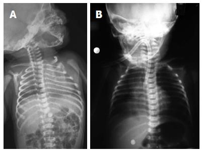
A one day old, full term, male child (2.7 kg) was referred from a peripheral Hospital as a case of imperforate anus with cleft lip and palate. At the peripheral hospital a 7 Fr nasogastric tube was inserted which had bilious aspirate. A chest X-ray showed the nasogastric tube in the stomach (Figure 1A). The baby had excessive oral secretions and bilateral chest crepitations along with cleft lip and palate. Prone crosstable X-ray was suggestive of high anorectal malformation. The patient was taken for diverting colostomy.
During intubation the anesthetist noticed that the nasogastric tube was passing through the trachea. Findings were reconfirmed by laryngoscopy and laryngotracheoesophagial cleft was ruled out. We tried to pass a number 10 stiff red rubber catheter through esophagus but were unable to pass beyond 10 cm. A diagnosis of esophageal atresia (EA) with tracheooesophageal fistula (TEF) was suspected and X-ray chest was repeated with the red rubber catheter in situ which confirmed the diagnosis (Figure 1B). Right posterolateral thoracotomy was done. The patient had tracheoesophageal fistula type III with a Wide fistula, short upper pouch and a long gap. The fistula was ligated and a Left sided cervical oseophagostomy with feeding gastrostomy was done. Transverse colostomy was done for imperforate anus.
The baby was shifted on ventilator and was successfully weaned off at post operative day 5. The baby is now 4 mo old and is doing well on follow up. He is awaiting definitive management for anorectal malformation, esophageal replacement, cleft lip and cleft palate repair.
Esophageal atresia with TEF occurs in 1 in 3500 live births[1]. Anorectal malformation is found to be associated with 14% of TEF[1]. TEF classically presents as excessive frothing from the mouth and regurgitation, choking and coughing after feed. There is a routine practice of passing a 5Fr or 6Fr infant feeding tube through the nose in patients of imperforate anus to decompress the obstructed intestinal tract and also to rule out associated esophageal atresia. If the radiograph shows a coiled catheter in the upper esophageal pouch one can suspect esophageal atresia.
Rarely gastric position of the feeding tube in a case of TEF can be obtained delaying the diagnosis of TEF[2,3]. In this case we did not suspect an esophageal atresia as the patient came with the IFT in the stomach with bilious aspirate and this had been confirmed radio graphically. The feeding tube could reach the stomach from the upper pouch then into the tracheal route and then through the TEF. A peculiar pathological anatomy and a weak cough reflex made this occurrence possible. Only three such cases have been reported so far in literature[2,3]. If an esophageal atresia is suspected on clinical grounds the ideal test would be to pass a stiff red rubber catheter through the mouth and note the resistance. Radiographs should be taken with a red rubber catheter in situ which will show the position of the catheter tip. Barium esophagography is not usually advised due to the risk of aspiration pneumonitis. Instead a small amount of air can be used as contrast[1].
In a conclusion, all neonates with excessive frothing and respiratory distress should be evaluated for TEF. Similar clinical situations can be avoided by using a stiff rubber catheter instead of a soft feeding tube for the diagnosis of EA and TEF.
Authors’ came across a uncommon situation, a neonate refered from peripheral hospital with nasogastric tube passed through tracheo-esophageal fistula into the stomach.
The baby had excessive oral secretions and bilateral chest crepitations along with cleft lip and palate.
Prone crosstable X-ray was suggestive of high anorectal malformation.
The case report is interesting and well written, the field of the report is focused on pediatric.
P- Reviewer: Lisotti A S- Editor: Wen LL L- Editor: A E- Editor: Wu HL
| 1. | Carroll MH, Arnold GC. Congenital anomalies of Esophagus. Pediatric Surgery, 7th Edition. Philadelphia: Elsevier Saunders 2012; 893-918. |
| 2. | Hombalkar NN, Dhanawade S, Hombalkar P, Vaze D. Esophageal atresia with tracheo-esophageal fistula: Accidental transtracheal gastric intubation. J Indian Assoc Pediatr Surg. 2009;14:224-225. [RCA] [PubMed] [DOI] [Full Text] [Cited by in Crossref: 4] [Cited by in RCA: 6] [Article Influence: 0.4] [Reference Citation Analysis (0)] |
| 3. | Rani RS, Shenoi A, Nagesh NK, Ramachandra C. Inadvertent passage of infant feeding tube into the stomach through a tracheo-esophageal fistula. Indian Pediatr. 2000;37:96-97. [PubMed] |


