Published online Jul 16, 2014. doi: 10.12998/wjcc.v2.i7.289
Revised: April 17, 2014
Accepted: May 16, 2014
Published online: July 16, 2014
Processing time: 236 Days and 4.3 Hours
Surgical treatment for cervical kyphotic deformity is still controversial. Circumferential approach has been well described in the literature but long terms outcomes are not well reported. Important to decide the correct treatment option is the preoperative radiological exams to value the type of deformity (flexible or fixed). We report the case of a 67-year-old woman affected by a severe cervical kyphotic deformity who underwent combined anterior/posterior surgical approach, getting a good reduction of the deformity and an optimal stability in a long term follow up.
Core tip: The choice of the treatment for cervical kyphotic deformity takes into account preoperative radiological exams which allow the classification of the deformity in flexible or fixed.
- Citation: Landi A, Marotta N, Mancarella C, Dugoni DE, Tarantino R, Delfini R. 360° fusion for realignment of high grade cervical kyphosis by one step surgery: Case report. World J Clin Cases 2014; 2(7): 289-292
- URL: https://www.wjgnet.com/2307-8960/full/v2/i7/289.htm
- DOI: https://dx.doi.org/10.12998/wjcc.v2.i7.289
The treatment option for correcting a cervical kyphotic deformity is currently controversial. Lots of studies examined the one/stage combined anterior-posterior treatment, although the rate of fusion and the long term follow up controls are rarely mentioned in the literature[1-7]. We present the case of a 67-year-old woman affected by a severe cervical kyphosis. We performed a one-step combined anterior/posterior approach to correct the deformity, getting a good reduction of kyphosis and good stability in a long term follow up. We enclosed that the evidence of motility by dynamic X-rays permits a good anterior decompression and reduction only by discectomy, fusion and plating, without need of multiple corpectomy. Treatment must be completed with posterior fixation and fusion. These strategies could be performed in one step, and shows a good reduction and optimal stability in a long term follow-up. Immobilizing with hard collar and neurophysiological monitoring remains fundamental for the safe and efficacy of this treatment.
A 67-year-old woman affected by a severe cervical kyphotic deformity, came to our attention complaining 4 mohistory of bilateral cervicobrachialgia. She didn’t have any significant medical diseases; she denied to have ever suffered of ankylosing spondylitis, osteogenesis imperfecta, rheumatoid arthritis or Larsen syndrome.
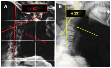
Neurological examination showed moderate upper limbs weakness, confirmed by signs of radicular suffering on the ElectroMioGraphy (EMG). The patient performed standard and flexion/extension cervical spine X-ray demonstrating a severe cervical kyphosis [preop. Ishihara index 64.18% and (8) preop. Angle of Jackson +62°] apparently fixed on the dynamic X-ray. Performed, then, cervical spine computed tomography (CT) and Magnetic Resonance Imaging showing important myeloradicular compression at C5-C6 and C4-C5.
The immediate preoperative exam that the patient had to perform was a cervical spine X-ray on the bed with a pillow under the shoulders in prevision to apply a traction system. This exam showed a good reduction of kyphosis (+23° according to Jackson) due to the motor unit C4-C5 mobility (Figure 1). It was therefore decided not to apply traction and to proceed with combined anterior/posterior surgical approach using neurophysiological monitoring SomatoSensory evoked potentials (SSEP), EMG and motor evoked potentials (MEP).
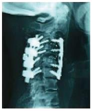
The first surgical step was the anterior approach, with hyperextension of the neck of the patient. The reduction status of the kyphosis was assessed under fluoroscopic visualization; patient underwent left anterior presternocleidomastoid-precarotid approach, anterior decompression through microdiscectomy C3-C4 and C4-C5, followed by interbody fusion using a carbon fiber cage in lordosis and anterior plate fixed on C3-C4-C5-C6. The second step was represented by the posterior approach, so the patient was placed in prone position. A C3/C6 posterior stabilization according to magerl was performed followed by posterolateral fusion at all levels. At the end of the procedure a Philadelphia brace was applied. The postoperative CT and X-ray control (Figure 2) and after 3 and 6 mo showed a good reduction of kyphosis (postop. Ishihara index 32.38 % and postop angle of Jackson + 19° with reduction of kyphosis of 31.8% according to Ishihara and 43° according to Jackson ), and a good anterior and posterolateral arthrodesis (Figure 3). The patient presented a complete regression of the upper limbs deficit and of the cervicobrachialgia. Six months later the patient is symptoms free.
Cervical kyphosis can be classified into two different groups: type 1 (flexible cervical kyphosis) and type 2 (fixed cervical kyphosis). The treatment for flexible cervical kyphosis (type 1) posturally reducible is usually a posterior stabilization with fusion to guarantee the stability of the cervical spine[6,7]. Alternatively, some authors have reported the use of anterior only surgery for flexible cervical kyphosis as discectomy and corporectomy. This approach is useful for anterior column load sharing however it is not required for deformity correction. Fixed cervical kyphosis characterized by postural rigidity needs circumferential approach[8-10]. The circumferential approach for the correction of cervical kyphotic deformity is well described in the literature although the long term controls are not always diriment on the real fusion of the correction[11,12]. The debate is not whether or not to perform circumferential correction, but if it is more useful to perform a multiple anterior discectomy or multiple corpectomy. In the literature it is described as the execution of multiple discectomies has a greater potential for correction of kyphosis in relation to: (1) a greater kyphosis correction due to the possibility of including more lordotic cages at multiple levels, so to restore a greater degree of lordosis; and (2) a greater possibility of fusion because of a larger cage-bone interface compared with the use of a Harms mesh or an expansion cage.
In cases of multilevel cervical stenosis, the choice of surgical technique (discectomy vs corpectomy) mainly depends on the location of the stenosis. In the case of a kyphotic deformity however, the choice depends on the mobility or less of the bodies involved in the deformity[10-16]. It is also important to note that such deformities occur slowly over the time and are frequently the product of a degenerative process that affects the patient for many years: this include a wrong postural attitude that causes a compensatory hypertrophy of the supporting muscles of the neck, which may hide a metameric mobility of kyphosis. In our case, in fact, the dynamic exam in the upright position showed that the kyphosis appeared fixed and cannot be reduced[10].
The X-ray performed in the bed with a pillow under the shoulder, allowed us to appreciate how such kyphosis was actually not fixed on two vertebrae. This made us choose a multiple discectomy and not corpectomy with consequent greater angular correction of kyphosis. Another important aspect in the evaluation of the motor unit motility is the reactive ankylosis of the articular processes. In the severe kyphosis the fusion occurs both at the level of the disc and of the articular masses, thus preventing a good correction of kyphosis after performing the anterior approach. When the articular masses are ankylotic it is necessary a 3-step surgery. The first step is represented by a posterior approach to release the ankylotic articular masses in order to allow the reduction of kyphosis. The second step is the anterior approach with discectomy, and the third step is represented by the posterior approach again to fix and to make arthrodesis[17-20].
To this end, it is crucial to recognize accurately the real motility of the vertebral body, and to do that it is important to perform a dynamic exam without load, to eliminate the reactive contracture of the muscles supporting the neck. Useful for this purpose is to perform radiographic examinations in supine position with supports placed at the base of the neck which put the cervical spine in hyperextension eliminating the analgesic muscle contracture. Another aspect to highlight is the use of intraoperative neurophysiological monitoring, in particular SEPP and MEP; these allow a direct observation of the function of the spinal cord during the entire procedure. The neurophysiological monitoring is important especially during the correction of the spinal deformities because, as these constituted and organized by time, may have led to spinal cord adaptations that may break with the correction maneuvers, resulting in severe neurological deficits. Their use avoid for such eventuality.
In a conclusion, we enclosed that the evidence of motility by dynamic X-rays permits a good anterior decompression and reduction only by discectomy, fusion and plating, without need of multiple corpectomy. Treatment have to be completed with posterior fixation and fusion. These strategies could be performed in one step, and shows a good reduction and optimal stability in a long term follow-up. Immobilizing with hard collar and neurophysiological monitoring remains fundamental for the safe and efficacy of these treatment.
The patient complained a 4-mo history of bilateral cervicobrachialgia.
At the neurological examination the patient presented moderate upper limb weakness and severe cervical kyphotic deformity.
Through radiological exams we put cervical kyphotic deformity due to degenerative process in differential diagnosis with neoplastic and infective pathologies.
The patient underwent computed tomography scan, magnetic resonance imaging and dynamic X-ray. The most important preoperative exam was X-ray performed in supine position with a pillow under the shoulder.
The patient suffered severe cervical kyphotic deformity.
The authors performed combined anterior\posterior surgical approach using neurophysiological monitoring SomatoSensory Evoked Potentials, ElectroMioGraphy and Motor Evoked Potentials.
The choice of surgical treatment depends on the mobility or less of the bodies involved in the deformity.
Cervical kyphosis is a progressive deformity; circumferential approach means one step combined anterior/posterior approach.
It is important to recognize the real motility of the vertebral body, and to do that it is necessary to perform a dynamic exam without load, to eliminate the reactive contracture of the muscles supporting the neck.
The author introduces an efficient surgical treatment for severe cervical spine deformity, and to improve the quality of life.
P- Reviewers: Tong C, Zhan RY S- Editor: Wen LL L- Editor: A E- Editor: Wu HL
| 1. | Abumi K, Shono Y, Taneichi H, Ito M, Kaneda K. Correction of cervical kyphosis using pedicle screw fixation systems. Spine (Phila Pa 1976). 1999;24:2389-2396. [PubMed] |
| 2. | Kanter AS, Wang MY, Mummaneni PV. A treatment algorithm for the management of cervical spine fractures and deformity in patients with ankylosing spondylitis. Neurosurg Focus. 2008;24:E11. [PubMed] |
| 3. | Ferch RD, Shad A, Cadoux-Hudson TA, Teddy PJ. Anterior correction of cervical kyphotic deformity: effects on myelopathy, neck pain, and sagittal alignment. J Neurosurg. 2004;100:13-19. [PubMed] |
| 4. | Zdeblick TA, Bohlman HH. Cervical kyphosis and myelopathy. Treatment by anterior corpectomy and strut-grafting. J Bone Joint Surg Am. 1989;71:170-182. [PubMed] |
| 5. | Lin D, Zhai W, Lian K, Kang L, Ding Z. Anterior versus posterior approach for four-level cervical spondylotic myelopathy. Orthopedics. 2013;36:e1431-e1436. [PubMed] |
| 6. | Spivak J, Giordano CP. Cervical kyphosis. The Textbook of Spinal Surgery. 2nd ed. Philadelphia: Lippincott-Raven 1997; 1027–1038. |
| 7. | Ganju A, Ondra SL, Shaffrey CI. Cervical kyphosis. Tech Orthop. 2003;17:345–354. [RCA] [DOI] [Full Text] [Cited by in Crossref: 17] [Cited by in RCA: 17] [Article Influence: 0.7] [Reference Citation Analysis (0)] |
| 8. | Herman JM, Sonntag VK. Cervical corpectomy and plate fixation for postlaminectomy kyphosis. J Neurosurg. 1994;80:963-970. [PubMed] |
| 9. | Steinmetz MP, Kager CD, Benzel EC. Ventral correction of postsurgical cervical kyphosis. J Neurosurg (Spine 2). 2002;97:1–7. |
| 10. | Batzdorf U, Batzdorff A. Analysis of cervical spine curvature in patients with cervical spondylosis. Neurosurgery. 1988;22:827-836. [PubMed] |
| 11. | McAfee PC, Bohlman HH, Ducker TB, Zeidman SM, Goldstein JA. One-stage anterior cervical decompression and posterior stabilization. A study of one hundred patients with a minimum of two years of follow-up. J Bone Joint Surg Am. 1995;77:1791-1800. [PubMed] |
| 12. | Mummaneni PV, Dhall SS, Rodts GE, Haid RW. Circumferential fusion for cervical kyphotic deformity. J Neurosurg Spine. 2008;9:515-521. [PubMed] |
| 13. | Takeshita K, Murakami M, Kobayashi A, Nakamura C. Relationship between cervical curvature index (Ishihara) and cervical spine angle (C2--7). J Orthop Sci. 2001;6:223-226. [PubMed] |
| 14. | Chang SW, Kakarla UK, Maughan PH, DeSanto J, Fox D, Theodore N, Dickman CA, Papadopoulos S, Sonntag VK. Four-level anterior cervical discectomy and fusion with plate fixation: radiographic and clinical results. Neurosurgery. 2010;66:639-646; discussion 646-647. [PubMed] |
| 15. | Chibbaro S, Benvenuti L, Carnesecchi S, Marsella M, Pulerà F, Serino D, Gagliardi R. Anterior cervical corpectomy for cervical spondylotic myelopathy: experience and surgical results in a series of 70 consecutive patients. J Clin Neurosci. 2006;13:233-238. [PubMed] |
| 16. | Hussain M, Nassr A, Natarajan RN, An HS, Andersson GB. Corpectomy versus discectomy for the treatment of multilevel cervical spine pathology: a finite element model analysis. Spine J. 2012;12:401-408. [PubMed] |
| 17. | Kawakami M, Tamaki T, Iwasaki H, Yoshida M, Ando M, Yamada H. A comparative study of surgical approaches for cervical compressive myelopathy. Clin Orthop Relat Res. 2000;129-136. [PubMed] |
| 18. | Konya D, Ozgen S, Gercek A, Pamir MN. Outcomes for combined anterior and posterior surgical approaches for patients with multisegmental cervical spondylotic myelopathy. J Clin Neurosci. 2009;16:404-409. [PubMed] |
| 19. | Lin Q, Zhou X, Wang X, Cao P, Tsai N, Yuan W. A comparison of anterior cervical discectomy and corpectomy in patients with multilevel cervical spondylotic myelopathy. Eur Spine J. 2012;21:474-481. [PubMed] |
| 20. | Jiang SD, Jiang LS, Dai LY. Anterior cervical discectomy and fusion versus anterior cervical corpectomy and fusion for multilevel cervical spondylosis: a systematic review. Arch Orthop Trauma Surg. 2012;132:155-161. [PubMed] |


