Published online Jun 16, 2014. doi: 10.12998/wjcc.v2.i6.232
Revised: February 11, 2014
Accepted: April 9, 2014
Published online: June 16, 2014
Processing time: 176 Days and 2.7 Hours
Meckel’s diverticulum is a very common congenital anomaly of the gastrointestinal tract but many cases remain asymptomatic and are diagnosed incidentally during laparoscopic or other surgical procedures. Cases of femoral hernia involving Meckel’s diverticulum are rare, with less than 50 cases reported in the literature since Littre published the first description of this coincident condition over 300 years ago. While all true “Littre’s hernias” contain a Meckel’s diverticulum, the involved anatomical sites are various, the most common being the inner groin (inguinal), the outer groin (femoral), and the belly button (umbilical). Complications of Littre’s hernias include incarceration, strangulation, necrosis, and perforation. Herein, we describe a case of Littre’s hernia that involved an incarcerated Meckel’s diverticulum in a femoral hernia that was diagnosed upon investigation of symptomology manifesting from perforation and was successfully managed by surgical resection with stapler devices.
Core tip: Meckel’s diverticulum is most commonly diagnosed congenital anomaly of the gastrointestinal tract. Any hernia containing a Meckel’s diverticulum is designated as a Littre’s hernia. Although rare in overall incidence, the most common complications of Littre’s hernias are perforation, bowel obstruction secondary to strangulation, and incarceration within the hernial sac. In this case study, we present and share the diagnosis and successfully management of a case of incarcerated Meckel’s diverticulum in a femoral hernia (Littre’s hernia) with perforation.
- Citation: Yagmur Y, Akbulut S, Can MA. Gastrointestinal perforation due to incarcerated Meckel’s diverticulum in right femoral canal. World J Clin Cases 2014; 2(6): 232-234
- URL: https://www.wjgnet.com/2307-8960/full/v2/i6/232.htm
- DOI: https://dx.doi.org/10.12998/wjcc.v2.i6.232
The most commonly diagnosed congenital abnormality of the small intestine, Meckel’s diverticulum, occurs when a portion of the vitelline duct fails to properly regress into the antimesenteric border of the terminal ileum at the end of the seventh week of gestation. As such, a Meckel’s diverticulum contains all layers of the intestinal wall and is classified as a true diverticulum[1-6]. Estimates of the incidence of this condition have ranged from 0.3% to 3%, but its frequent asymptomatic nature may belie its true rates among the general population. Meckel’s diverticula can be diagnosed as incident findings in laparoscopic or laparotomic examinations for unexplained symptoms or other conditions that have manifested clinical symptoms. Any hernia containing a Meckel’s diverticulum is designated as a Littre’s hernia. The most common sites of these coincident anatomical abnormalities are the inner groin (inguinal hernia, approximately 50%), the outer groin (femoral hernia, approximately 20%), and the belly button (umbilical hernia, approximately 20%). Although rare in overall incidence, the most common complications of Littre’s hernias are perforation, bowel obstruction secondary to strangulation, and incarceration within the hernial sac[1]. Herein, we report the diagnosis and successfully management of a case of incarcerated Meckel’s diverticulum in a femoral hernia (Littre’s hernia) with ileal perforation.
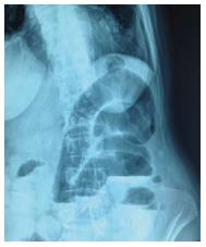
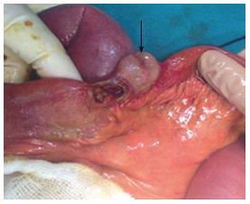
A 73-year-old female presented to our Emergency Department with severe abdominal pain, nausea, and vomiting; the symptoms had begun one week previous, but had increased in severity over the last two days. The patient also reported the last instances of flatulence and defecation being three days previous. Results of standard laboratory blood tests were normal, hemoglobin: 13.2 g/dL (normal range, NR: 12.5-16.0), blood urea nitrogen: 22 mg/dL (NR: 10-50 mg/dL), and creatinine: 0.5 mg/dL (NR: 0.4-1.2 mg/dL), with the exception of a high white blood cell count [16000/μL (NR: 4100-11200/μL)] and neutrophil ratio [89% (NR: 50%-78%)]. In physical examination, auscultation revealed obstructive and hyperactive bowel sounds and palpation revealed serious tenderness with rebound pain that was particularly robust in the right lower quadrant. Abdominal radiographic examination showed air-fluid levels (Figure 1). Nasogastric decompression was performed in the Emergency Department. The collected clinical symptoms and findings from biochemical analysis and radiological examination suggested acute mechanical small bowel obstruction possibly related to internal herniation or perforation. The patient was prepared for exploration. Laparotomy with a midline incision revealed a Meckel’s diverticulum with an ileal segment incarcerated in the right femoral canal and which had perforated the antimesenteric border of the ileum (Figure 2). The abnormality was managed by first removing the diverticulum through the femoral canal, suturing the femoral canal closed, resecting the partial ileal segment that included the perforated ileal segment and Meckel’s diverticulum (15 cm in length), and creating a side-to-side anastomosis with stapler devices (Endo-GIA Stapling System; Covidian, Dublin, Ireland). Post-operative recovery was uncomplicated and the patient was discharged to home. Pathological analysis of the resected tissues showed no signs of gastric or pancreatic metaplasia.
In the first five weeks of normal gestational development, the Meckel’s diverticulum is the intestinal portion of the omphaloenteric duct through which the midgut communicates with the umbilical vesicle. Located at the antimesenteric border of the ileum, about 30 to 90 cm from the ileocecal valve, it can measure between 3 and 6 cm in length and is usually approximately 2 cm in diameter[2]. Failure of the tissue to regress by gestational week 7 is not fatal, and the congenital abnormality may remain asymptomatic (and undetected) throughout life. The lifetime complication rate estimated to be approximately 4%, with a higher incidence in males[4], and the most common complications are bleeding, inflammation, and obstruction[5].
Cases of femoral hernia involving Meckel’s diverticulum are rare, with less than 50 cases reported in the literature since Littre published the first description of this coincident condition over 300 years ago[2]. A true Littre’s hernia contains a Meckel’s diverticulum alone, but cases of mixed Littre’s hernia containing ileum or other abdominal viscera have been reported[3]. Perforation of the Meckel’s diverticulum may occur due to peptic ulceration or compromised circulation and luminal patency at the narrow neck of the hernia[3].
Preoperative diagnosis of a strangulated Littre’s hernia is unlikely, as the presenting signs and symptoms are subtler and evolve more slowly than those of strangulated small intestine. Fever, pain, and signs of intestinal obstruction occur late or not at all. Moreover, there may be no specific sign of bowel involvement, other than local inflammation surrounding the hernia until an enterocutaneous fistula develops[5]. The septuagenarian case described herein presented with signs of obstruction (i.e., serious abdominal tenderness, rebound pain, and air-fluid levels).
In Littre’s hernia, obstruction can occur if the base of the diverticulum is broad enough to cause narrowing of the intestinal lumen. Presenting symptoms are tender mass proximal to a hernial orifice with nausea, vomiting, abdominal pain, local groin pain and swelling. Pyrexia is normally associated with strangulation[5]. Repair of the Littre’s hernia consists of local diverticulum resection and herniorrhaphy. In cases of perforation, care must be taken to not contaminate the hernia field[2], and resection of the diverticulum ileal loop with end-to-end or side-to-side anastomosis is the recommended treatment.
A 73-year-old female presented to our Emergency Department with severe abdominal pain, nausea, and vomiting; the symptoms had begun one week previous, but had increased in severity over the last two days.
Auscultation revealed obstructive and hyperactive bowel sounds and palpation revealed serious tenderness with rebound pain that was particularly robust in the right lower quadrant.
Abdominal radiographic examination showed air-fluid levels. Laparotomy with a midline incision revealed a Meckel’s diverticulum with an ileal segment incarcerated in the right femoral canal and which had perforated the antimesenteric border of the ileum
Pathological analysis of the resected tissues showed no signs of gastric or pancreatic metaplasia.
Care must be taken to not contaminate the hernia field, and resection of the diverticulum ileal loop with end-to-end or side-to-side anastomosis is the recommended treatment.
The authors described the case of gastrointestinal perforation due to incarcerated Meckel’s diverticulum in right femoral canal. The authors also demonstrated the figure of air-fluid levels detected by abdominal radiography.
P- Reviewers: Contini S, Lin JK, Masaki T, Pellicano R, Sadik R S- Editor: Gou SX L- Editor: A E- Editor: Wu HL
| 1. | Perlman JA, Hoover HC, Safer PK. Femoral hernia with strangulated Meckel’s diverticulum (Littre’s hernia). Am J Surg. 1980;139:286-289. [RCA] [PubMed] [DOI] [Full Text] [Cited by in Crossref: 34] [Cited by in RCA: 24] [Article Influence: 0.5] [Reference Citation Analysis (0)] |
| 2. | Skandalakis PN, Zoras O, Skandalakis JE, Mirilas P. Littre hernia: surgical anatomy, embryology, and technique of repair. Am Surg. 2006;72:238-243. [PubMed] |
| 3. | Jacob TJ, Gaikwad P, Tirkey AJ, Rajinikanth J, Raj JP, Muthusami JC. Perforated obturator Littre hernia. Can J Surg. 2009;52:E77-E78. [PubMed] |
| 4. | Dumper J, Mackenzie S, Mitchell P, Sutherland F, Quan ML, Mew D. Complications of Meckel’s diverticula in adults. Can J Surg. 2006;49:353-357. [PubMed] |
| 5. | Mirza MS. Incarcerated Littre’s femoral hernia: case report and review of the literature. J Ayub Med Coll Abbottabad. 2007;19:60-61. [PubMed] |
| 6. | Emre A, Akbulut S, Yilmaz M, Kanlioz M, Aydin BE. Double Meckel’s diverticulum presenting as acute appendicitis: a case report and literature review. J Emerg Med. 2013;44:e321-e324. [RCA] [PubMed] [DOI] [Full Text] [Cited by in Crossref: 13] [Cited by in RCA: 11] [Article Influence: 0.9] [Reference Citation Analysis (0)] |


