Published online May 16, 2014. doi: 10.12998/wjcc.v2.i5.133
Revised: January 24, 2014
Accepted: April 11, 2014
Published online: May 16, 2014
Processing time: 161 Days and 17 Hours
AIM: To evaluate endometrioma located at cesarean scatrix.
METHODS: Medical data of 6 patients who presented to our institution with abdominal wall endometrioma were evaluated retrospectively and reviewed literature in this case series. The diagnostic approaches and treatment is discussed.
RESULTS: All patients had a painful mass located at abdominal scars with history of cesarean section. The ages ranged from 31 to 34 and Doppler ultrasonography (US) detected hypoechoic mass with a mean diameter of 30 mm. Initial diagnosis was endometrioma in 4 and incisional hernia in 2 of 6 patients. Treatment was achieved with surgical excision in 5 patients, and one is followed by hormone suppression therapy with gonadotropin.
CONCLUSION: Malignant or benign tumors of abdominal wall and incisional hernias should be kept in mind for diagnosis of endometrioma. Imaging methods like doppler US, computed tomography and magnetic resonance imaging should be used for differential diagnosis. Definitive diagnosis can only be made histopathologically. The treatment should be complete surgical excision and take care against intraoperative auto-inoculation of endometrial tissue in order to prevent recurrences.
Core tip: This is a case series about endometrioma located at cesarean scatrix. We present 6 patients who have painful abdominal mass of endometrioma. Medical data of 6 patients who admitted to our institution with abdominal wall endometrioma were evaluated respectively in this study. The diagnostic approaches and treatment is discussed and recommended.
- Citation: Çöl C, Yilmaz EE. Cesarean scar endometrioma: Case series. World J Clin Cases 2014; 2(5): 133-136
- URL: https://www.wjgnet.com/2307-8960/full/v2/i5/133.htm
- DOI: https://dx.doi.org/10.12998/wjcc.v2.i5.133
Endometriosis is defined as extrauterine localization of ectopic functional endometrial gland and stroma. Cystic or solid tumoral masses caused by endometriosis are named as endometrioma. Although this pathologic condition is mostly encountered in ligaments of uterus, ovaries, Douglas pouch and pelvic peritoneum; endometriosis has also been reported in nose, breast, lung, spleen, gastrointestinal tractus, kidney, abdominal wall, but scar endometrioma is extremely rare[1,2]. Endometriosis is seen in 8%-15% of young fertile women with regular menstrual cycles, and it’s frequency varies between 0.03%-1.08% on incision scar following gynecologic operations and caesarean sections (C/S)[3-5]. Endometrioma is a disease with symptoms such as a cyclic painful mass. Endometrioma should be remembered in patients who have the history of C/S and a painful mass on scar tissue and in whom both pain and mass size differ during menstrual period and imaging methods like doppler ultrasonography (US), computed tomography (CT), magnetic resonance imaging (MRI) and fine needle aspiration cytology should be benefited for diagnosis.
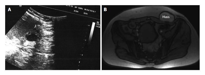
This is a small case series and retrospective study on patients with abdominal wall C/S endometrioma. In this study, we presented clinical and laboratory findings of six consecutive patients with scar endometrioma (Figure 1) who admitted to our departement between 2009 and 2012 and reviewed the literature. There were 28 patients with incisional hernias and 36 benign abdominal wall tumors like lipomas, etc. in this period. Medical data of the patients were evaluated, and presented our experience with scar endometrioma consequently by this case series.
Routine hematological and biochemical examinations were done following medical history and physical examination of patients who have a painful mass in their C/S site. Main complaint of our cases on admission was a palpable mass on incision site and cyclic pain. Abdominal pain was non-cyclic unusually in one patient. As you seen in Table 1, all patients were in third decade and aged between 31-34 years. Parity was 1 in three and 2 in other three patients and all deliveries were C/S’s. While caesarean incision was Pfannenstiel in 5 patients and there was a subumbilical midline incision in one. Duration between the last C/S and hospital admission varied between 2-7 years and duration between emergence of a painful mass on scar site and hospital admission varied between 6 mo and 4 years. There were not abdominal endometriosis and scar endometrioma in previous history. There were 28 patients with incisional hernias and 36 benign abdominal wall tumors like lipomas, etc. during three years period. Initial diagnosis were incisional hernia in four and endometrioma in two of our six patients. Definite diagnosis was made by histopathological evaluation of excised materials. Prediagnosis was irreducible incisional hernia in 4 out of 6 patients (66.6%) and endometrioma in 2 patients (33.3%). There were four patients diagnosed in first five years after C/S, and two patients more than five years. Doppler US was done to make a definite diagnosis succesfully in five patients. Final diagnosis was made with USG and abdominal CT in one patient. The weight of resected materials were min 48 g, max 70 g and ultrasonographical size of the palpable mass in scar tissue varied between 2 to 5 cm (Table 1).
| Age, yr | C/S | Recurrent disease | Years after C/S | Interval to symptoms onset, yr | Pain type | Weight of lesion (g) | Size (cm) | Initial diagnosis |
| 34 | 1 | No | 3 | 1 | Cyclic | 54 | 3 × 2 | Incisional hernia |
| 32 | 2 | No | 7 | 4 | Non-cyclic | 60 | 3 × 3 | Incisional hernia |
| 32 | 1 | No | 3 | 1/2 | Cyclic | 58 | 3 × 3 | Incisional hernia |
| 31 | 2 | No | 3 | 1 | Cyclic | 48 | 2 × 2 | Incisional hernia |
| 33 | 2 | No | 4 | 1 | Cyclic | 70 | 4 × 4 | Endometrioma |
| 31 | 1 | No | 6 | 2 | Cyclic | 62 | 3 × 5 | Endometrioma |
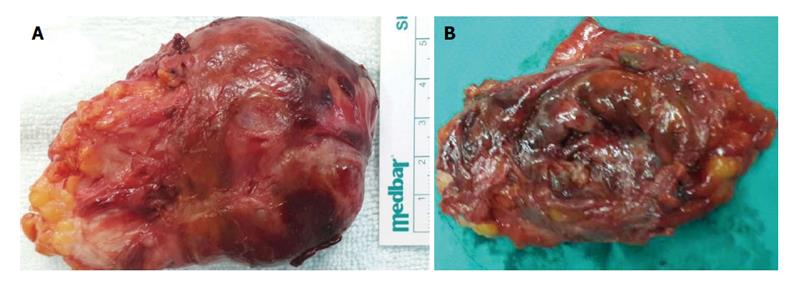
Treatment was achieved with complete surgical resection in 5 of six patients. One patient did not accept surgical intervention and she was followed up with medical treatment by using hormone suppressor drugs. Painful scar endometrioma were excised with undamaged borders and without making endometrial tissue implantation into the neighboring tissue in five patients who accepted surgical treatment. The glandular structure of endometrium and stroma were seen microscopically in fibrocollagenous tissue and that the mass belonged to endometrioma (Figure 2).
Common use of laparoscopy has enabled more frequent detection of intraabdominal endometriosis. Dysmenorrhea, irregular menstrual cycles and infertility are the most common symptoms seen in endometriosis. A palpable mass on C/S incision site and a cyclic pain are pathognomonic in patients with endometrioma[2-6].
Two different theories are available to explain endometrioma development. According to the first hypothesis, multipotent mesenchymal cells differentiate to endometrial tissues in their site after puberty and show physiopathological changes, like proliferation, hemorrhage as response to hormonal functions. And second hypothesis; endometrial cells are transported to extrauterine areas in some instances and similarly endometrioma develops by being affected from hormonal changes[5]. Endometrioma development in C/S scar tissue on abdominal wall seems more consistent with the second hypothesis. An increase has been reported in endometrioma frequency in parallel with the increase in number of C/S in recent years[7,8]. Procedure responsible for endometrioma development on incision site is iatrogenic inoculation of endometrial tissue into incision site[9]. Three of our cases underwent one C/S, three cases twice C/S. Endometrioma usually emerges during a ten year period following C/S[10].
Scar endometrioma are not frequently met more than 5 years after C/S, but we have two patients with relatively late diagnosed scar endometrioma in our series. Basic and the most commonly detected complaints of the patients were a palpable mass in C/S scar tissue and cyclic pain[11]. These symptoms have begun within a 6 mo-4 year period following C/S operation. Abdominal pain or painfull mass were seen cyclic usually, but there were one patient with non-cyclic painfull endometrioma in our case series.
Definitive diagnosis of endometrioma is not possible without histopathological examination. They are some published parameters on clinical and ultrasonographic findings for differential diagnosis of abdominal wall endometrioma near cesarean section[12]. Lipomas , sebaceous cysts, hemangiomas and lymphangiomas, desmoid tumors among common benign tumors of abdominal wall[13,14]. It is recommended to keep in mind that these tumors may be a metastatic lesion arising from a malignant focus and to make diagnosis of endometrioma definite through US-guided fine needle aspiration cytology in patients who do not accept surgical treatment[5,12,13]. Imaging methods like doppler US, CT and MRI should absolutely be benefited for differential diagnosis of suspected masses. Definitive diagnosis can only be made histopathologically.
In a conclusion, the soft tissue tumors and endometrioma are probable diagnosis besides incisional hernia in patients who have a palpable mass on C/S scar following some obstetric and gynecologic interventions. A detailed medical history, physical examination findings and imaging methods in suspected cases are significant diagnostic tools to investigate the features of the pain and relationship with menstrual cycle. Radical treatment should be complete surgical excision for patients who receive prediagnosis of endometrioma and one should take care against intraoperative auto-inoculation of endometrial tissue in order to prevent recurrences. Combined oral contraceptives, progestagens and hormone suppression therapy with gonadotropin releasing hormone analogues should be used for medical treatment of patients who don’t want surgery and whose diagnosis of endometrioma was verified through fine needle aspiration cytology.
Endometriosis and/or endometrioma is defined as ectopic occurrence of endometrium. Cystic or solid tumoral masses located at extrauterine organs caused by endometriosis are named as endometrioma.
Abdominal endometriosis was seen often but cesarean scars endometrioma is rare and difficult to diagnose preoperatively.
This manuscript is worth to publish for increasing the awareness of incisional endometrioma possibility among patients with endometriosis.
P- Reviewers: Kahyaoglu S, Wang CC S- Editor: Wen LL L- Editor: A E- Editor: Liu SQ
| 1. | Akbulut S, Sevinc MM, Bakir S, Cakabay B, Sezgin A. Scar endometriosis in the abdominal wall: a predictable condition for experienced surgeons. Acta Chir Belg. 2010;110:303-307. [PubMed] |
| 2. | Bektaş H, Bilsel Y, Sari YS, Ersöz F, Koç O, Deniz M, Boran B, Huq GE. Abdominal wall endometrioma; a 10-year experience and brief review of the literature. J Surg Res. 2010;164:e77-e81. [RCA] [PubMed] [DOI] [Full Text] [Cited by in Crossref: 94] [Cited by in RCA: 104] [Article Influence: 6.9] [Reference Citation Analysis (0)] |
| 3. | Teng CC, Yang HM, Chen KF, Yang CJ, Chen LS, Kuo CL. Abdominal wall endometriosis: an overlooked but possibly preventable complication. Taiwan J Obstet Gynecol. 2008;47:42-48. [RCA] [PubMed] [DOI] [Full Text] [Cited by in Crossref: 17] [Cited by in RCA: 22] [Article Influence: 1.3] [Reference Citation Analysis (0)] |
| 4. | Gunes M, Kayikcioglu F, Ozturkoglu E, Haberal A. Incisional endometriosis after cesarean section, episiotomy and other gynecologic procedures. J Obstet Gynaecol Res. 2005;31:471-475. [RCA] [PubMed] [DOI] [Full Text] [Cited by in Crossref: 54] [Cited by in RCA: 54] [Article Influence: 2.7] [Reference Citation Analysis (0)] |
| 5. | Hensen JH, Van Breda Vriesman AC, Puylaert JB. Abdominal wall endometriosis: clinical presentation and imaging features with emphasis on sonography. AJR Am J Roentgenol. 2006;186:616-620. [RCA] [PubMed] [DOI] [Full Text] [Cited by in Crossref: 147] [Cited by in RCA: 134] [Article Influence: 7.1] [Reference Citation Analysis (0)] |
| 6. | Chang Y, Tsai EM, Long CY, Chen YH, Kay N. Abdominal wall endometriomas. J Reprod Med. 2009;54:155-159. [PubMed] |
| 7. | Brimienė V, Brimas G, Strupas K. Differential diagnosis between chronic pancreatitis and pancreatic cancer: a prospective study of 156 patients. Medicina (Kaunas). 2011;47:154-162. [PubMed] |
| 8. | Zhu Z, Al-Beiti MA, Tang L, Liu X, Lu X. Clinical characteristic analysis of 32 patients with abdominal incision endometriosis. J Obstet Gynaecol. 2008;28:742-745. [RCA] [PubMed] [DOI] [Full Text] [Cited by in Crossref: 17] [Cited by in RCA: 16] [Article Influence: 1.0] [Reference Citation Analysis (0)] |
| 9. | Horton JD, Dezee KJ, Ahnfeldt EP, Wagner M. Abdominal wall endometriosis: a surgeon’s perspective and review of 445 cases. Am J Surg. 2008;196:207-212. [RCA] [PubMed] [DOI] [Full Text] [Cited by in Crossref: 196] [Cited by in RCA: 223] [Article Influence: 13.1] [Reference Citation Analysis (0)] |
| 10. | Nominato NS, Prates LF, Lauar I, Morais J, Maia L, Geber S. Caesarean section greatly increases risk of scar endometriosis. Eur J Obstet Gynecol Reprod Biol. 2010;152:83-85. [RCA] [PubMed] [DOI] [Full Text] [Cited by in Crossref: 54] [Cited by in RCA: 66] [Article Influence: 4.4] [Reference Citation Analysis (0)] |
| 11. | Lipscomb GH, Givens VM, Smith WE. Endometrioma occurring in abdominal wall incisions after cesarean section. J Reprod Med. 2011;56:44-46. [PubMed] |
| 12. | Francica G. Reliable clinical and sonographic findings in the diagnosis of abdominal wall endometriosis near cesarean section scar. World J Radiol. 2012;4:135-140. [PubMed] |
| 13. | Savelli L, Manuzzi L, Di Donato N, Salfi N, Trivella G, Ceccaroni M, Seracchioli R. Endometriosis of the abdominal wall: ultrasonographic and Doppler characteristics. Ultrasound Obstet Gynecol. 2012;39:336-340. [RCA] [PubMed] [DOI] [Full Text] [Cited by in Crossref: 49] [Cited by in RCA: 53] [Article Influence: 4.1] [Reference Citation Analysis (0)] |
| 14. | Ozel L, Sagiroglu J, Unal A, Unal E, Gunes P, Baskent E, Aka N, Titiz MI, Tufekci EC. Abdominal wall endometriosis in the cesarean section surgical scar: a potential diagnostic pitfall. J Obstet Gynaecol Res. 2012;38:526-530. [RCA] [PubMed] [DOI] [Full Text] [Cited by in Crossref: 40] [Cited by in RCA: 46] [Article Influence: 3.5] [Reference Citation Analysis (0)] |


