Published online Oct 16, 2025. doi: 10.12998/wjcc.v13.i29.110454
Revised: June 27, 2025
Accepted: July 24, 2025
Published online: October 16, 2025
Processing time: 83 Days and 2.5 Hours
Oil-based iodinated contrast media have excellent contrast properties and are widely used for hysterosalpingographic evaluation of female infertility. On abdominal radiography and computed tomography (CT) scans, their radiodensity is similar to that of metallic objects, which can sometimes lead to diagnostic confusion in the postoperative settings. In this case, retained oil-based contrast medium was observed on an abdominal radiograph following a cesarean section, making it difficult to differentiate from an intraperitoneal foreign body from surgery.
The patient was a 37-year-old pregnant woman who was referred to our hospital at 32 weeks and 1 day of pregnancy due to complete placenta previa for mana
When intraperitoneal foreign bodies are suspected on postoperative radiographs, the possibility of oil-based iodinated contrast medium retention should be considered.
Core Tip: Iodine oil contrast agents have good contrast properties and are widely used in hysterosalpingography for evaluation of infertility. However, due to their slow absorption, they can remain in the body for an extended period. Therefore, when a postoperative plain abdominal X-ray reveals a suspected intraperitoneal foreign body, it is important to consider the possibility of residual iodine oil contrast agent when providing treatment.
- Citation: Morita A, Kakinuma T, Segawa A, Harada S, Takae S, Tamura M, Suzuki N. Prolonged retention of oil-based iodinated contrast medium observed on plain abdominal radiograph after cesarean section: A case report. World J Clin Cases 2025; 13(29): 110454
- URL: https://www.wjgnet.com/2307-8960/full/v13/i29/110454.htm
- DOI: https://dx.doi.org/10.12998/wjcc.v13.i29.110454
Oil-based iodinated contrast media have excellent contrast properties and allow for prolonged contrast, making them widely used in hysterosalpingography. However, when used in this context. These agents are absorbed slowly by the body and, in some cases, can remain in the abdominal cavity for an extended period[1]. In particular, encapsulated oil-based iodinated contrast materials in the body may appear as hyperdense foci on plain radiographs or computed tomography (CT) scans, with radiodensity similar to that of metals. In patients with a history of surgery, such findings may be difficult to distinguish from metallic objects, potentially leading to diagnostic confusion[2-5]. In this report, we present a case in which residual oil-based contrast medium was detected on plain abdominal radiograph immediately following a cesarean section. The imaging appearance closely mimicked that of a retained intraperitoneal foreign body, posing a diagnostic challenge.
The patient reported no symptoms, and there were no notable clinical complaints.
The patient conceived via frozen-thawed blastocyst transfer at another facility and received antenatal care at a different hospital from the early stage of pregnancy. At 32 weeks and 1 day of gestation, she was referred to our hospital for further management of the pregnancy and delivery due to a diagnosis of complete placenta previa.
The patient had no relevant personal medical history.
The patient had no relevant family medical history.
No abnormal physical findings were observed.
Routine laboratory tests revealed no abnormalities.
Transvaginal ultrasound examination revealed the presence of complete placenta previa.
The radiological interpretation of the plain abdominal CT scan indicated that the foreign body detected on the initial abdominal X-ray image was located in an area consistent with the right side of the Douglas pouch. Additionally, the CT value measured approximately 7000 Hounsfield units (HU), which is similar to that of metal. However, upon further inquiry with the initial examining physician, it was revealed that the patient had undergone hysterosalpingography using iodine oil contrast 8 years prior.
Based on the clinical and radiological findings, retention of oil-based contrast medium was strongly suspected, and conservative management with close observation was initiated.
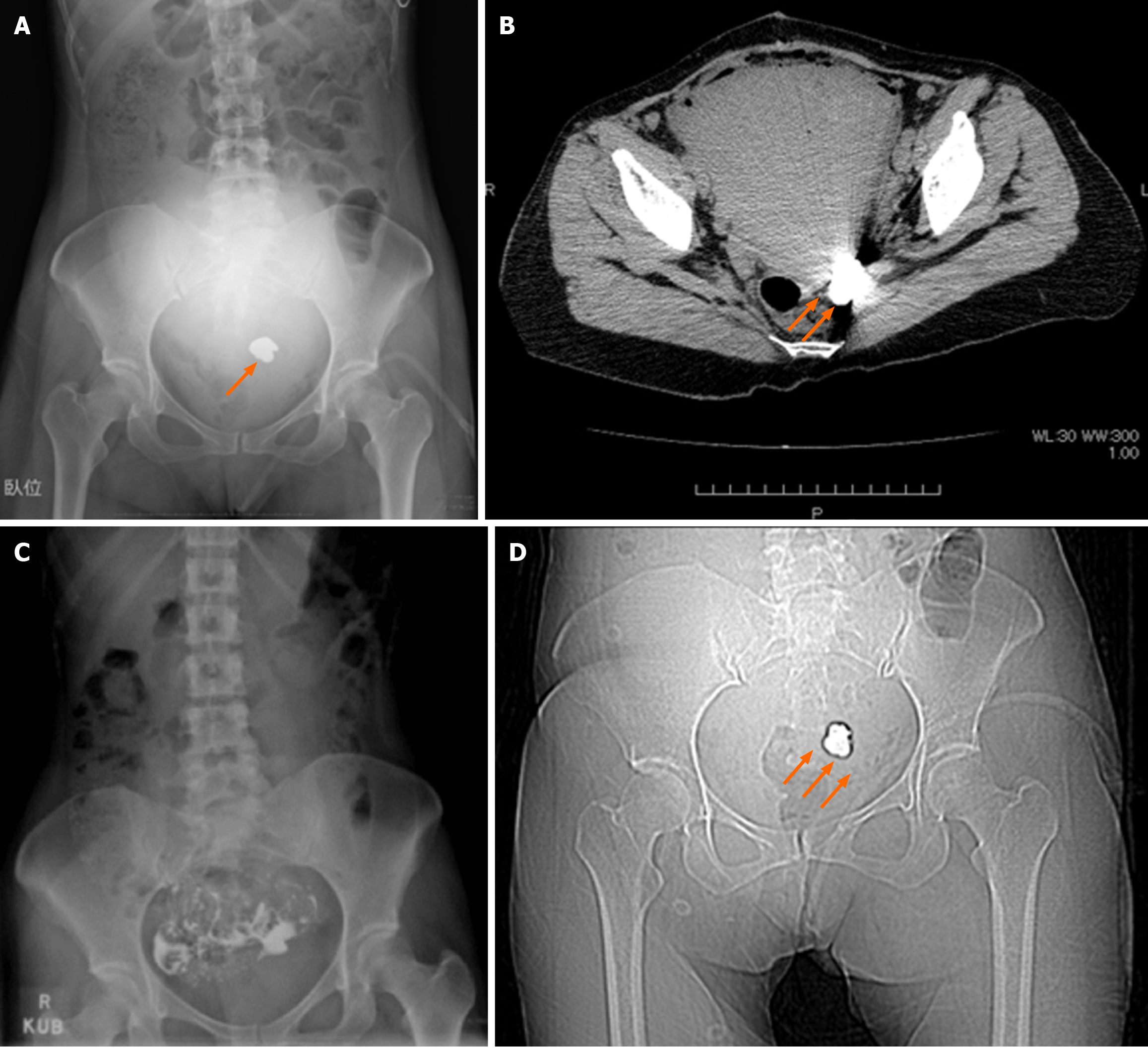
The pregnancy progressed without complications, and an elective cesarean section was performed at 37 weeks and 3 days of gestation. A midline abdominal incision followed by a transverse incision in the lower uterine segment was utilized to deliver a healthy male infant in cephalic presentation. The placenta was manually removed. Intraoperative palpation of the uterus, including the fundus and posterior wall revealed no significant adhesions. Following confirmation of hemostasis and absence of retained foreign material, the surgery was completed. A routine postoperative plain abdominal radiograph was performed immediately after surgery to check for any retained surgical instruments in the body, which revealed a near-round, homogeneously hyperdense lesion with a regular margin in the pelvic cavity (Figure 1A). Surgical counts of instruments and gauze were accurate, and no instrument damage was noted. Therefore, a plain abdominal CT scan was performed for further assessment (Figure 1B), which identified hyperdense object on the left side of the pouch of Douglas, with attenuation values measured at 7000 HU, which is similar to that of metals. However, no metallic foreign body was felt through palpitation on pelvic or rectal examination. Subsequently, a more detailed patient interview revealed that hysterosalpingography was performed by her initial physician. Upon further inquiry, it was confirmed that an oil-based iodinated contrast agent had been used 8 years prior (Figure 1C). Accordingly, contrast medium retention was strongly suspected, and a decision was made to continue observation. At 3 months post-operatively, a follow-up plain abdominal radiograph showed deformation and reduction in size of the previously visualized hyperdense lesion (Figure 1D), confirming the diagnosis of retained oil-based iodinated contrast medium, and conservative management was continued.
At the 3-month post-operative follow-up, a plain abdominal radiograph showed significant deformation and shrinkage of the previously observed intra-pelvic mass-like lesion (Figure 1D). These findings conclusively confirmed the diagnosis of retained oil-based iodinated contrast medium.
Hysterosalpingography is a widely used modality in the evaluation of female infertility, particularly for the assessment of fallopian tube patency, intrauterine adhesions, and uterine anomalies. Oil-based iodinated contrast agents possess a high viscosity, resistance to dilution by body fluids, and slow diffusion and absorption. These properties make them suitable for use in hysterosalpingography, sialography, lymphangiography, and in the preparation of epirubicin hydrochloride for hepatic arterial infusion chemotherapy. Although oil-based iodinated contrast agents are absorbed slowly and may remain in the body for an extended period, they are generally expected to be eliminated from the lymphatic system within several months[2-5].
Postoperative foreign bodies retained in the body can cause infection, organ dysfunction, the need for reoperation, and significant physical, psychological, and social burdens for the patient. Furthermore, with the increasing advancement in medical devices and miniaturization of surgical instruments, greater caution is required to prevent instrument damage and retention. In addition, surgical counts of gauzes and instruments, postoperative radiographs are useful and are routinely performed in Japan to detect retained foreign bodies. In the present case, a plain abdominal radiograph was also performed immediately after the cesarean section, which revealed a mass-like shadow in the pelvic cavity, raising suspicion of a retained foreign body.
The differential diagnosis for mass-like abnormal shadows on postoperative plain abdominal radiographs generally includes retained surgical instruments, urinary calculi, gallstones, phleboliths, fecaliths, calcified uterine myomas, teeth within dermoid cysts, peritoneal loose bodies, or ingested foreign materials such as teeth; however, retained oil-based contrast media should be considered[2,4]. In general, calcification and bones have CT values of 80-1000 HU, contrast-enhanced vessels have 200-300 HU, and metals exhibit 2000 HU or more. The radiodensity of oil-based iodinated contrast agents on plain radiographs and CT scans can mimic that of metals. When encapsulated, these agents may form mass-like structures that closely resemble metallic foreign bodies and may cause diagnostic uncertainty[2-5]. On magnetic resonance imaging scans, oil-based contrast agents show signal characteristics similar to those of adipose tissue, facilitating differentiation from metallic objects. However, magnetic resonance imaging cannot be performed when the possibility of a metallic object cannot be definitely ruled out[3].
The patient in this case had no prior surgical history before the cesarean section, and instrument and gauze counts were unremarkable. However, the observed radiographic prompted further investigation using plain abdominal CT, considering the possibility of a calcified lesion in the abdominal cavity or an accidentally ingested foreign body. CT imaging revealed a mass located in the pouch of Douglas, with an attenuation value of 7000 HU, an artificially high density inconsistent with calcification or other pathological processes.
Subsequently, a more detailed interview with the patient revealed a history of hysterosalpingography performed 8 years earlier, with the use of an oil-based contrast medium in the hysterosalpingography documented in the patient’s medical records by another physician, leading to a diagnosis of retained oil-based iodinated contrast medium. A follow-up plain abdominal radiograph taken approximately 3 months after discharge showed deformation and reduction in size of the intra-pelvic mass-like shadow, presumably due to gradual leakage and absorption of the ruptured encapsulated contrast agent within the pouch of Douglas following the cesarean section. This confirmed the diagnosis of retained oil-based iodinated contrast medium.
Potential adverse effects of retained oil-based iodinated contrast agent include inflammatory reactions and granulation tissue formation in the epithelial tissue or peritoneum[6,7]. While several reports have been published in the field of otolaryngology and ophthalmology, adverse events related to retention of these agents in the abdominal cavity are lacking[8]. However, there are case reports describing inflammatory granuloma formation after migration of contrast medium into an inguinal hernia requiring surgical removal and extension of granulation tissue extending into the retroperitoneum, resulting in ureteral compression, hydronephrosis, and urinary tract infection[7,9]. Thus, we opted for close postoperative follow-up.
With the recent trend toward delayed marriage and increased maternal age at first pregnancy, the number of patients undergoing infertility treatment has risen. Consequently, the prevalence of hysterosalpingography is also expected to increase. In clinical practice, it is therefore important to obtain a detailed patient history and consider the possibility of retained contrast medium in the differential diagnosis when unexplained radiopaque findings are observed. Additionally, physicians performing hysterosalpingography should inform patients about the use of oil-based iodinated contrast medium and their potential for retention. Furthermore, for patients with a known history of hysterosalpingography or other procedures involving oil-based contrast media, a plain abdominal radiograph may be warranted as part of preoperative screening before abdominal surgery.
In this report, we presented a case in which retained oil-based iodinated contrast medium was initially misinterpreted as an intraperitoneal foreign body following cesarean section. When suspected intraperitoneal foreign bodies are observed on postoperative plain abdominal radiographs, the possibility of retained oil-based iodinated contrast medium should be considered in the differential diagnosis.
| 1. | Roest I, Rosielle K, van Welie N, Dreyer K, Bongers M, Mijatovic V, Mol BW, Koks C. Safety of oil-based contrast medium for hysterosalpingography: a systematic review. Reprod Biomed Online. 2021;42:1119-1129. [RCA] [PubMed] [DOI] [Full Text] [Cited by in Crossref: 6] [Cited by in RCA: 19] [Article Influence: 4.8] [Reference Citation Analysis (0)] |
| 2. | Wakabayashi Y, Hashimura N, Kubouchi T. Retained lipiodized oil misdiagnosed as residual metallic material. Radiat Med. 2004;22:362-363. [PubMed] |
| 3. | Lindequist S, Justesen P, Larsen C, Rasmussen F. Diagnostic quality and complications of hysterosalpingography: oil- versus water-soluble contrast media--a randomized prospective study. Radiology. 1991;179:69-74. [RCA] [PubMed] [DOI] [Full Text] [Cited by in Crossref: 54] [Cited by in RCA: 48] [Article Influence: 1.4] [Reference Citation Analysis (0)] |
| 4. | Hayashida H, Furuya K, Kurahashi H, Yamashita S, Chang Y, Tsubouchi H, Shikado K, Ogita K. Incidental detection of retained oil-based hysterosalpingography contrast medium on postoperative postpartum radiography: A case report. Clin Case Rep. 2022;10:e05925. [RCA] [PubMed] [DOI] [Full Text] [Full Text (PDF)] [Cited by in RCA: 2] [Reference Citation Analysis (0)] |
| 5. | Takeyama K, Ishikawa R, Nakayama K, Suzuki T. Intraperitoneal residual contrast agent from hysterosalpingography detected following cesarean section. Tokai J Exp Clin Med. 2014;39:69-71. [PubMed] |
| 6. | Eisenberg AD, Winfield AC, Page DL, Holburn GE, Schifter T, Segars JH. Peritoneal reaction resulting from iodinated contrast material: comparative study. Radiology. 1989;172:149-151. [RCA] [PubMed] [DOI] [Full Text] [Cited by in Crossref: 10] [Cited by in RCA: 11] [Article Influence: 0.3] [Reference Citation Analysis (0)] |
| 7. | Miyazaki Y, Yamamoto T, Hyakudomi R, Taniura T, Hirayama T, Takai K, Hirahara N, Tajima Y. Case of inflammatory granuloma in inguinal hernia sac after hysterosalpingography with oily contrast medium. Int J Surg Case Rep. 2020;72:215-218. [RCA] [PubMed] [DOI] [Full Text] [Full Text (PDF)] [Cited by in Crossref: 5] [Cited by in RCA: 4] [Article Influence: 0.8] [Reference Citation Analysis (0)] |
| 8. | Shigetaka Y, Masatsugu S, Yoshikuni F, Yoshihiro T. Parotid and pterygomaxillary lipogranuloma caused by oil-based contrast medium used for sialography: report of a case. J Oral Maxillofac Surg. 1996;54:350-353. [RCA] [PubMed] [DOI] [Full Text] [Cited by in Crossref: 14] [Cited by in RCA: 16] [Article Influence: 0.6] [Reference Citation Analysis (0)] |
| 9. | Munehisa G. Ureteral stenosis caused by pelvic lipogranuloma following the administration of oil-based contrast media for hysterosalpigography. A case report. Jpn J Urol Surg. 2002;15:1063-1065. |


