Published online Jun 26, 2025. doi: 10.12998/wjcc.v13.i18.101545
Revised: January 23, 2025
Accepted: February 17, 2025
Published online: June 26, 2025
Processing time: 161 Days and 18.2 Hours
Correcting skeletal class III malocclusion with anterior crossbite in adolescents using only orthodontic treatment poses challenges. This report highlights a novel approach leveraging improved superelastic Ni–Ti alloy wire (ISW) to address these conditions effectively.
A 17-year-old male patient presented with the chief complaint of an underbite. The patient was given a diagnosis of skeletal class III malocclusion and anterior crossbite. The orthodontic treatment plan was implemented and did not require teeth extractions or orthognathic surgery. Key interventions involved the app
The application of ISW for treating skeletal class III malocclusion with anterior crossbite in a 17-year-old male patient resulted in exceptional outcomes. The treatment led to a marked improvement in the patient’s facial profile and to proper overjet, overbite, and midline alignment. These results were maintained over a one-year follow-up, indicating that a minimally invasive orthodontic approach can effectively address complex skeletal discrepancies in adolescent patients. This case illustrates that with the careful use of advanced orthodontic techniques, major skeletal challenges can be resolved without resorting to surgical procedures.
Core Tip: Correcting skeletal class III malocclusion with anterior crossbite in adolescents can be challenging. In this case, a 17-year-old male with an underbite was successfully treated using improved superelastic Ni–Ti alloy wire (ISW), intermaxillary elastics, and ISW unilateral multi-bend edgewise archwire. These techniques corrected the anterior crossbite, aligned the midline, and achieved proper overjet and overbite. The treatment lasted approximately one year and five months, with a stable outcome observed after one year of follow-up. This case demonstrates that skeletal Class III malocclusion in adolescents can be addressed effectively with minimally invasive orthodontic methods.
- Citation: Chang YHS, Chen YH, Yu JH. Improved superelastic Ni–Ti alloy wire for treating skeletal class III malocclusion combined with anterior crossbite: A case report. World J Clin Cases 2025; 13(18): 101545
- URL: https://www.wjgnet.com/2307-8960/full/v13/i18/101545.htm
- DOI: https://dx.doi.org/10.12998/wjcc.v13.i18.101545
Skeletal class III malocclusion, often characterized by a forward-positioned lower jaw leading to an anterior crossbite, presents a major challenge in orthodontic treatment, particularly in adolescent patients. The traditional approaches frequently involve teeth extractions or orthognathic surgery to correct these skeletal discrepancies. Invasive procedures carry risks and have long recovery periods, making them unappealing for some patients and their families.
Less invasive treatments have been developed. For example, aligner-based treatments, intermaxillary elastics (IME) combined with skeletal anchorage, and improved superelastic Ni–Ti alloy wire (ISW) treatments are effective and less invasive alternatives to traditional treatments for skeletal class III malocclusion[1-3]. Biomechanical innovations such as ISW have further enhanced the potential of non-surgical methods by achieving efficient tooth movement with minimal patient discomfort[4]. This report illustrates the successful management of a 17-year-old male adolescent with skeletal class III malocclusion and anterior crossbite, managed without teeth extraction or surgery. The strategic use of ISW, along with IME and a unilateral multi-bend edgewise archwire (ISW MEAW), resulted in substantial improvements in occlusion, facial profile, and dental alignment, highlighting the potential of minimally invasive techniques in addressing complex malocclusions.
A 17-year-old male patient sought orthodontic treatment at our clinic with the chief complaint of an underbite, he also expressed concerns about his dental alignment and overall facial profile. The patient had no notable medical issues or temporomandibular joint symptoms at the time of his visit.
The patient reported no current medical conditions or symptoms at the time of evaluation.
The patient had no significant medical history, nor had undergone any previous orthodontic treatments.
The patient’s family history was unremarkable for any dental or orthodontic conditions.
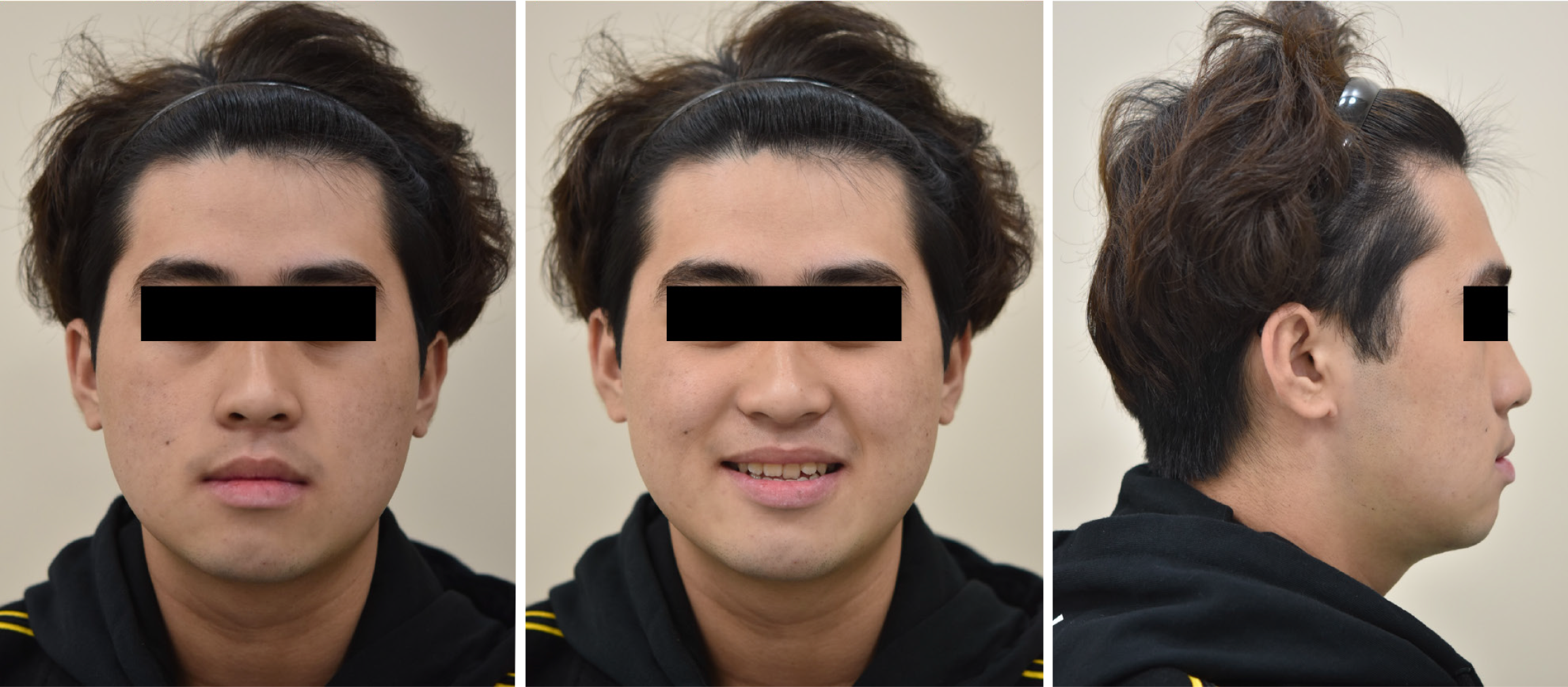
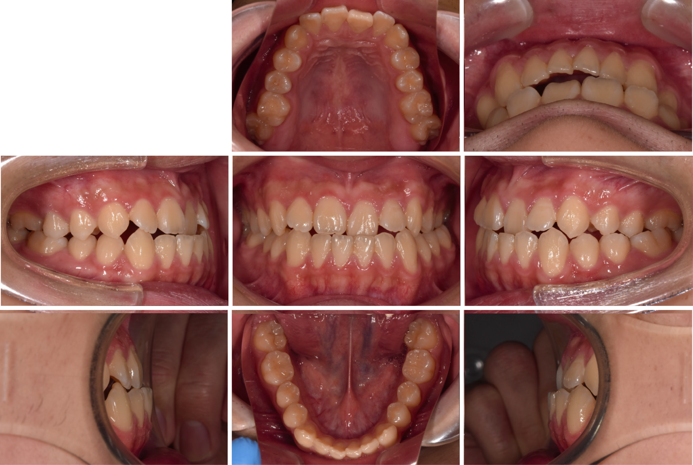
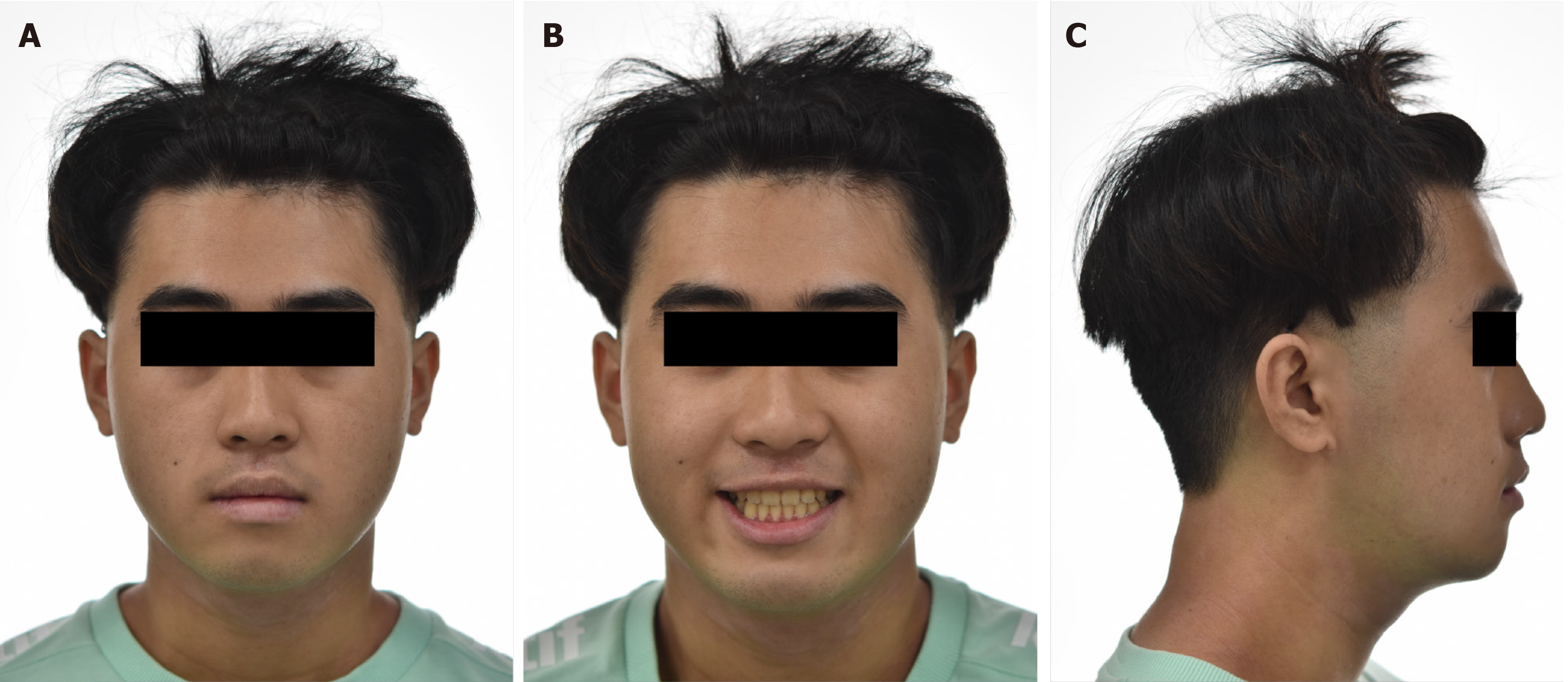
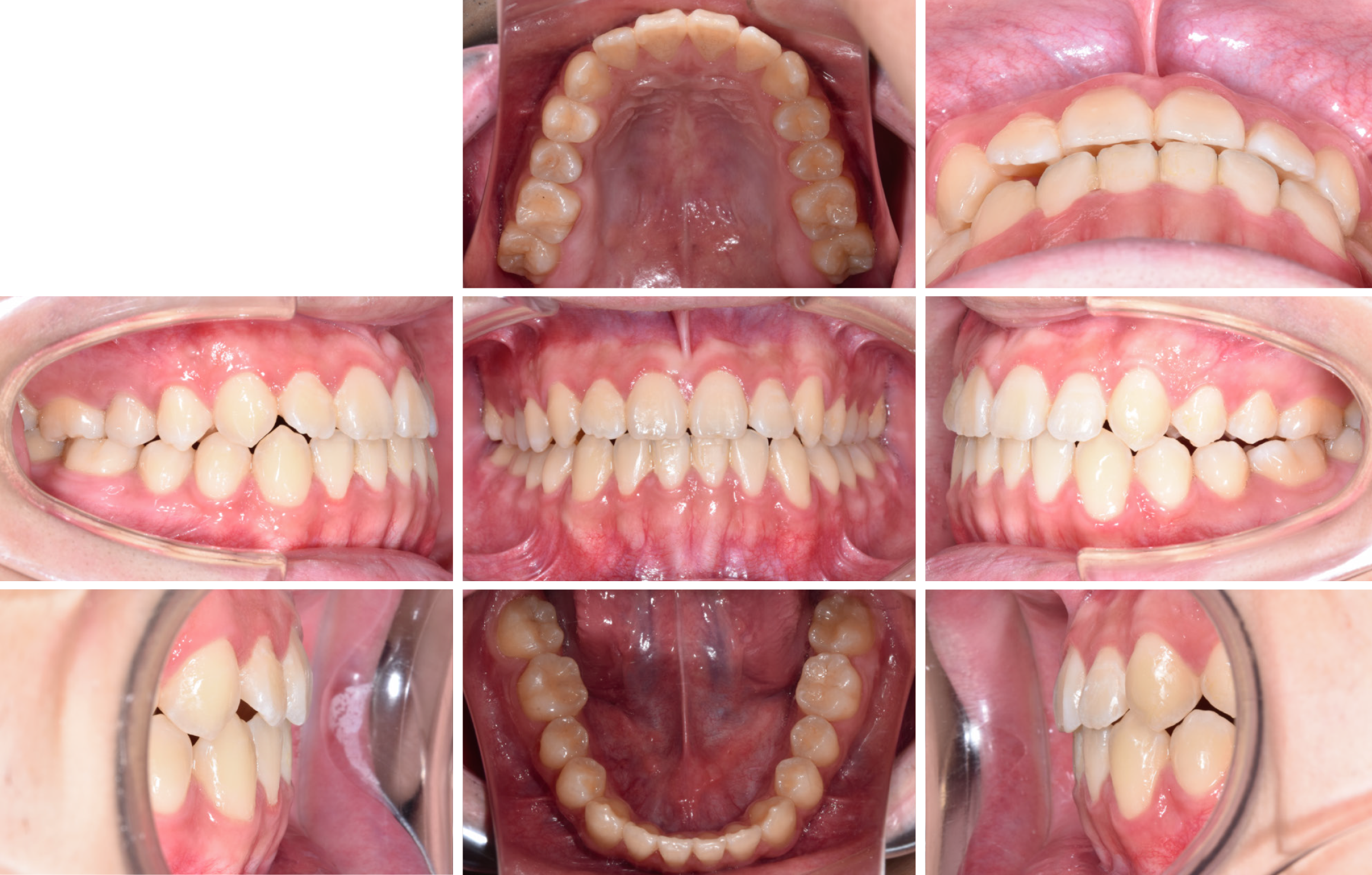
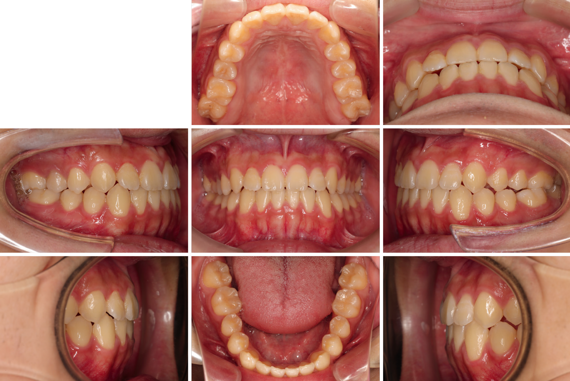
Initial facial photographs showed a prominent chin without any noticeable deviation when viewed from the front. The maxillary midline was shifted 1.0 mm from the facial midline, while the mandibular midline displayed a slight deviation toward the left side (Figure 1). Intraoral images revealed an anterior crossbite extending from the right lateral incisor to the left canine, alongside a bilateral class III molar relationship. Mild anterior crowding was also observed in both the upper and lower arches (Figure 2).
The analysis indicated a skeletal class III discrepancy, characterized by an ANB angle of ≤ 0° and a bilateral class III molar relationship with mesially positioned lower molars relative to the upper molars. No notable facial asymmetry was detected, and other clinical parameters were within normal ranges.
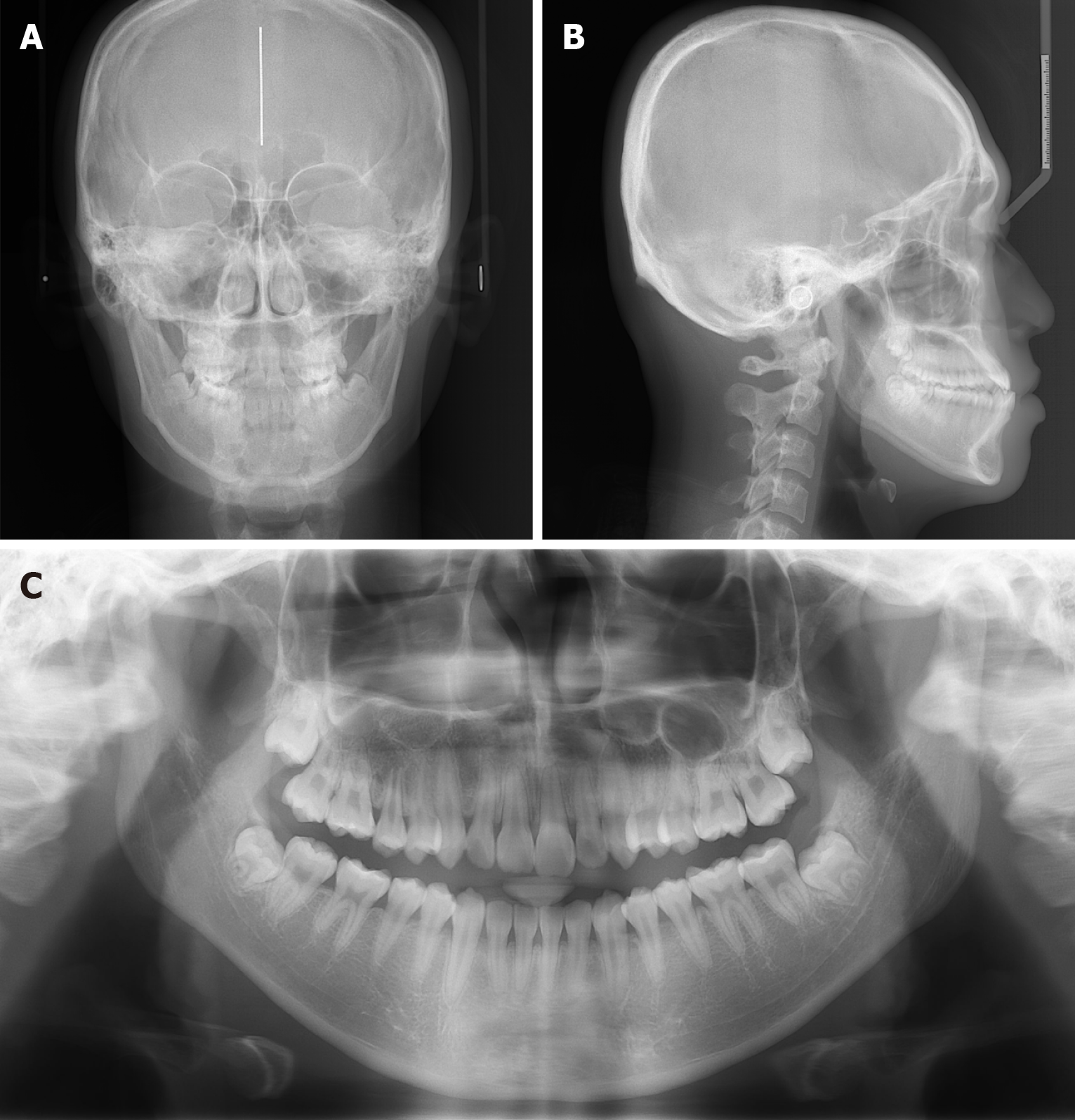
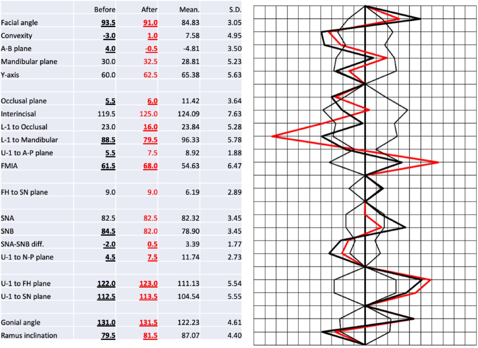
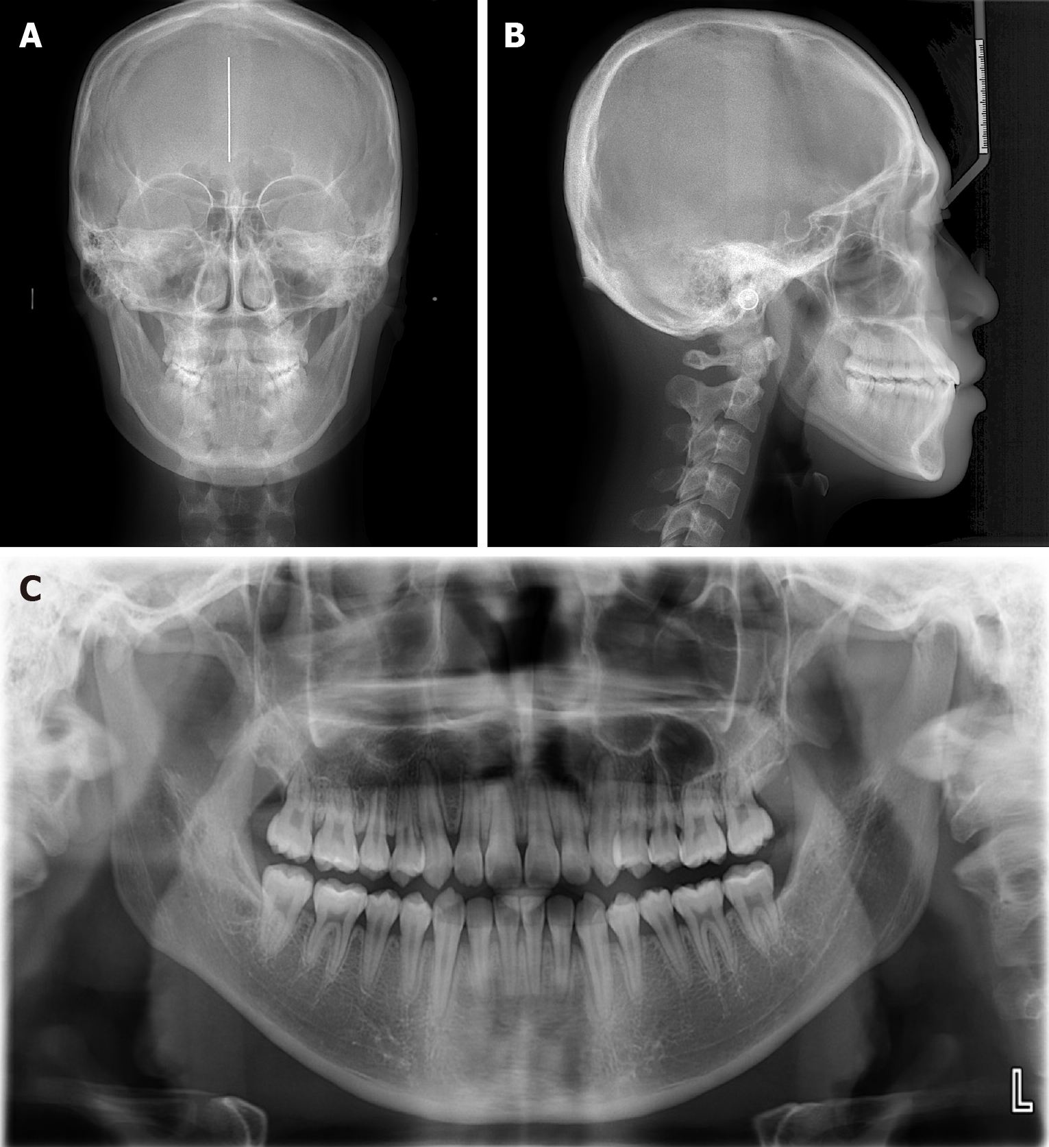
Panoramic X-rays confirmed the presence of all teeth, including the third molars in both jaws. Lateral cephalometric analysis (conducted using Romexis 3.2.0, Helsinki, Finland) demonstrated skeletal class III malocclusion with an ANB angle of –2.0°, a mandibular plane angle of 30.0°, and retroclination of the lower incisors at 88.5° relative to the mandibular plane. Posteroanterior radiographs showed no significant mandibular asymmetry or deviation from the mid-sagittal line (Figure 3).
The patient was given a diagnosis of skeletal class III malocclusion, anterior crossbite, mild anterior crowding, and slight leftward mandibular midline deviation, with a balanced vertical facial pattern.
The patient exhibited skeletal class III malocclusion, an anterior crossbite, and mild anterior crowding. The primary goals of treatment were: (1) To correct the anterior crossbite; (2) To relieve the crowding among the anterior teeth; (3) To achieve optimal dental alignment; (4) To establish proper overjet and overbite relationships; and (5) To enhance the patient's facial aesthetics.
Two potential treatment plans were discussed. The first option involved orthognathic surgery to address the skeletal discrepancy. The second, preferred by the patient, was non-surgical orthodontic treatment using ISW and IME to manage the malocclusion.
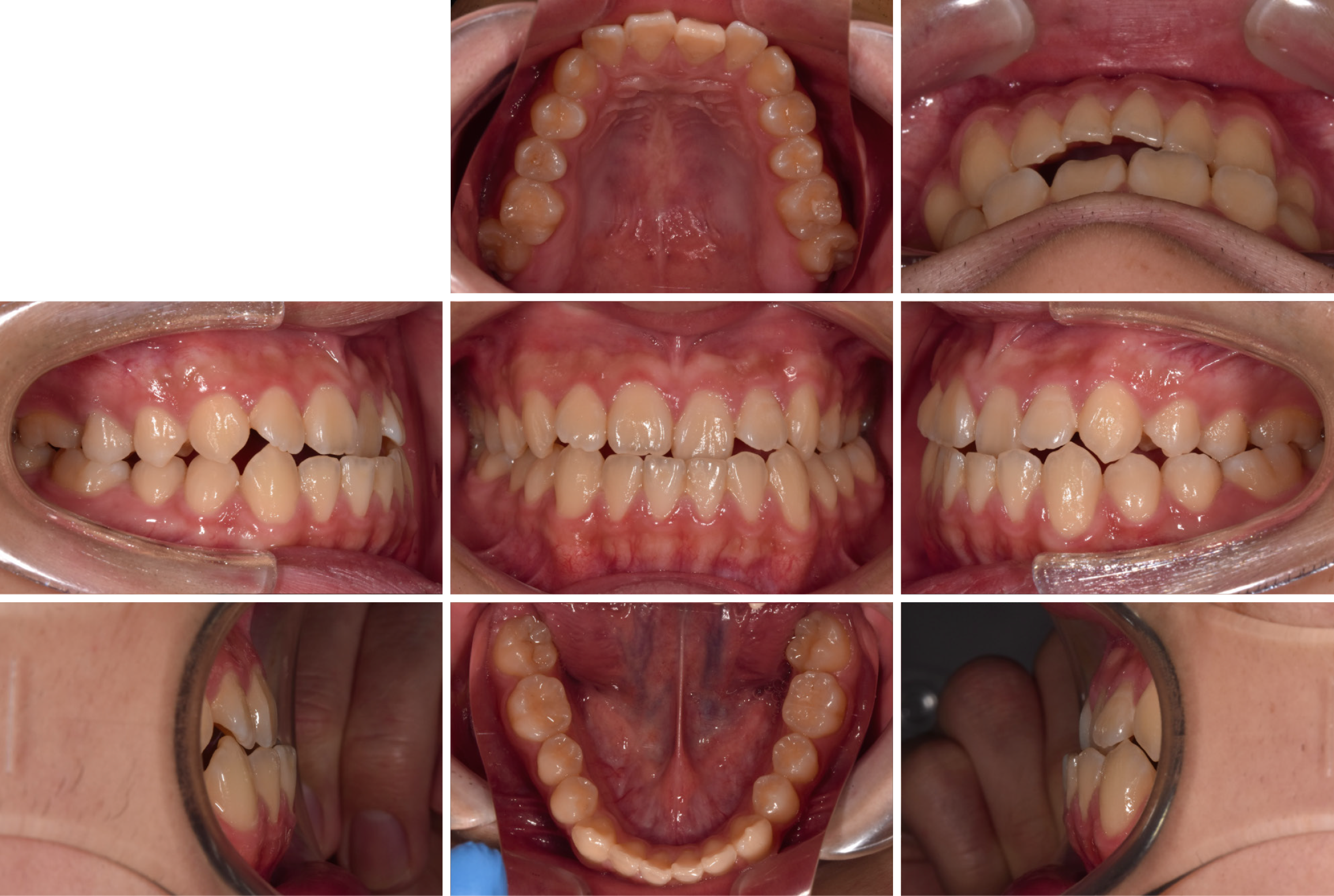
The orthodontic treatment commenced with the application of a full-mouth direct bonding system, which refers to the technique of bonding brackets directly onto the tooth surface, followed by leveling with 0.016 × 0.022-inch ISW (Figure 4). ISW MEAW with long class III elastics (16–43 and 26–33) was used to correct the anterior crossbite (Figure 4).
The patient's midline and crowding issues were resolved without surgery (Figure 4). After 13 months of active orthodontic treatment, the fixed appliances were removed, and retainers were provided. The patient was instructed to wear the retainers full-time for the first three months, followed by nighttime-only wear for maintenance.
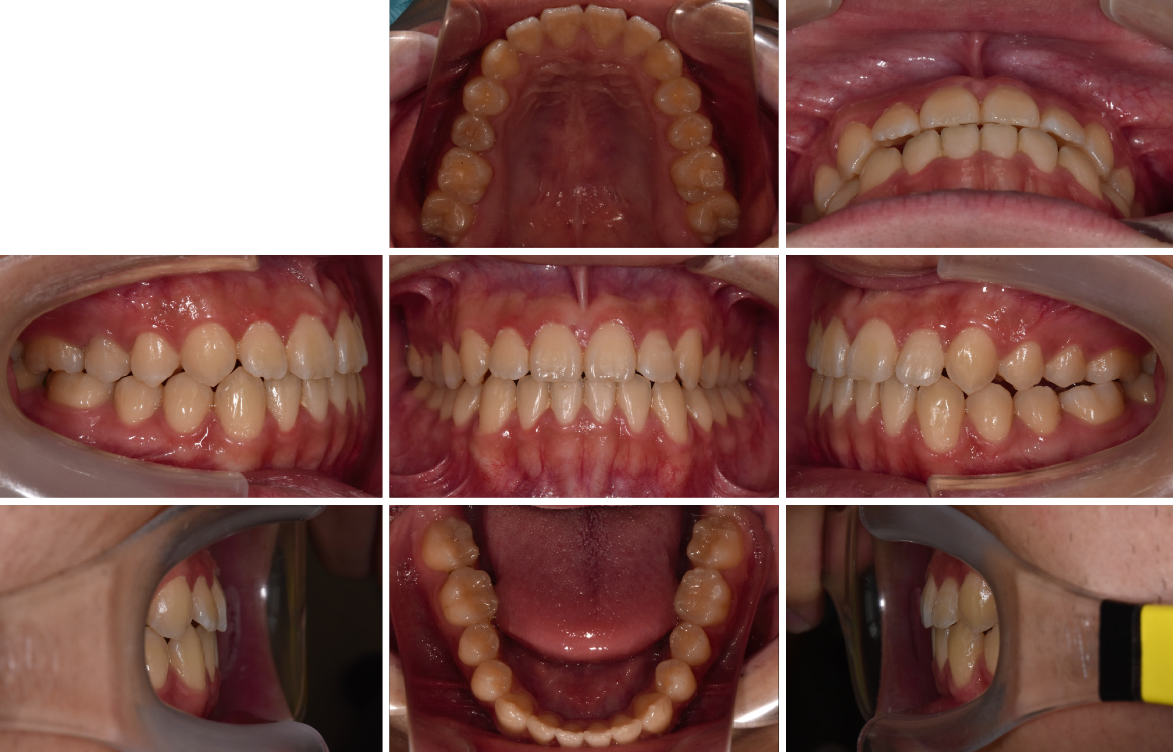
The patient's anterior crossbite was successfully corrected using the ISW MEAW technique (Figure 4) and wire + aligner (WA) orthodontics (Figure 5); a well-aligned arch and stable occlusion were achieved. The final records (Figures 6, 7, 8, 9, and 10) demonstrated proper alignment of the dental midline, while panoramic radiographs (Figure 10C) confirmed parallel root positioning without any evidence of root resorption. Additionally, the one-year follow-up records (Figure 11) showed that the occlusal corrections remained stable and that proper overjet, overbite, and midline alignment were preserved, confirming the long-term success of the treatment.
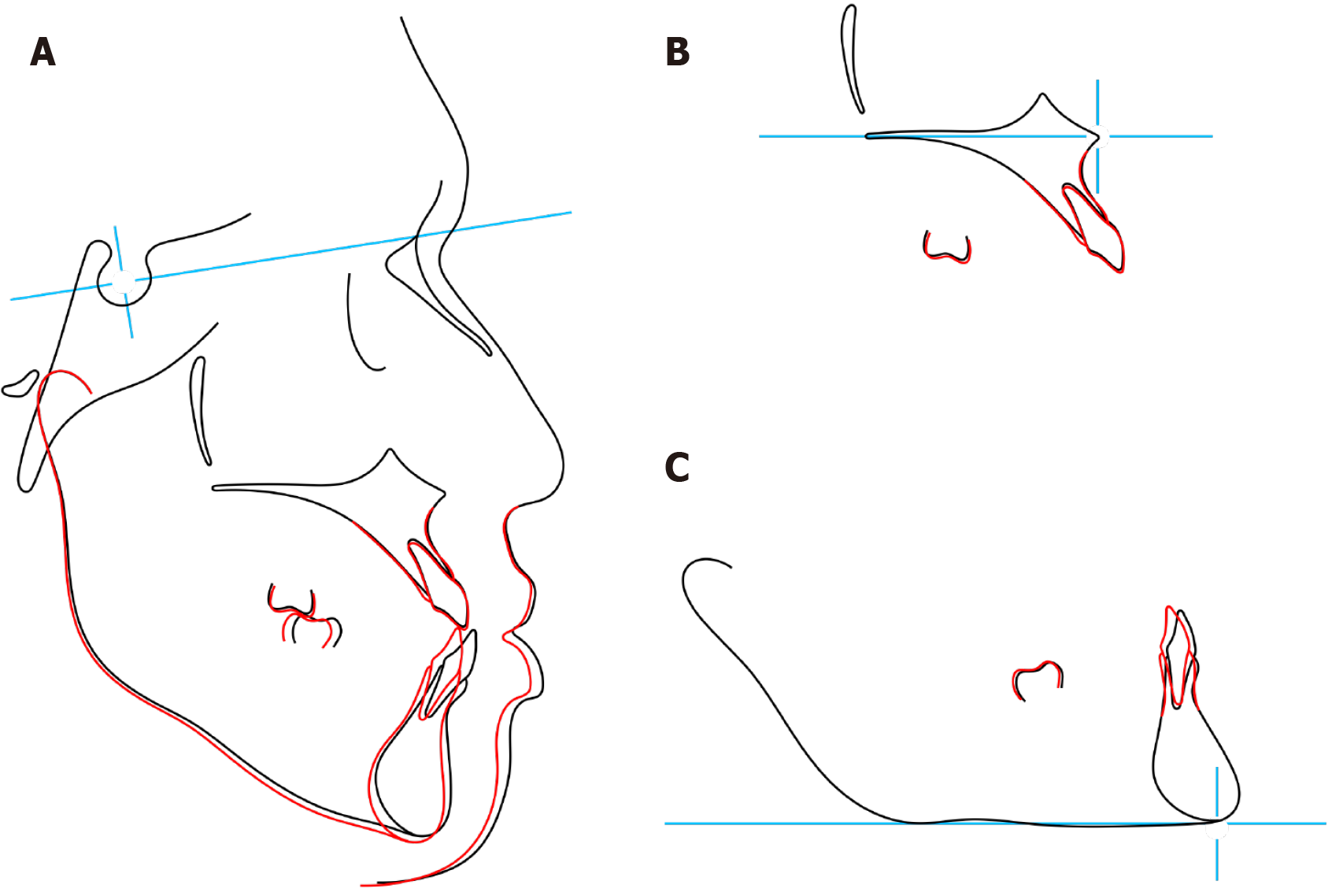
Superimposition images (Figure 12) revealed the distal movement of the mandibular molars and the mesial shift of the maxillary molars, which contributed to the establishment of proper overjet and overbite.
The following section discusses the efficacy of ISW MEAW approach for the correction of anterior crossbite and the method of WA orthodontics. Proper arch coordination was achieved in our case. The treatment was a success.
In this case, the efficacy of ISW MEAW for the correction of anterior crossbite and midline deviation, along with the method of WA orthodontics, were discussed. Proper arch coordination, midline correction, and profile improvement were achieved, leading to a satisfactory treatment result.
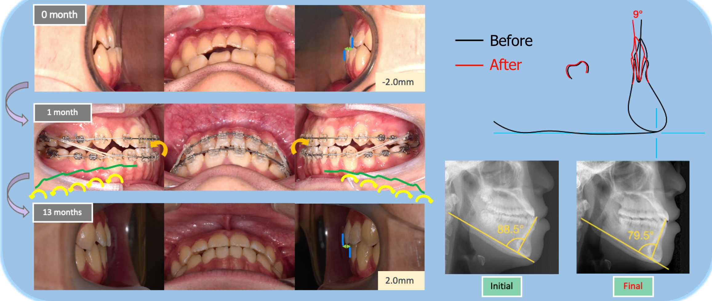
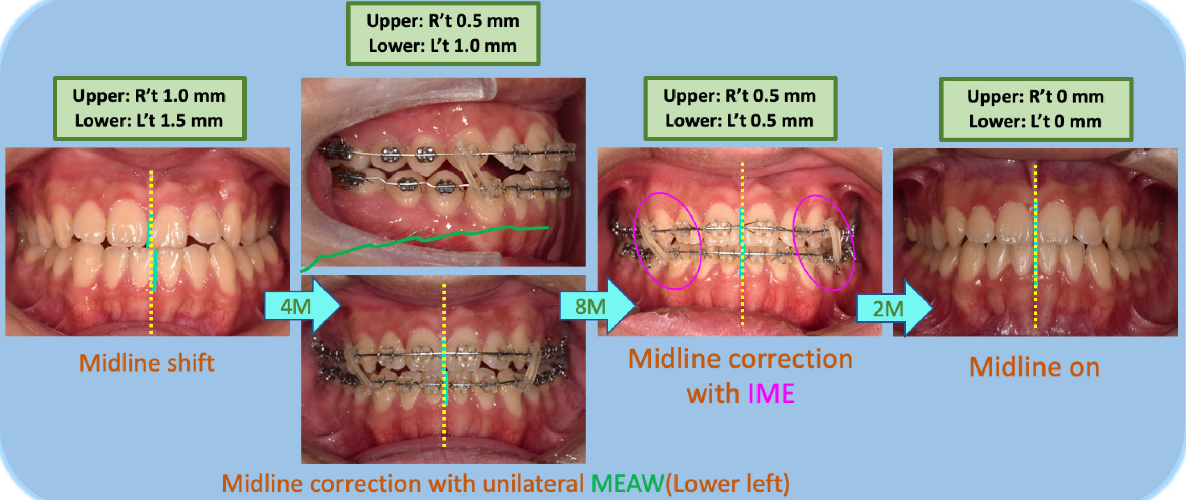
The ISW MEAW technique was employed to correct the patient's anterior crossbite by using step and slope bends for controlled teeth movement, considerably improving dental alignment. Retroclination of the mandibular incisors played a crucial role in establishing an ideal overjet and overbite, improving both the patient's occlusal function and facial aesthetics. In this case, the mandibular anterior teeth were retroclined by 9°, further contributing to the overall outcome. The technique’s ability to achieve precise arch coordination made it possible to conservatively treat the skeletal class III malocclusions without the need for surgical intervention[5]. Additionally, the midline correction was achieved through the controlled application of a unilateral ISW MEAW and IME, improving the overall balance of the dentition (Figure 13).
Despite the careful use of anchorage control mechanisms, midline deviation occurred during treatment. To address this, a unilateral ISW MEAW and IME were applied to teeth (13–43 and 23–34) (Figure 14). This combination enabled precise correction of the midline and the simultaneous achievement of the optimal overjet and overbite. Use of the ISW MEAW facilitated distal tipping of the teeth while minimizing unwanted side effects, making it possible to achieve excellent results without the need for a more invasive anchorage technique, such as miniscrews[6]. One limitation of the approach is the long duration of intermaxillary elastic wear, which can lead to increased proclination of the upper incisors (U1-SN angle) and steepening of the mandibular plane angle. This highlights the need for careful monitoring during treatment to prevent unfavorable side effects. More severe class III cases may require alternative strategies such as the use of mini-screws or molar extraction should be considered. These approaches can provide more effective anchorage control and facilitate the correction of major skeletal discrepancies. For instance, Nguyen et al[7] demonstrated the successful application of miniscrew-assisted rapid palatal expansion combined with lingual appliances to address skeletal class III malocclusion in adults. This method is a non-invasive alternative that is suitable for managing severe cases. It improves dental alignment, and maintains esthetics during treatment[7]. Similarly, Ngoc et al[8] reported on the effectiveness of asymmetric molar extraction with lingual appliances for treating skeletal class III malocclusion with mandibular deviation. This approach provided sufficient space for retraction and improved the torque control of the lower incisors, leading to favorable facial profile changes and occlusal outcomes[8].
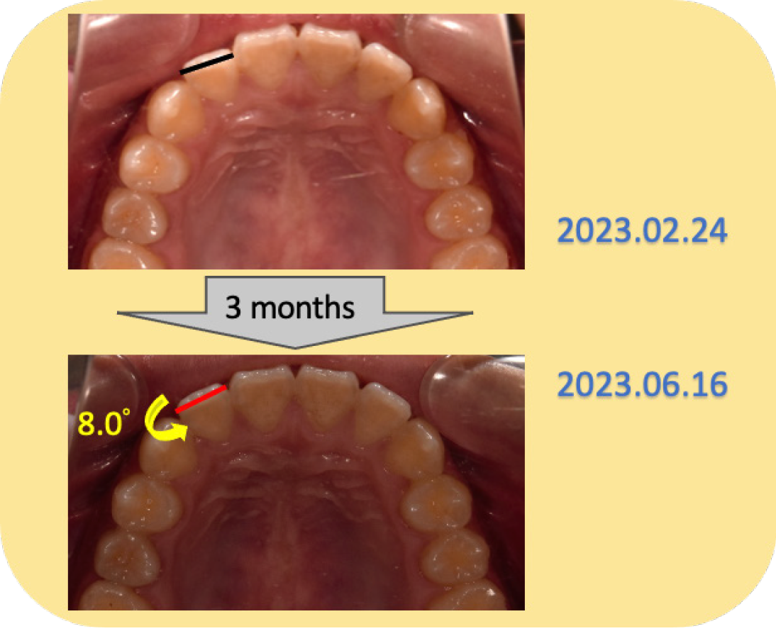
WA orthodontics, a hybrid approach combining wires and aligners, played an essential role in the finishing stages of treatment for our patient. After wire treatment, aligners were used to meet the patient’s preference for early debonding. The aligners provided precise control of smaller movements, such as the derotation of tooth (No. 12) (Figure 15), which would have been difficult to achieve with wires alone. Fixed appliances were initially insufficient to fully correct the rotation of tooth (No. 12); therefore clear aligners were introduced after debonding. This approach successfully refined the alignment, as confirmed during follow-up visits (Figure 11), and improved the patient's compliance, oral hygiene, and comfort, contributing to the overall success of the treatment.
Additionally, skeletal class III malocclusion may have a genetic component, which can influence long-term stability. Even after successful correction, there is a potential for relapse because of inherent genetic factors. Thus, long-term retention is essential to maintain treatment outcomes. In this case, retainers were prescribed for full-time wear for the first three months, followed by nighttime wear, ensuring the preservation of overjet, overbite, and midline correction. Addressing genetic predispositions in future treatment planning may provide insights into optimizing long-term stability strategies.
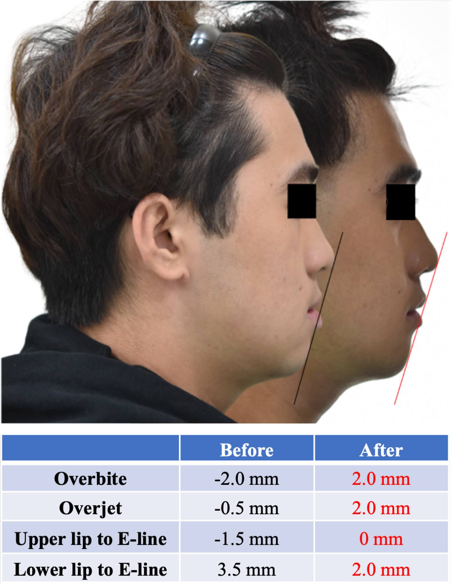
Finally, the combination of the ISW MEAW technique with WA orthodontics produced considerable improvements in the patient’s facial profile (Figure 16). The posttreatment ANB angle was 0.5° (Figure 9). This result reflects the increased mandibular plane angle and clockwise rotation of the mandible during treatment, contributing to a more balanced skeletal relationship (Figure 12). Controlled retroclination of the lower anterior teeth helped reduce the patient's protrusive lower lip, resulting in a more harmonious and aesthetically balanced profile. Although torque control of the lower incisors resulted in slight root prominence, this was an acceptable compromise because of the patient's preference to avoid extraction and surgical procedures. The patient's periodontal condition was closely monitored and no major issues were discovered during follow-up visits (Figure 11). Retainers were prescribed for full-time wear for the first three months after treatment, which was followed by nighttime wear to maintain the overjet, overbite, and midline correction achieved during active treatment and ensure long-term stability.
In the treatment of skeletal class III malocclusion with anterior crossbite, the ISW MEAW technique combined with WA orthodontics proved to be an effective and minimally invasive approach. Through controlled teeth movement and IME, we achieved an improved overjet, overbite, and midline correction. Additionally, the patient’s facial aesthetics showed noticeable enhancement, with the nonsurgical approach successfully meeting both functional and cosmetic objectives. While the treatment yielded favorable results, further optimization could be achieved by enhancing the torque control of the lower incisors and considering alternative biomechanical techniques to improve treatment efficiency and accuracy. In this challenging case, a satisfactory result was achieved using a conservative approach.
| 1. | Inchingolo AD, Patano A, Coloccia G, Ceci S, Inchingolo AM, Marinelli G, Malcangi G, Di Pede C, Garibaldi M, Ciocia AM, Mancini A, Palmieri G, Rapone B, Piras F, Cardarelli F, Nucci L, Bordea IR, Scarano A, Lorusso F, Giovanniello D, Costa S, Tartaglia GM, Di Venere D, Dipalma G, Inchingolo F. Treatment of Class III Malocclusion and Anterior Crossbite with Aligners: A Case Report. Medicina (Kaunas). 2022;58:603. [RCA] [PubMed] [DOI] [Full Text] [Full Text (PDF)] [Cited by in Crossref: 5] [Cited by in RCA: 13] [Article Influence: 4.3] [Reference Citation Analysis (0)] |
| 2. | Lim LI, Choi JY, Ahn HW, Kim SH, Chung KR, Nelson G. Treatment outcomes of various force applications in growing patients with skeletal Class III malocclusion. Angle Orthod. 2021;91:449-458. [RCA] [PubMed] [DOI] [Full Text] [Cited by in RCA: 5] [Reference Citation Analysis (0)] |
| 3. | Mehta S, Chen PJ, Upadhyay M, Yadav S. Intermaxillary elastics on skeletal anchorage and MARPE to treat a class III maxillary retrognathic open bite adolescent: A case report. Int Orthod. 2021;19:707-715. [RCA] [PubMed] [DOI] [Full Text] [Cited by in Crossref: 1] [Cited by in RCA: 1] [Article Influence: 0.3] [Reference Citation Analysis (0)] |
| 4. | Alnefaie M, Han WJ, Ahn YS, Baik WK, Choi SH. Orthopedic and Nonsurgical Orthodontic Treatment of Adolescent Skeletal Class III Malocclusion Using Bone-Anchored Maxillary Protraction and Temporary Anchorage Devices: A Case Report. Children (Basel). 2022;9:683. [RCA] [PubMed] [DOI] [Full Text] [Full Text (PDF)] [Reference Citation Analysis (0)] |
| 5. | Chen P, Chen Y, Wang I, Yu J. Improved Super-elastic Ti-Ni alloy wire (ISW) for Treatment of Adult Angle Class III with Anterior Crossbite. Taiwan J Orthod. 2023;35. [DOI] [Full Text] |
| 6. | Lo YC, Chen YH, Chiang HH, Yu JH. Use Improved Super-elastic Ti–Ni Alloy Wire to Correct Class II Division 2 Malocclusion with Unstable Mandibular Position. Taiwan J Orthod. 2018;30. [DOI] [Full Text] |
| 7. | Nguyen VA, Nguyen NA, Doan HL, Pham TH, Doan BN. Management of anterior and posterior crossbites with lingual appliances and miniscrew-assisted rapid palatal expansion: A case report. Medicine (Baltimore). 2024;103:e40832. [RCA] [PubMed] [DOI] [Full Text] [Reference Citation Analysis (0)] |
| 8. | Ngoc VTN, Phuong NTT, Anh NV. Skeletal Class III Malocclusion with Lateral Open Bite and Facial Asymmetry Treated with Asymmetric Lower Molar Extraction and Lingual Appliance: A Case Report. Int J Environ Res Public Health. 2021;18:5381. [RCA] [PubMed] [DOI] [Full Text] [Full Text (PDF)] [Reference Citation Analysis (0)] |


