Published online May 26, 2025. doi: 10.12998/wjcc.v13.i15.101884
Revised: December 31, 2024
Accepted: January 9, 2025
Published online: May 26, 2025
Processing time: 113 Days and 1.6 Hours
Orthodontic treatment for open bite and crossbite cases is always challenging. In this paper, we demonstrate a skeletal class III patient with anterior open bite and crossbite whose problem was successfully corrected with improved super-elastic Ti-Ni alloy wire (ISW).
A 19 years old male came to our clinic with a chief complaint of anterior open bite and crossbite and not able to chew food well. Clinical examination revealed an angle class III malocclusion with anterior open bite, crossbite and spaced arch. Ra
In a case of class III angular malocclusion with open bite and crossbite in the ante
Core Tip: The orthodontic treatment for patients with skeletal class III is usually difficult, and the treatment usually requires combination with orthognathic surgery, which not only involves higher costs but also requires a significant amount of recovery time post-surgery. However, by using improved super-elastic Ti-Ni alloy wire, we successfully corrected the skeletal class III patient with anterior crossbite and open bite without surgical intervention. Patient is satisfied with the result of the treatment.
- Citation: Fan Y, Yu JH, Chen YH. Improved super-elastic Ti-Ni alloy wire for the angle class III patient with anterior open bite: A case report. World J Clin Cases 2025; 13(15): 101884
- URL: https://www.wjgnet.com/2307-8960/full/v13/i15/101884.htm
- DOI: https://dx.doi.org/10.12998/wjcc.v13.i15.101884
A class III malocclusion is a type of dental misalignment that refers to a situation resulting from skeletal problems, dental alveolar anomalies, or both. Its clinical features typically present as insufficient maxillary development and an elongated mandible[1]. Anterior open bite and crossbite are also commonly associated clinical manifestations. This may lead to difficulties for the patient in chewing and speaking, as well as negatively impact their confidence and social interactions. When treating class III patients, especially those with severe skeletal discrepancies, orthodontic treatment often needs to be combined with orthognathic surgery to correct the anomalies[1]. However, due to various factors, including the pa
The 19-year-old male complained of an anterior open bite, crossbite, spaced arch, and difficulty chewing food.
The patient did not have any current illness or symptoms.
The patient has no past medical history.
There is no family history of dental or orthodontic issues in the patient.
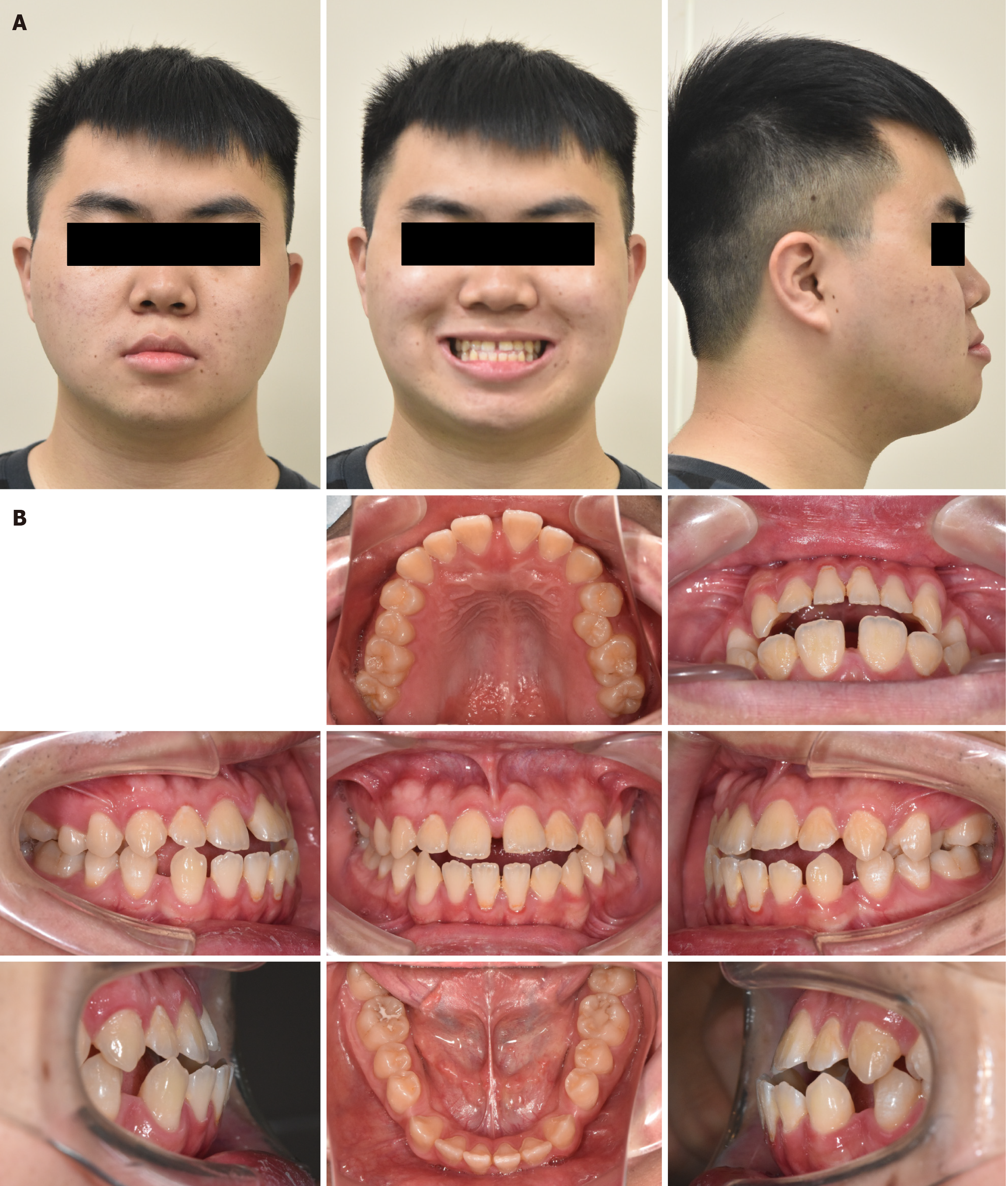
The pretreatment lateral facial photograph showed a protruding chin. In the frontal view, no obvious chin deviation was observed, and a 1.5 mm diastema was noted in the maxillary dentition (Figure 1A). Intraoral photographs revealed a bilateral class III molar and class III canine relationship, as well as an anterior open bite and crossbite. Additionally, space was noted in both the maxillary and mandibular dentitions (Figure 1B).
All examination data were within normal limits.
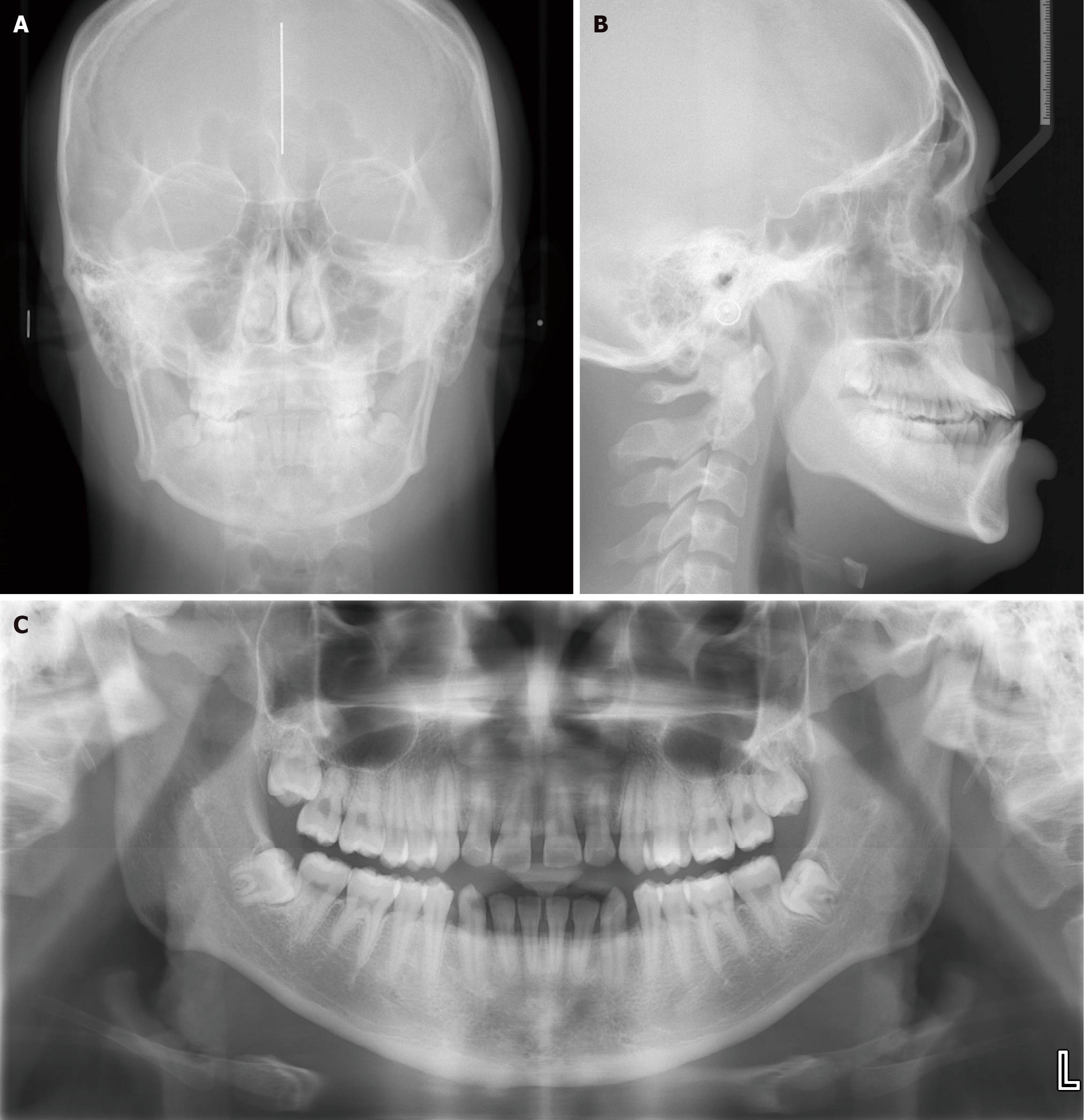
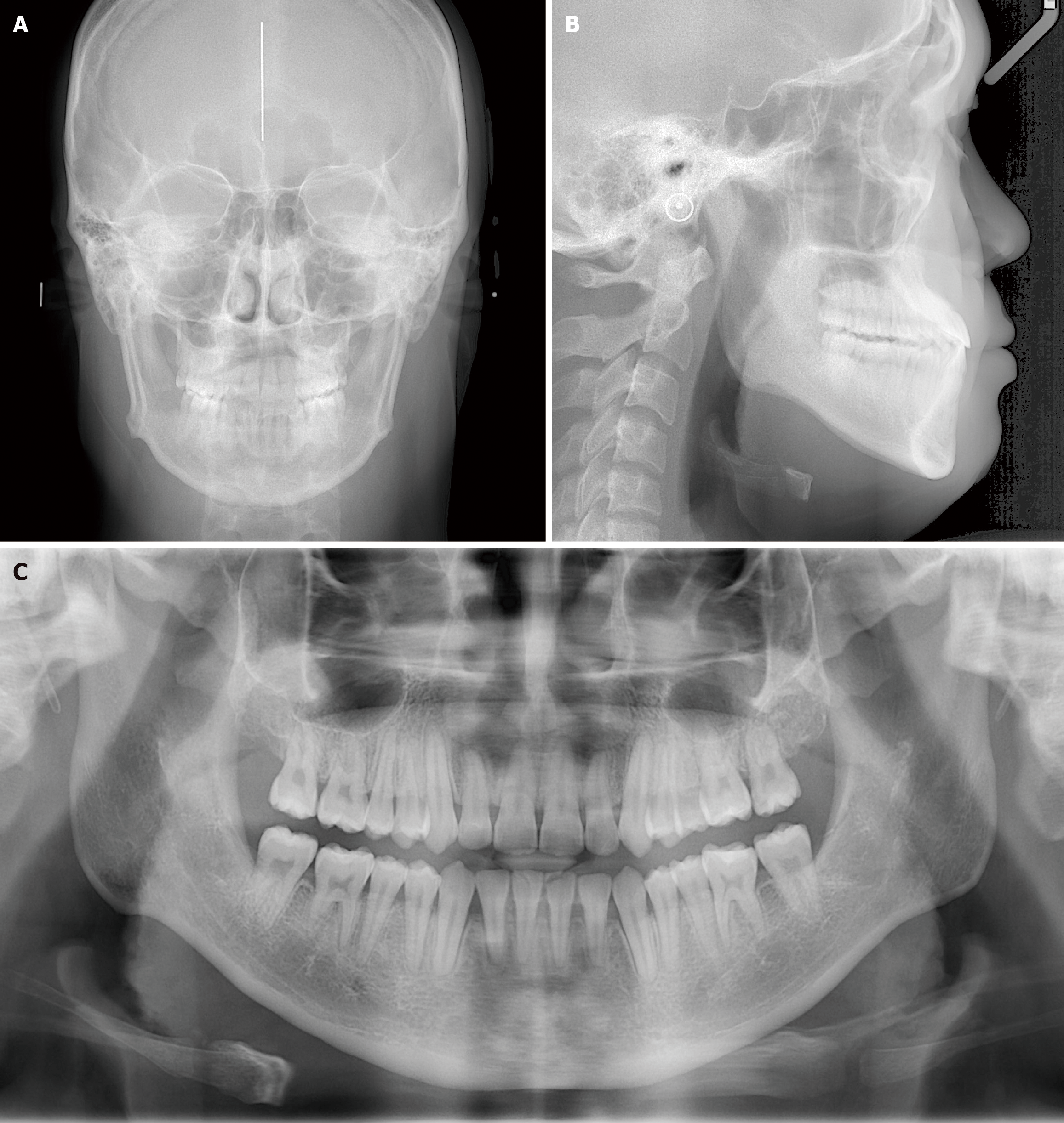
Panoramic radiographs revealed the presence of all teeth, including four wisdom teeth. Initial lateral cephalometric ana
The patient was diagnosed with skeletal class III malocclusion, anterior open bite, crossbite, and spaced dentitions in both the maxillary and mandibular arches.
The patient presented with skeletal class III malocclusion, anterior open bite, crossbite, and spaced maxillary and mandi
Two treatment options were proposed. The first involved orthognathic surgery to correct the skeletal discrepancy. The second option was non-surgical orthodontic treatment using improved super-elastic ISW and IME to manage the malo
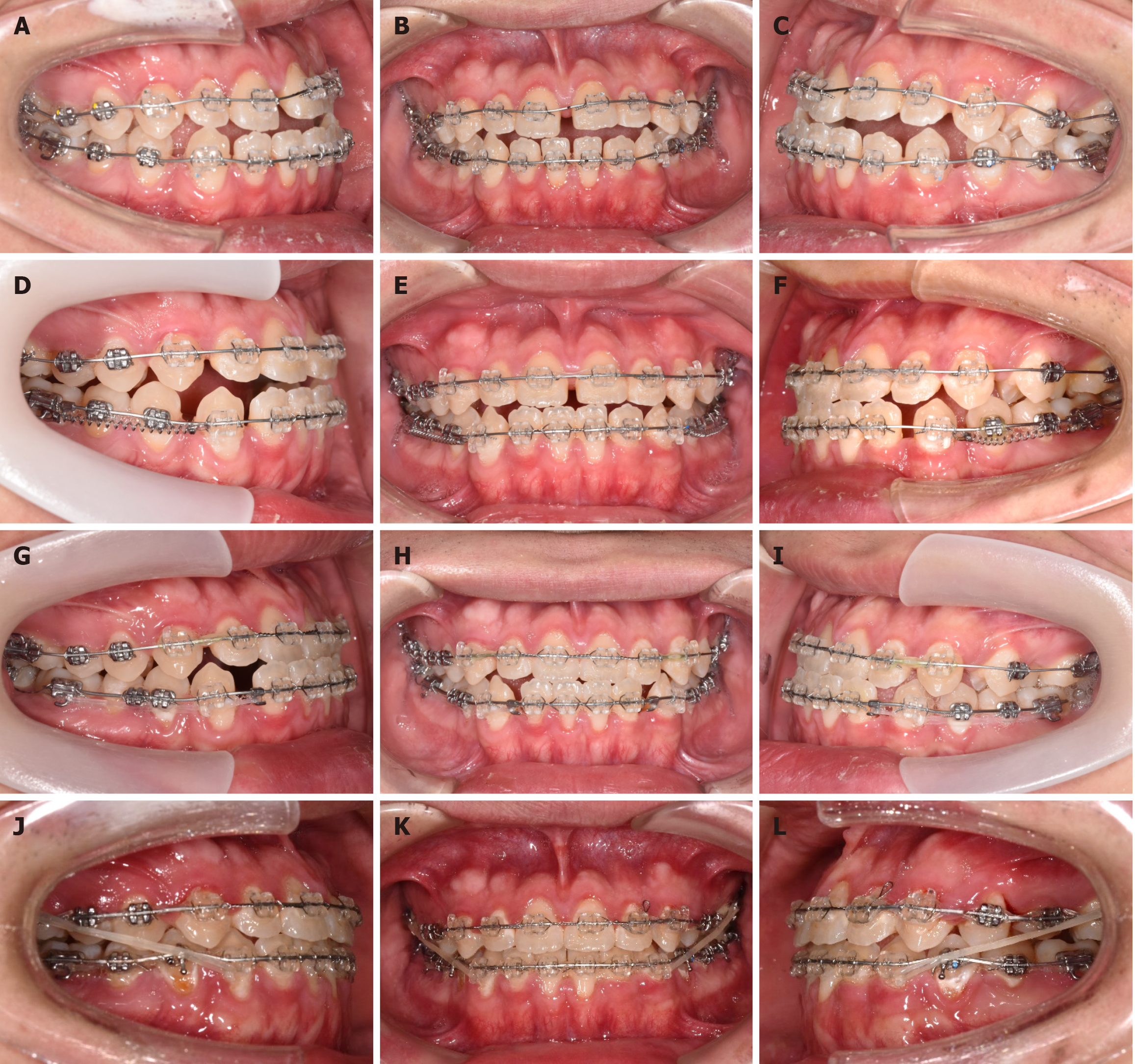
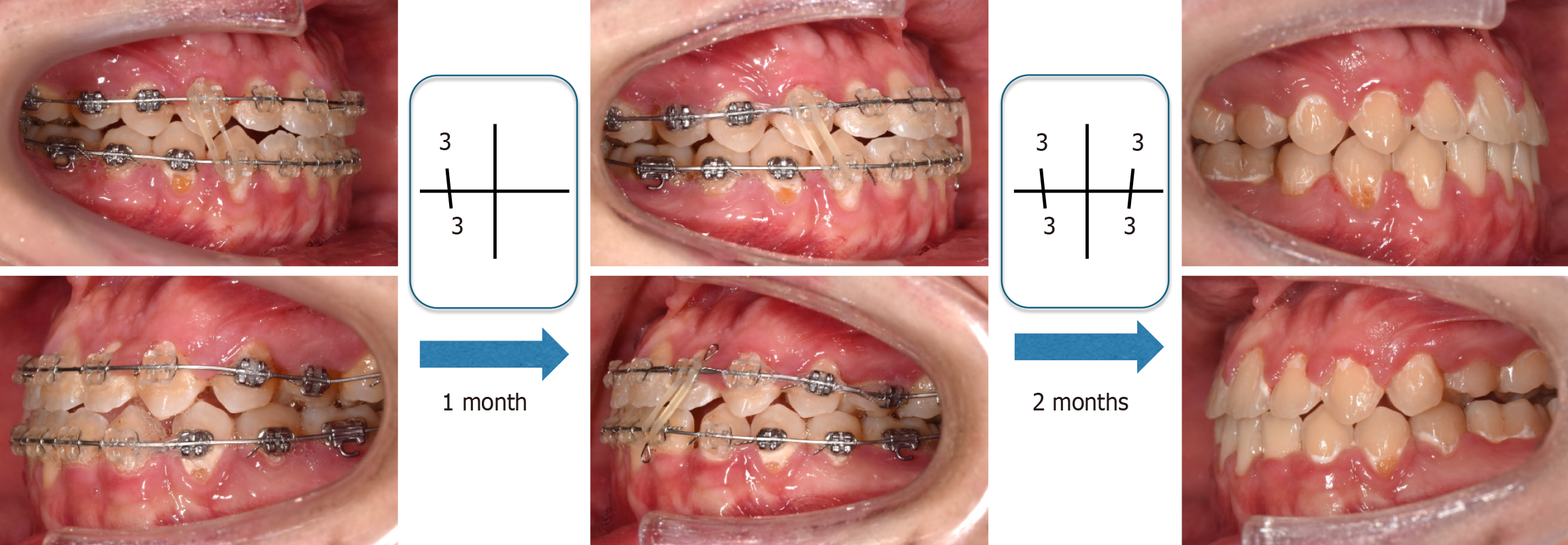
Treatment began with full-mouth direct bonding system and leveling using 0.016-inch × 0.022-inch ISW (Figure 3A-C). As the treatment progressed, we took photographs and X-rays to document and monitor whether the results aligned with our expectations. Adjustments were made as needed to address any issues. The anterior crossbite was corrected by lower canine distal drive with a closed coil spring and anterior retraction (32nd-42nd) with a power chain (Figure 3D-I). Class III IME were used to achieve normal overbite and overjet by the 20 months (Figure 3J-L). After 26 months of active treat
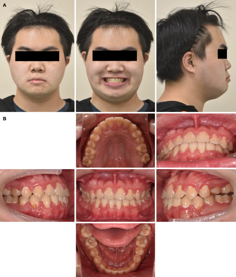
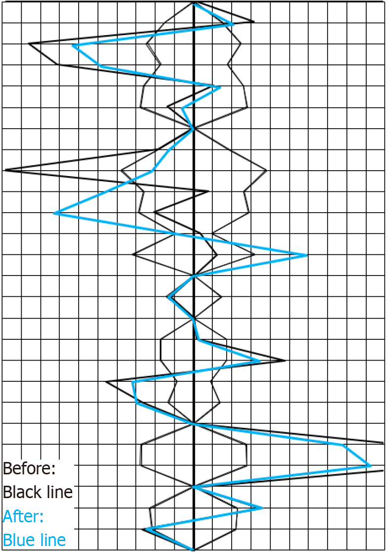
The anterior open bite and crossbite were successfully corrected through the retraction of the lower anterior teeth (Figure 3D-I) and class III IME (Figure 3J-L), achieving a well-aligned arch and stable occlusion. Final records (Figures 4A and B) showed that the dental midline was properly aligned, polygons of patients before and after active treatment (Figure 5 and Table 1), and panoramic radiographs (Figure 6) confirmed well-aligned parallel roots with no signs of root resorption. Compared to the postoperative recovery time and high cost associated with orthognathic surgery, the non-surgical orthodontic treatment, chosen after thorough discussion, successfully addressed the patient’s main concerns. Throughout the treatment, the patient experienced no significant discomfort, and both the patient and their family were extremely satisfied with the results. Superimposition images (Figure 7) demonstrated the retraction and retroclination of the upper and lower incisors, successfully correcting the anterior open bite and crossbite. The patient’s occlusion re
| Characteristics | Before | After | mean ± SD |
| Facial angle | 91.0 | 89.5 | 84.83 ± 3.05 |
| Convexity | -10.0 | -8.0 | 7.58 ± 4.95 |
| A-B plane | 10.0 | 7.0 | -4.81 ± 3.50 |
| Mandibular plane | 31.0 | 32.0 | 28.81 ± 5.23 |
| Y-axis | 62.5 | 64.0 | 65.38 ± 5.63 |
| Occlusal plane | 7.5 | 8.5 | 11.42 ± 3.64 |
| Interincisal | 104.0 | 120.0 | 124.09 ± 7.63 |
| L-1 to occlusal | 25.0 | 14.0 | 23.84 ± 5.28 |
| L-1 to mandibular | 92.0 | 81.0 | 96.33 ± 5.78 |
| U-1 to A-P plane | 9.5 | 8.0 | 8.92 ± 1.88 |
| FMIA | 57.0 | 67.0 | 54.63 ± 6.47 |
| FH to SN plane | 4.0 | 4.0 | 6.19 ± 2.89 |
| SNA | 82.5 | 83.0 | 82.32 ± 3.45 |
| SNB | 88.5 | 86.5 | 78.90 ± 3.45 |
| ANB | -6.0 | -3.5 | 3.39 ± 1.77 |
| U-1 to N-P plane | 6.5 | 5.5 | 11.74 ± 2.73 |
| U-1 to FH plane | 133.0 | 127.5 | 111.13 ± 5.54 |
| U-1 to SN plane | 129.5 | 123.5 | 104.54 ± 5.55 |
| Gonial angle | 129.0 | 131.0 | 122.23 ± 4.61 |
| Ramus inclination | 81.5 | 82.0 | 87.07 ± 4.40 |
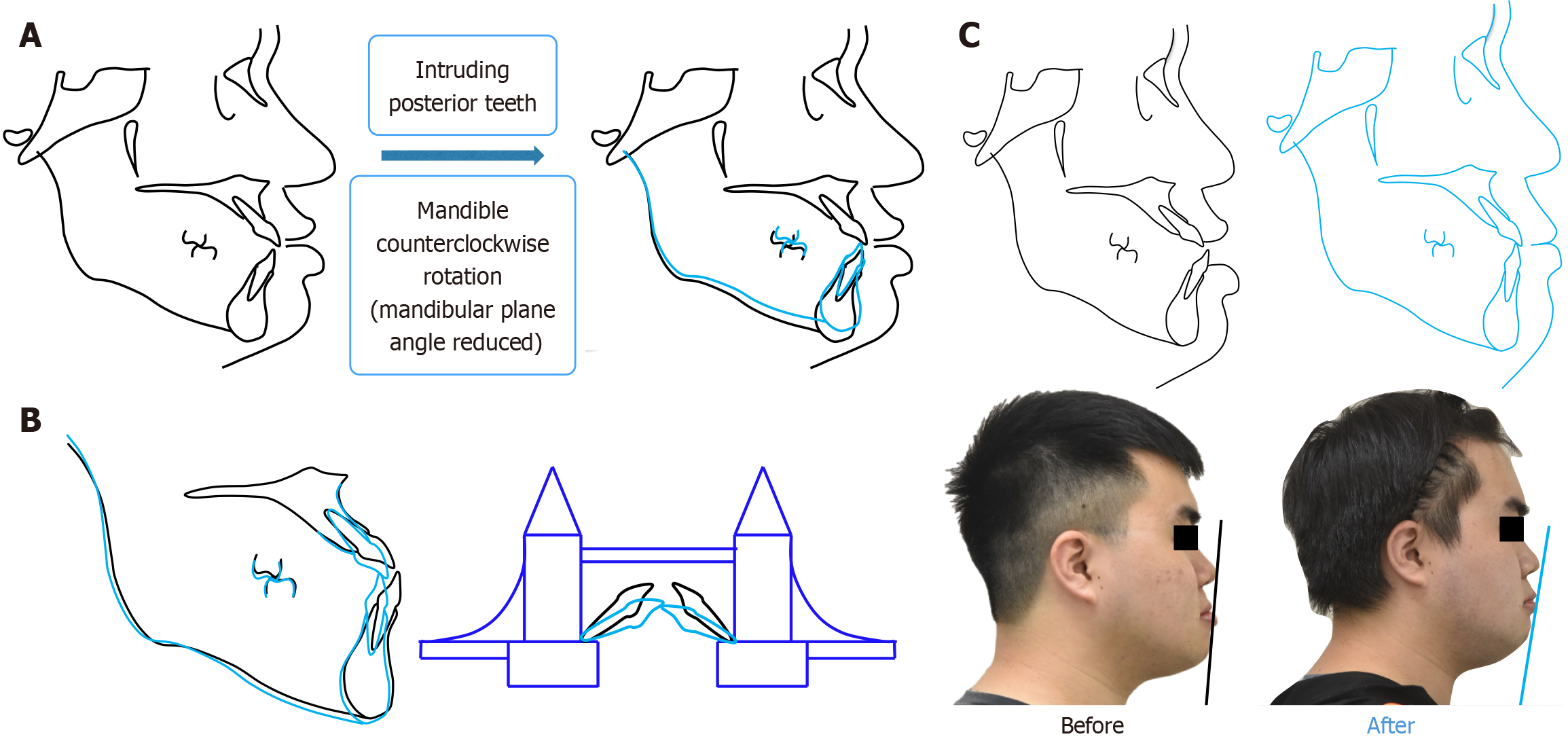
In this case, we chose a non-extraction strategy, utilizing ISW leveling and IME to correct the anterior open bite, crossbite, and inter-jaw relationship. After achieving adequate overbite and overjet, we found that the patient’s arch interdigitation was still insufficient. We advised the patient to continue wearing IME to improve interdigitation. After several months, a desirable occlusion with adequate overbite and overjet was achieved (Figure 8). In order to correct an anterior open bite, we need to increase the overbite. This can be achieved by intruding the posterior teeth or extruding the anterior teeth. Intrusion of the posterior teeth is applicable in high-angle open bite cases and results in counterclockwise rotation of the mandible, reducing the mandibular plane angle (Figure 9A). Extrusion of the anterior teeth can be applied in low-angle open bite cases (Figure 9B).
In our case, the initial A point-nasion-B point angle was -6.0°, indicating that our patient had a severe skeletal problem. If we had chosen a strategy such as intruding the posterior teeth to increase the anterior overbite, it would have resulted in a more pronounced skeletal class III pattern. Therefore, the strategy we selected to address the problem was to extrude the anterior teeth, also known as the drawbridge effect, to correct the open bite (Figure 9B). Before orthodontic treatment, we observed a skeletal class III pattern and a prominent chin. Therefore, we retracted the anterior teeth. After treatment, the patient’s facial profile improved significantly, and his chin appeared less prominent (Figure 9C and Table 2).
| Characteristics | Before | After |
| Nasolabial angle | 85° | 95° |
| U-1 to FH plane | 133.0 | 127.5 |
| Mandibular plane | 31.0° | 32.0° |
| Chin | Prominent | Less prominent |
In this case of skeletal class III malocclusion accompanied by anterior open bite and crossbite, we discussed various options with the patient. Ultimately, the patient chose to undergo pure orthodontic treatment instead of orthodontic treatment combined with orthognathic surgery, which proved to be an effective and minimally invasive approach. Following the active treatment, the patient’s facial profile showed significant enhancement, and the treatment outcomes successfully met both functional and aesthetic goals without the need for surgical intervention. This result satisfied the needs and expectations of both the patient and the doctor, demonstrating the viability of conservative orthodontic me
| 1. | Ngan P, Moon W. Evolution of Class III treatment in orthodontics. Am J Orthod Dentofacial Orthop. 2015;148:22-36. [RCA] [PubMed] [DOI] [Full Text] [Cited by in Crossref: 97] [Cited by in RCA: 102] [Article Influence: 10.2] [Reference Citation Analysis (0)] |
| 2. | Miura F, Mogi M, Okamoto Y. New application of superelastic NiTi rectangular wire. J Clin Orthod. 1990;24:544-548. [PubMed] |
| 3. | Miura F, Mogi M, Ohura Y, Hamanaka H. The super-elastic property of the Japanese NiTi alloy wire for use in orthodontics. Am J Orthod Dentofacial Orthop. 1986;90:1-10. [RCA] [PubMed] [DOI] [Full Text] [Cited by in Crossref: 311] [Cited by in RCA: 235] [Article Influence: 6.0] [Reference Citation Analysis (0)] |
| 4. | Iramaneerat K, Hisano M, Soma K. Dynamic analysis for clarifying occlusal force transmission during orthodontic archwire application: difference between ISW and stainless steel wire. J Med Dent Sci. 2004;51:59-65. [PubMed] |


