Published online May 6, 2025. doi: 10.12998/wjcc.v13.i13.100822
Revised: November 29, 2024
Accepted: December 20, 2024
Published online: May 6, 2025
Processing time: 136 Days and 20.9 Hours
The numbers of mandibular first premolar roots and root canals vary, and the incidence of three roots and three canals is 0.09%.
In this article, we review the root and root canal conditions for the mandibular first premolar and report the case of a mandibular left first premolar with three roots and three canals in a male patient, with suggestions for clinical diagnosis and treatment. The patient was referred by an orthodontist for the extraction of the tooth. Preoperative cone-beam computed tomography examination revealed that it had three roots. Under local anesthesia, the extraction socket was carefully expanded, and the tooth was successfully removed intact using forceps. The procedure was uneventful, with no root fractures, postoperative bleeding, or sensory abnormality observed.
The mandibular first premolar is characterized by multiple roots and canal variations that can increase the difficulty of treatment.
Core Tip: This article highlights a rare case of the orthodontic extraction of a mandibular left first premolar with three roots and three canals. It offers practical clinical insights and summarizes recent global incidence rates for this unique anatomical variation.
- Citation: Lin CY, Yu YY. Mandibular left first premolar with three roots and three canals: A case report. World J Clin Cases 2025; 13(13): 100822
- URL: https://www.wjgnet.com/2307-8960/full/v13/i13/100822.htm
- DOI: https://dx.doi.org/10.12998/wjcc.v13.i13.100822
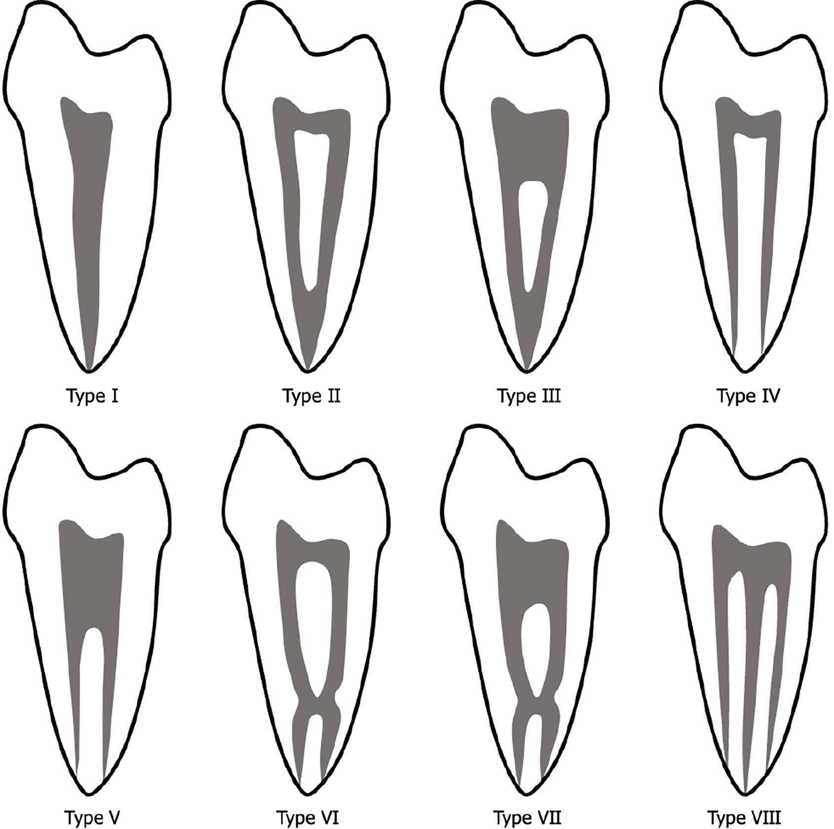
Mandibular first premolars with three roots and three canals are rarely seen in clinical practice. Cleghorn et al[1] reported that 97.9% of 4462 mandibular first premolars had single roots, 1.8% had double roots, and 0.2% had triple roots. The incidences of these different root numbers vary among ethnic groups[2]. The root canal systems of mandibular first premolars also vary, with 74%-80% of these teeth having single canals, approximately 22% having two canals, and 0.1% having three canals[3-5]. A cone-beam computed tomography (CBCT) analysis indicated that the incidence of three roots with three canals (type VIII in the Vertucci root canal classification) (Figure 1) in the Chinese population is 0.09%[6]. Here, we report the case of a male patient with a three-rooted and three-canalled mandibular left first premolar.
A 13-year-old male presented to our hospital's orthodontic department due to irregular dentition.
He was diagnosed with angle class II division 1 malocclusion with a skeletal class II pattern. The patient’s treatment plan included extractive orthodontic camouflage therapy, and his orthodontist referred him to our department for the extraction of the mandibular left first premolar.
The patient had previously been in good health. The patient's family denies any history of significant systemic diseases, infectious diseases, major surgeries, blood transfusions, or allergies to food or medications.
The patient denied any family history.
Oral examination revealed scattered spaces in the upper dentition and mild crowding in the lower dentition. Tooth 12 was hypodontic, and teeth 27 and 37 presented with positive scissor bite. The patient had degree I overbite and degree I overjet. The right canine and molars had a distal relationship. The lower anterior teeth had obvious labial inclination. Maxillary protrusion and mandibular retrusion were noted. Tooth 34 had erupted.
The laboratory tests showed no significant abnormalities.
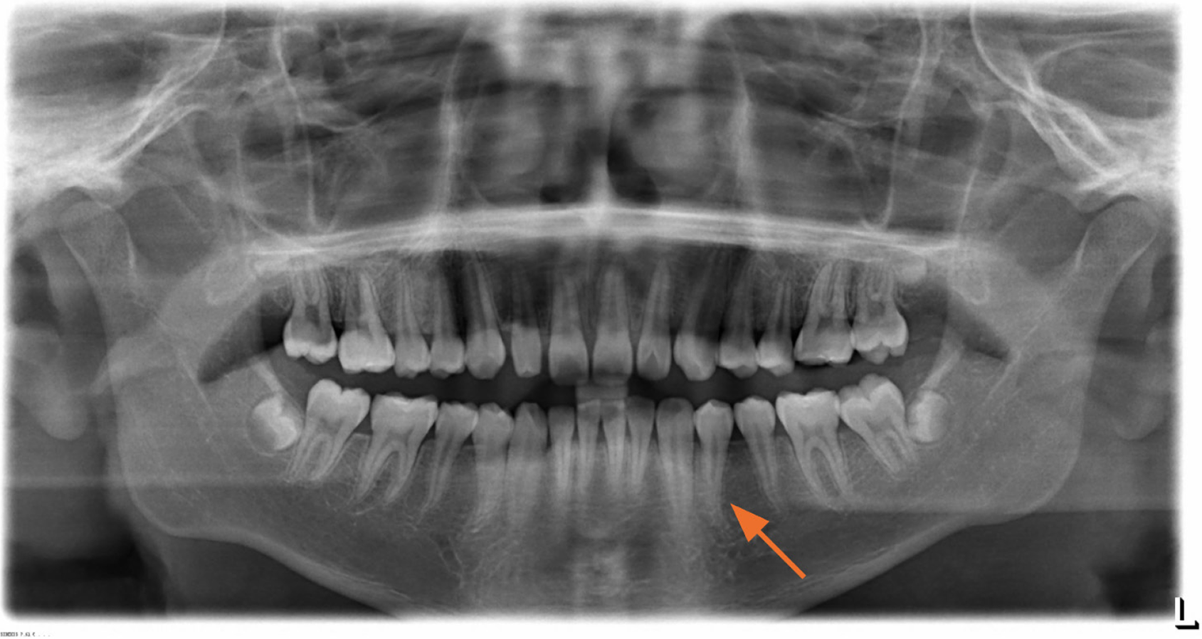
X-ray image showed that it had two roots, with the bifurcation into mesial and distal roots occurring near the apical third of the root and both roots curving distally by approximately 40°. Image overlap on the mesial root was noted (Figure 2).
CBCT imaging revealed that tooth 34 had three root canals (mesiolingual, distolingual, and buccal) corresponding to its roots (Figure 3).
Combined with the patient’s medical history, the final diagnosis was irregular dentition.
For the extraction of tooth 34, 0.9 mL 4% articaine hydrochloride was infiltrated supraperiosteally into the mucobuccal fold below the apex, followed immediately by the injection of 0.9 mL anesthetic onto the lingual aspect of the tooth. Five minutes later, the tooth was extracted using lower universal forceps. The forceps were adapted to the tooth as apically as possible, beneath the cervical line, with the beaks oriented parallel to the long axis of the tooth. Initially, gentle movements were made with the application of buccal and lingual pressure. After the tooth had been slightly mobilized, the force was increased gradually and the final extraction movement was buccal. The expanded buccolingual plates were compressed back to their original configuration. Initial control of hemorrhage was achieved by placing moistened gauze over the extraction socket.
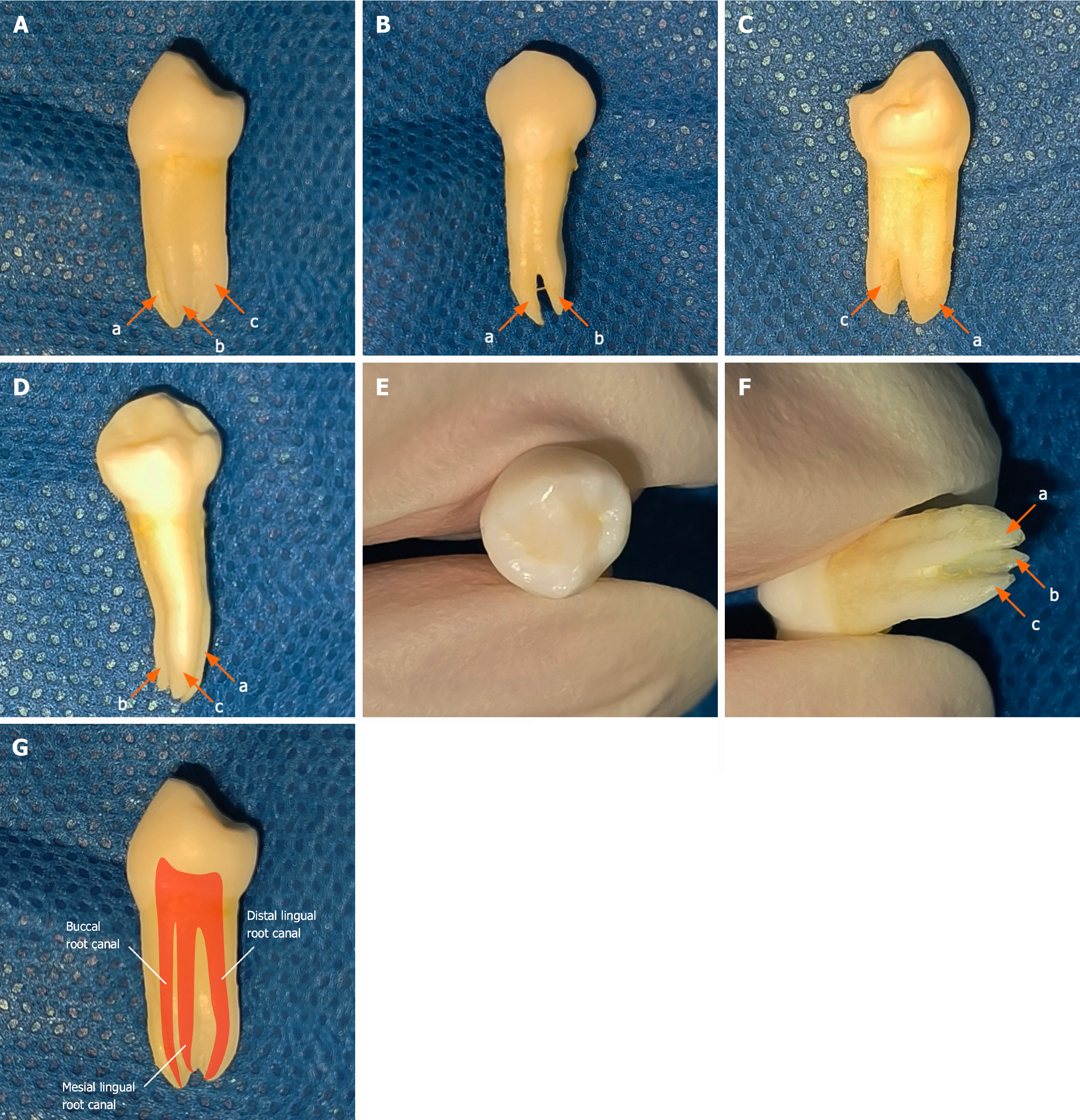
The tooth was observed to have three roots (mesiolingual, distolingual, and buccal) with clear contours (Figure 4). On the mesial surface, the mesiolingual root was thin and shorter than the other two roots, and its bifurcation into an independent root occurred at the middle third of the root. On the buccal surface, the mesiolingual root was connected to the upper part of the buccal root, with the bifurcation occurring approximately at the apical root third. On the distal surface, the distolingual and buccal roots bifurcated at the middle third of the root, near the apex, with the buccal root exhibiting a shallow distal groove and both roots being relatively thick. On the lingual surface, the distolingual root was located lingual to the mesiolingual root. On the occlusal surface, distinct mesial and distal fossae were visible, with a prominent transverse ridge, and the lingual cusp was approximately half the length of the buccal cusp (Figure 4).
One week after surgery, the patient reported that he was in good overall condition, with no significant bleeding at the extraction site, no sensory disturbance, and no indication of dry socket.
Textbooks describe the mandibular first premolar as typically having a flat, slender single root, often exhibiting bifurcation traces in the apical region of the mesial surface[7]. Clinicians often overlook the considerable possibility that this tooth has multiple roots and/or complex root-canal configurations, increasing the likelihood of missed diagnoses and untreated canals[8].
The underfilling of root canals in mandibular first premolars, the reported incidence of which is 34.6%, is a common cause of root canal treatment failure. Canals are missed in up to 14.1% of cases, another leading cause of treatment failure[9]. These oversights significantly elevate the risk of infection, which can progress to pulpitis and lead to complications such as periapical periodontitis, often causing severe pain for patients.
Zhang et al[10] reported a case of a mandibular first premolar with five root canals, in which the fifth canal was discovered only after the other four had been filled. Penukonda et al[11] described a taurodontic mandibular first premolar with two roots and four canals. Similarly, Nouroloyouni et al[12] reported a case with two roots and five canals. Li et al[13] documented a mandibular first premolar with a C-shaped root canal and taurodontism. Jain et al[14] described a mandibular first premolar with three roots and three canals, similar to the present case, in a patient who presented with secondary caries as the chief complaint.
In all of these cases, the root canal variations were identified during the treatment of irreversible pulpitis and periapical periodontitis. Recent reports predominantly describe mandibular first premolars with two roots and more common variations[15,16]. The discovery of a mandibular first premolar with three roots and three canals during orthodontic extraction is extraordinarily rare, making this finding of significant clinical value.
Before the extraction or root canal treatment of the mandibular first premolar, careful assessment is required to confirm the number of root canals present. Various radiographic images, including bitewing, periapical, and panoramic X-rays, should be used to evaluate the canal number, location, and length, and to identify any narrowing or accessory canals. Such thorough assessment helps to avoid misdiagnosis and underfilling. In particular, the possible presence of multiple roots and canals should be strongly considered when root visualization is blurred on a panoramic radiograph. When images are unclear, the acquisition of additional X-rays from different angles may be necessary for confirmation[17]. However, this approach can be complex.
With the widespread availability of CBCT, root variations in the mandibular first premolar can be identified much more clearly and easily[18]. The use of this modality avoids the need to acquire X-rays from multiple angles; most variations can be confirmed with the simple adjustment of the sectional axis direction.
Due to the variant roots of the mandibular first premolar, significant force is required to extract it, which may risk the fracturing of the root tips. To prevent this outcome, initial movements made during extraction should be gentle and minimal, with the gradual increase of buccopalatal pressure, particularly on the buccal side, where resistance is lesser. Rotational movements should be avoided due to the tooth’s anatomical structure.
If a tooth root fractures during extraction and remains embedded in the bone—particularly if the root is long or bulky and requires removal—a flap-and-bone removal technique can be employed. In the minimally invasive approach described by Fragiskos[19], a buccal flap is created and the buccal bone is fenestrated at the level of the root apex. Once the root apex is exposed, controlled force is applied with instruments, facilitating the smooth elevation and removal of the root fragment.
Root canal variations in mandibular left and right first premolars have been reported to be symmetrical in 51.9% and 66.7% of cases, respectively[20,21]. In another study of 349 mandibular first premolars, the prevalence of symmetrical root canal variation did not differ significantly between the left and right sides[3]. Thus, when one mandibular first premolar has multiple-root canals, the presence of a similar variation in the contralateral tooth is highly likely. For instance, the cross-sectional CBCT image from the present case suggests that the mandibular right first premolar also has the three-root, three-canal variation (Figure 3B).
Recent data on the incidence of Vertucci type-VIII variations in representative regions are provided in Table 1[22-27]. This incidence is higher in Europe and East Asia, and lower in Africa, Southwest Asia, and South America. To our knowledge, no recent report describes the incidence of mandibular first premolar variations in a North American population. Thus, oral clinicians in Europe and East Asia should exercise heightened caution when performing mandibular first premolar treatments.
| Ref. | Region | Country | Sample size | Type-VIII variation (n) | Incidence | Roots/tooth (n) |
| Wu et al[22], 2020 | East Asia | China | 1280 | 6 | 0.47% | 1 |
| Thanaruengrong et al[23], 2021 | South Asia | Thailand | 621 | 0 | 0 | / |
| Algarni et al[24], 2021 | Southwest Asia | Saudi Arabia | 216 | 0 | 0 | / |
| Sierra-Cristancho et al[25], 2021 | South America | Chile | 186 | 0 | 0 | / |
| Reda et al[26], 2022 | Europe | Italy | 380 | 2 | 0.53% | / |
| Buchanan et al[27], 2022 | Africa | South Africa | 386 | 1 | 0.26% | 3 |
Due to the rarity of Vertucci type-VIII variations in mandibular first premolars, extensive and comprehensive data are lacking, which may introduce bias. Thus, insights from the present case are clinically applicable only for healthy mandibular first premolars requiring extraction. In pathological cases involving such variations, the determination of the tooth’s anatomical structure and relationship to surrounding tissues is challenging, and the potential complications of extraction remain unclear, highlighting the need for further research.
Additionally, this case description focuses on the anatomical appearance of the mandibular first premolar, aided by panoramic and CBCT images. As no pulp opening or root canal treatment was performed, the details of the root canal structure could not be determined definitively. Additional canals or accessory canals not clearly visible on the images may have been present.
The mandibular first premolar may present with variations, including multiple roots and/or canals, potentially complicating treatment procedures. These variations are highly symmetrical, and the careful evaluation of root conditions before treatment is required. When anomalies are suspected, additional imaging should be performed for confirmation, the alveolar fossa should be fully enlarged, and excessive rotational force should be avoided during surgical procedures.
| 1. | Cleghorn BM, Christie WH, Dong CC. The root and root canal morphology of the human mandibular first premolar: a literature review. J Endod. 2007;33:509-516. [RCA] [PubMed] [DOI] [Full Text] [Cited by in Crossref: 95] [Cited by in RCA: 106] [Article Influence: 5.9] [Reference Citation Analysis (0)] |
| 2. | Zhang D, Chen JH, Lan GH, Xu Q, Yang JJ, Liu ZH, Wen XJ, Liu LC, Deng MJ, Stomatology DO. [Research on root and root canal morphology in mandibular first premolars of Chinese]. Di-San Junyi Daxue Xuebao. 2016;38:1188-1194. [DOI] [Full Text] |
| 3. | Arayasantiparb R, Banomyong D. Prevalence and morphology of multiple roots, root canals and C-shaped canals in mandibular premolars from cone-beam computed tomography images in a Thai population. J Dent Sci. 2021;16:201-207. [RCA] [PubMed] [DOI] [Full Text] [Full Text (PDF)] [Cited by in Crossref: 4] [Cited by in RCA: 10] [Article Influence: 2.0] [Reference Citation Analysis (0)] |
| 4. | Bürklein S, Heck R, Schäfer E. Evaluation of the Root Canal Anatomy of Maxillary and Mandibular Premolars in a Selected German Population Using Cone-beam Computed Tomographic Data. J Endod. 2017;43:1448-1452. [RCA] [PubMed] [DOI] [Full Text] [Cited by in Crossref: 48] [Cited by in RCA: 66] [Article Influence: 8.3] [Reference Citation Analysis (0)] |
| 5. | Yang H, Tian C, Li G, Yang L, Han X, Wang Y. A cone-beam computed tomography study of the root canal morphology of mandibular first premolars and the location of root canal orifices and apical foramina in a Chinese subpopulation. J Endod. 2013;39:435-438. [RCA] [PubMed] [DOI] [Full Text] [Cited by in Crossref: 37] [Cited by in RCA: 43] [Article Influence: 3.6] [Reference Citation Analysis (0)] |
| 6. | Wang JZ, Wang ZW, Xu LQ, Yang Y, Shi ZP, Zhou H, Xu P. [Study on root and canal morphology of mandibular first premolar by cone beam CT]. Zhonghua Laonian Kouqiang Yixue Zazhi. 2016;14:36-40. [DOI] [Full Text] |
| 7. | Nelson SJ, Ash MM. Wheeler's Dental Anatomy, Physiology, and Occlusion. 9th ed. United Kingdom: Elsevier Health Sciences, 2009: 164. |
| 8. | Wang P, Wang Y, Fan XM, Zhao RN, Qu RL. [The root canals in mandibular first premolars failed to be treated]. Yati Yasui Yazhoubingxue Zazhi. 2004;14:505-507. [DOI] [Full Text] |
| 9. | Nascimento EHL, Nascimento MCC, Gaêta-Araujo H, Fontenele RC, Freitas DQ. Root canal configuration and its relation with endodontic technical errors in premolar teeth: a CBCT analysis. Int Endod J. 2019;52:1410-1416. [RCA] [PubMed] [DOI] [Full Text] [Cited by in Crossref: 11] [Cited by in RCA: 17] [Article Influence: 2.8] [Reference Citation Analysis (0)] |
| 10. | Zhang M, Xie J, Wang YH, Feng Y. Mandibular first premolar with five root canals: a case report. BMC Oral Health. 2020;20:253. [RCA] [PubMed] [DOI] [Full Text] [Full Text (PDF)] [Cited by in Crossref: 4] [Cited by in RCA: 4] [Article Influence: 0.8] [Reference Citation Analysis (0)] |
| 11. | Penukonda R, Pattar H, Siang Lin GS, Kacharaju KR. Cone-beam computed tomography diagnosis and nonsurgical endodontic management of a taurodontic mandibular first premolar with two roots and four canals: A rare case report. J Conserv Dent. 2021;24:634-639. [RCA] [PubMed] [DOI] [Full Text] [Reference Citation Analysis (0)] |
| 12. | Nouroloyouni A, Lotfi M, Milani AS, Nouroloyouni S. Endodontic Management of a Two-rooted Mandibular First Premolar with Five Root Canals with Cone-beam Computed Tomography: A Case Report. J Dent (Shiraz). 2021;22:225-228. [RCA] [PubMed] [DOI] [Full Text] [Full Text (PDF)] [Cited by in RCA: 3] [Reference Citation Analysis (0)] |
| 13. | Li D, Zhang JP, Zhang C, Hou BX, Su Z. [Mandibular first premolar with hyper-taurodont and C3 root canal: a case report]. Zhonghua Kou Qiang Yi Xue Za Zhi. 2022;57:1173-1176. [RCA] [PubMed] [DOI] [Full Text] [Reference Citation Analysis (0)] |
| 14. | Jain R, Mala K, Shetty N, Bhimani N, Kamath PM. Endodontic Management of Mandibular anterior teeth and premolars with Vertucci's Type VIII canal morphology: A Rare Case. J Conserv Dent. 2022;25:197-201. [RCA] [PubMed] [DOI] [Full Text] [Reference Citation Analysis (0)] |
| 15. | Mathew M, Nayak V, Nettemu SK, Ong TYD. Rare root morphology of mandibular first premolars. BMJ Case Rep. 2024;17:e260402. [RCA] [PubMed] [DOI] [Full Text] [Reference Citation Analysis (0)] |
| 16. | Bodrumlu E, Dinger E. Treatment of anatomic canal variations in premolar teeth: Five case reports. Int Dent Res. 2021;11:279-284. [DOI] [Full Text] |
| 17. | Liang CY, Chen WX. [Diversity of root canal morphology in mandibular first premolars and its clinical strategies]. Zhonghua Kou Qiang Yi Xue Za Zhi. 2023;58:92-97. [RCA] [PubMed] [DOI] [Full Text] [Reference Citation Analysis (0)] |
| 18. | Naitoh M, Hiraiwa Y, Aimiya H, Ariji E. Observation of bifid mandibular canal using cone-beam computerized tomography. Int J Oral Maxillofac Implants. 2009;24:155-159. [PubMed] |
| 19. | Fragiskos D. Oral Surgery. Germany: Physica-Verlag, 2007: 109. |
| 20. | Dai DH, Chen JX, Wei W, He GQ. [Cone beam computed tomography study of root canal morphology of mandibular premolars]. Yati Yasui Yazhoubingxue Zazhi. 2016;26:301-305. [DOI] [Full Text] |
| 21. | Liao Q, Han JL, Xu X. [Analysis of canal morphology of mandibular first premolar]. Shanghai Kouqiang Yixue. 2011;20:517-521. |
| 22. | Wu D, Hu DQ, Xin BC, Sun DG, Ge ZP, Su JY. Root canal morphology of maxillary and mandibular first premolars analyzed using cone-beam computed tomography in a Shandong Chinese population. Medicine (Baltimore). 2020;99:e20116. [RCA] [PubMed] [DOI] [Full Text] [Full Text (PDF)] [Cited by in Crossref: 5] [Cited by in RCA: 8] [Article Influence: 1.6] [Reference Citation Analysis (0)] |
| 23. | Thanaruengrong P, Kulvitit S, Navachinda M, Charoenlarp P. Prevalence of complex root canal morphology in the mandibular first and second premolars in Thai population: CBCT analysis. BMC Oral Health. 2021;21:449. [RCA] [PubMed] [DOI] [Full Text] [Full Text (PDF)] [Cited by in Crossref: 1] [Cited by in RCA: 16] [Article Influence: 4.0] [Reference Citation Analysis (0)] |
| 24. | Algarni YA, Almufarrij MJ, Almoshafi IA, Alhayaza HH, Alghamdi N, Baba SM. Morphological variations of mandibular first premolar on cone-beam computed tomography in a Saudi Arabian sub-population. Saudi Dent J. 2021;33:150-155. [RCA] [PubMed] [DOI] [Full Text] [Full Text (PDF)] [Cited by in Crossref: 1] [Cited by in RCA: 6] [Article Influence: 1.5] [Reference Citation Analysis (0)] |
| 25. | Sierra-Cristancho A, González-Osuna L, Poblete D, Cafferata EA, Carvajal P, Lozano CP, Vernal R. Micro-tomographic characterization of the root and canal system morphology of mandibular first premolars in a Chilean population. Sci Rep. 2021;11:93. [RCA] [PubMed] [DOI] [Full Text] [Full Text (PDF)] [Cited by in Crossref: 4] [Cited by in RCA: 15] [Article Influence: 3.8] [Reference Citation Analysis (0)] |
| 26. | Reda R, Zanza A, Bhandi S, Biase A, Testarelli L, Miccoli G. Surgical-anatomical evaluation of mandibular premolars by CBCT among the Italian population. Dent Med Probl. 2022;59:209-216. [RCA] [PubMed] [DOI] [Full Text] [Cited by in Crossref: 1] [Cited by in RCA: 2] [Article Influence: 0.7] [Reference Citation Analysis (0)] |
| 27. | Buchanan GD, Gamieldien MY, Fabris-Rotelli I, van Schoor A, Uys A. A study of mandibular premolar root and canal morphology in a Black South African population using cone-beam computed tomography and two classification systems. J Oral Sci. 2022;64:300-306. [RCA] [PubMed] [DOI] [Full Text] [Cited by in RCA: 18] [Reference Citation Analysis (0)] |


