Published online Mar 6, 2024. doi: 10.12998/wjcc.v12.i7.1365
Peer-review started: December 16, 2023
First decision: January 9, 2024
Revised: January 17, 2024
Accepted: February 3, 2024
Article in press: February 3, 2024
Published online: March 6, 2024
Processing time: 75 Days and 20.4 Hours
A fish spike stuck in the throat is a common ear, nose, and throat (ENT) emer
In the case presented here, the causative factor was dentures, but improper management aggravated the condition. In the case presented here, an elderly woman with a history of accidentally swallowing fish bones for 20 d had a sensation of foreign bodies in her throat. Eventually, computed tomography (CT) of the neck showed that the left side of the thyroid gland had a dense shadow in the form of a stripe.
If a fishbone foreign body is not visible during endoscopic examination but the patient has significant symptoms, the surgeon should be aware that the fishbone may be lodged in the thyroid. To avoid a misdiagnosis, ultrasound, CT, and other tests can be used to clarify the diagnosis. T The first step in treating a fish bone in the thyroid gland is to determine the position of the foreign body and the extent of the infection, and to develop a personalized surgical plan for its removal. At the same time, scientific information should be made available to the general public so that people know that if a fish bone is accidentally lodged, they should not force it to be swallowed or be spit out by inducing vomiting, which are incorrect methods and may aggravate the condition or even cause it to migrate outside the cavity, leading to serious complications, as in this reported case.
Core Tip: A fish spike stuck in the throat is a common ear, nose, and throat emergency. However, it is extremely rare for a fish spike to penetrate the thyroid tissue through the throat. This approach can be extremely dangerous and can lead to pharyngeal fistula, cervical abscess, mediastinal abscess, thyroid abscess, and other serious complications. Proper and timely management is crucial for reducing complications, particularly in elderly patients. Comprehensive scientific information must be provided to people to ensure that they seek proper and timely medical attention in a case of fish spike ingestion. PubMed-indexed cases can be analyzed to obtain this information and avoid serious complications.
- Citation: Li D, Zeng WT, Jiang JG, Chen JC. Translocation of a fish spike from the pharynx to the thyroid gland: A case report. World J Clin Cases 2024; 12(7): 1365-1370
- URL: https://www.wjgnet.com/2307-8960/full/v12/i7/1365.htm
- DOI: https://dx.doi.org/10.12998/wjcc.v12.i7.1365
Dentures are known to be a major risk factor for accidental ingestion of foreign bodies[1]. Elderly patients with missing teeth or dentures may be at greater risk. Having dentures reduces the perception of fish spines in the palate and tongue. These patients may swallow food without fully chewing. The fish bone may not be detected. Fish bones lodged in the throat are common ear, nose, and throat (ENT) emergencies. Fish bones that are found in the upper gastrointestinal tract are usually located in the palatine tonsils, the root of the tongue, the pyriform sinus and the esophagus[2]. However, the migration of fish spines into extracavernous soft tissues or organs can cause serious complications[3]. Therefore, to reduce morbidity and mortality from complications, immediate intervention to remove the fish bone is necessary.
A 70-year-old Chinese woman presented with a sensation of foreign body in the throat for 20 d.
The woman presented with a history of accidentally swallowing fish bones for 20 d. She had a foreign body sensation in her throat.
The patient had been fitted with dentures six months prior and had been experiencing discomfort, inability to chew flexibly and inability to discriminate inedible parts of food during swallowing.
After the fish bone became stuck, she tried to swallow it by swallowing vegetable leaves and rice balls.
On physical examination, her temperature was 37.3 °C, and there were no other positive physical signs.
Laboratory examinations: Laboratory tests showed an elevated white blood cell count of 9.34 × 10⁹/L, total eosinophils of 8.08 × 10⁹/L, C-reactive protein level of 2.97 mg/dL, centriolar percentage of 86.5%, lymphocyte percentage of 10.3%, and triiodothyronine concentration of 0.84 nmol/L.
Endoscopic examination (esophagoscopy and laryngoscopy) did not reveal any foreign bodies. Computed tomography (CT) of the neck showed that the left side of the thyroid gland had a dense shadow in the form of a stripe.
Thyroid foreign body and acute laryngitis.
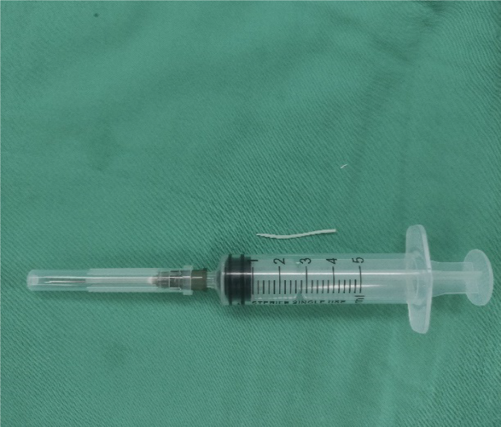
After careful consultation with the patient and her representative, surgery was performed to remove the foreign body. CT of the neck revealed that the left side of the thyroid had a dense shadow in the form of a stripe (Figure 1). Exploration of the neck was performed under general anesthesia. In the end, we found a fishhook sticking out of the esophagus into the thyroid, with one end in the thyroid and one end still in the esophagus (Figure 2). The fish spike was subsequently removed intact and was approximately 3 cm in length (Figure 3). The patient recovered well after the operation.
The patient recovered after surgery, was rechecked 3 months later and had no discomfort.
Fish bones stuck in the throat or esophagus are common emergencies, and mental illness and dentures are important risk factors. This is especially true for elderly patients, whose symptoms were not adapted to the fitting of dentures, leading to the accidental swallowing of a fish bone. However, it is very rare for fish bones to penetrate the cervical region of the esophagus and migrate to the thyroid gland. These fish bones tend to be relatively large, hard, and sharp. Several other factors are also attributed to the migration of fish skeletons into the thyroid gland, such as dislocation of the fish bone due to forceful swallowing of large amounts of food, the direction in which the fish bone is lodged, shrinking of the cricopharyngeal muscles during swallowing, contracting and relaxing the neck muscles when moving the neck., and local infection of the esophagus or pharynx. This condition is difficult to diagnose on the basis of common symptoms or by endoscopy, and prolonged fishbone impaction may increase the likelihood of perforation or migration, leading to serious complications.
In adults, the oropharynx and hypopharynx are the most common sites of impaction, followed by the oral cavity and esophagus[4]. The palatine tonsil, root of the tongue, pyriform fossa and esophagus are the most common sites of embolism. The three physiological stricture points are the most common sites in the adult esophagus. A correlation between the site of embolism and age has been reported in several studies evaluating adults with embolism[3]. Patients aged < 40 years had more foreign bodies in the oropharynx, whereas those aged > 40 years had more foreign bodies in the esophagus. A possible cause is a weakened swallowing mechanism, such as impaired pharyngeal muscle movement, epiglottis and swallowing dysfunction, and incomplete laryngeal closure, which is more common in older patients[5]. The characteristics of the spike site vary depending on the type of fish, shape of the spike, cooking method and diet. Flat or pointed shapes are more likely to cause oesophageal impaction, whereas straight bones are more often associated with pharyngeal impaction, and sharp, straight fish bones are more likely to result in local injury, including mucosal laceration and perforation, as well as penetration into adjacent tissues and migration into other tissues. The clinical presentation of symptoms following a fish skewer varies widely from asymptomatic patients to those with a foreign body sensation, sore throat, difficulty swallowing, painful swallowing, pain behind the breastbone and vomiting blood. When a fish bone becomes stuck in the esophagus, the usual early symptoms are severe pain and uncomfortable at rest. As the fish spike penetrates the esophageal wall, the Clinical symptoms rapidly diminish, and the only clinical signs are persistent neck pain and mild dysphagia. If clinical symptoms are not obvious, the patient will not be concerned about the disease, and the fish spike may be retained for a long time. Long-term containment of foreign substances can lead to non-typical chronic symptoms like painful difficulty swallowing, dysphagia, neck swelling, neck mass, fever, Serious systemic infection and other clinical phenomena. According to the reported cases[6,7], the main symptom was usually a neck abscess, with sore throat and neck pain as the main complaints. In the present case, the patient had a foreign body sensation only in the throat, and we were unable to palpate the mass; however, a foreign body was present on CT imaging.
The main methods for detecting external objects in the upper gastrointestinal (GI) tract include barium swallow, laryngoscopy, plain film radiography, color Doppler ultrasound, CT, and magnetic resonance imaging (MRI). Barium swallowing is the preferred and most widely used imaging method for diagnosing foreign bodies in the upper GI tract. However, laryngoscopy or esophagoscopy may be the preferred method for detecting smaller foreign bodies, such as fish spikes. Laryngoscopy can be classified as either direct or indirect laryngoscopy, both of which are most frequently used for examining foreign bodies in the pharynx. The radiographic opacity of fish spines varies between species. It is sometimes difficult to detect on plain radiographs. Ultrasound is a diagnostic method that can be used at the patient's bedside and has many advantages over other modalities. It is easily accessible and handheld, and images can be viewed in live time. In addition, MRI is a cheaper option and More non-invasive than other technologies. The sensitivity and specificity of CT scans for identifying fish spurs are greater than those of other methods, allowing good visualization of the foreign body; accurate localization of the foreign body; depiction of the size, shape, location and orientation of the foreign body; and its relationship to the surrounding tissues. Also, there is ability to determine the extent of injury and surrounding conditions with ultrasound[8]. For this reason, CT scanning is the preferred method when a fish spur is not detected via endoscopy[9].
Major complications of esophageal foreign bodies include esophageal perforation with perioesophagitis, paraesophageal abscess, mediastinitis, or vascular rupture, such as aortoesophageal fistula, anomalous esophageal fistula, and carotid artery rupture[10]. Extraluminal migration of esophageal foreign bodies is relatively rare and may occur in the lung, liver, subcutaneous neck, thyroid, vessels and pericardium. When extraluminal migration occurs, the method of removing the foreign body becomes more complicated. The importance of early diagnosis and treatment is highlighted by the increased risk of complications over time. A careful history of the presentation is essential for early diagnosis. Physical examination, blood tests and direct laryngoscopy are often necessary, especially within a short time of the onset of impaction. It is advisable to use a protective device (tube and rubber cover) to avoid mucosal injury when removing a sharp object such as a fish bone. In addition, to avoid unintentional movements that could lead to mucosal injury, deep sedation is important when removing sharp objects. Similarly, if there is a high risk of aspiration, tracheal intubation should be considered.
If a fishbone foreign body is not visible during endoscopic examination but the patient has significant symptoms, the surgeon should be aware that the fishbone may be lodged in the thyroid. To avoid a misdiagnosis, ultrasound, CT, and other tests can be used to clarify the diagnosis. The first step in treating a foreign body in the thyroid gland is to determine the position of the lesion and the degree of infection, and to develop a personalized surgical plan for removing the foreign body. At the same time, scientific information should be made available to the general public so that people know that if a fish bone is accidentally lodged, they should not force it to be swallowed or be spit out by inducing vomiting, which are incorrect methods and may aggravate the condition or even cause it to migrate outside the cavity, leading to serious complications, as in this reported case.
Provenance and peer review: Unsolicited article; Externally peer reviewed.
Peer-review model: Single blind
Specialty type: Surgery
Country/Territory of origin: China
Peer-review report’s scientific quality classification
Grade A (Excellent): 0
Grade B (Very good): B
Grade C (Good): 0
Grade D (Fair): 0
Grade E (Poor): 0
P-Reviewer: Swiha MM, Canada S-Editor: Liu JH L-Editor: A P-Editor: Zhao S
| 1. | Bunker PG. The role of dentistry in problems of foreign bodies in the air and food passages. J Am Dent Assoc. 1962;64:782-787. [RCA] [PubMed] [DOI] [Full Text] [Cited by in Crossref: 33] [Cited by in RCA: 31] [Article Influence: 0.5] [Reference Citation Analysis (0)] |
| 2. | Sasaki S, Nishikawa J, Saito M, Suenaga S, Uekitani T, Yokoyama Y, Sakaida I. Endoscopic Removal of a Fish Bone Migrating and Penetrating the Stomach. Am J Gastroenterol. 2018;113:1282. [RCA] [PubMed] [DOI] [Full Text] [Cited by in Crossref: 2] [Cited by in RCA: 2] [Article Influence: 0.3] [Reference Citation Analysis (0)] |
| 3. | Hong KH, Kim YJ, Kim JH, Chun SW, Kim HM, Cho JH. Risk factors for complications associated with upper gastrointestinal foreign bodies. World J Gastroenterol. 2015;21:8125-8131. [RCA] [PubMed] [DOI] [Full Text] [Full Text (PDF)] [Cited by in CrossRef: 54] [Cited by in RCA: 79] [Article Influence: 7.9] [Reference Citation Analysis (1)] |
| 4. | Conthe A, Payeras Otero I, Pérez Gavín LA, Baines García A, Usón Peiron C, Villaseca Gómez C, Herrera Fajes JL, Nogales Ó. Esophageal fish bone impaction: the importance of early diagnosis and treatment to avoid severe complications. Rev Esp Enferm Dig. 2022;114:660-662. [RCA] [PubMed] [DOI] [Full Text] [Cited by in Crossref: 1] [Cited by in RCA: 1] [Article Influence: 0.3] [Reference Citation Analysis (0)] |
| 5. | Shishido T, Suzuki J, Ikeda R, Kobayashi Y, Katori Y. Characteristics of fish-bone foreign bodies in the upper aero-digestive tract: The importance of identifying the species of fish. PLoS One. 2021;16:e0255947. [RCA] [PubMed] [DOI] [Full Text] [Full Text (PDF)] [Cited by in Crossref: 16] [Cited by in RCA: 15] [Article Influence: 3.8] [Reference Citation Analysis (0)] |
| 6. | Hendricks A, Meir M, Hankir M, Lenschow C, Germer CT, Schneider M, Wiegering A, Schlegel N. Suppurative thyroiditis caused by ingested fish bone in the thyroid gland: a case report on its diagnostics and surgical therapy. BMC Surg. 2022;22:92. [RCA] [PubMed] [DOI] [Full Text] [Full Text (PDF)] [Cited by in Crossref: 4] [Reference Citation Analysis (0)] |
| 7. | Huang HY, Wang CC. Migration of a Fish Bone From the Esophagus to the Thyroid Gland. Ear Nose Throat J. 2022;1455613221086032. [RCA] [PubMed] [DOI] [Full Text] [Cited by in Crossref: 1] [Reference Citation Analysis (0)] |
| 8. | Klein A, Ovnat-Tamir S, Marom T, Gluck O, Rabinovics N, Shemesh S. Fish Bone Foreign Body: The Role of Imaging. Int Arch Otorhinolaryngol. 2019;23:110-115. [RCA] [PubMed] [DOI] [Full Text] [Full Text (PDF)] [Cited by in Crossref: 25] [Cited by in RCA: 37] [Article Influence: 5.3] [Reference Citation Analysis (0)] |
| 9. | Hokama A, Uechi K, Takeshima E, Kobashigawa C, Iraha A, Kinjo T, Kishimoto K, Kinjo F, Fujita J. A fish bone perforation of the esophagus. Endoscopy. 2014;46 Suppl 1 UCTN:E216-E217. [RCA] [PubMed] [DOI] [Full Text] [Cited by in Crossref: 4] [Cited by in RCA: 5] [Article Influence: 0.5] [Reference Citation Analysis (0)] |
| 10. | Yorita K, Miike T, Sakaguchi K, Onaga M, Yao T, Sakugawa C, Kataoka H. Corrigendum: A Novel Case of an Unusual Esophageal Submucosal Tumor: An Esophageal Submucosal Gland Duct Hamartoma. Am J Gastroenterol. 2015;110:1634. [RCA] [PubMed] [DOI] [Full Text] [Cited by in Crossref: 1] [Cited by in RCA: 1] [Article Influence: 0.1] [Reference Citation Analysis (0)] |


