Published online Jan 26, 2024. doi: 10.12998/wjcc.v12.i3.643
Peer-review started: November 9, 2023
First decision: November 22, 2023
Revised: November 22, 2023
Accepted: January 3, 2024
Article in press: January 3, 2024
Published online: January 26, 2024
Processing time: 69 Days and 17.9 Hours
Elderly giant retrosternal thyroid goiter is a rare yet significant medical condition, often presenting clinical symptoms that can be confused with other diseases, posing diagnostic and therapeutic challenges. This study aims to delve into the characteristics and potential mechanisms of this ailment through pathological diagnosis and immunohistochemical analysis, providing clinicians with more precise diagnostic and treatment strategies.
A 77-year-old male, was admitted to hospital with the chief complaint of finding a goiter in the semilunar month during physical examination, accompanied by dyspnea. Locally protruding into the superior mediastinum, the adjacent structure was compressed, the trachea was compressed to the right, and the local lumen was slightly narrowed. The patient was diagnosed with giant retrosternal goiter. Considering dyspnea caused by trachea compression, our department planned to perform giant retrosternal thyroidectomy. Immunohistochemical results: Tg (+), TTF-1 (+), Calcitonin (CT) (I), Ki-67 (+, about 20%), CD34 (-). Retrosternal goiter means that more than 50% of the volume of the thyroid gland is below the upper margin of the sternum. As retrosternal goiter disease is a relatively rare disease, once the disease is diagnosed, it should be timely surgical treatment, and the treatment is more difficult, the need for professional medical team for comprehensive treatment.
The imaging manifestations of giant retrosternal goiter are atypical, histomorphology and immunohistochemistry can assist in its diagnosis. This article reviews the relevant literature of giant retrosternal goiter immunohistochemistry and shows that giant retrosternal goiter is positive for Tg, TTF-1, and Ki-67.
Core Tip: Elderly giant retrosternal thyroid goiter is a rare yet significant medical condition, often presenting clinical symptoms that can be confused with other diseases, posing diagnostic and therapeutic challenges. The diagnosis of elderly giant retrosternal thyroid goiter is difficult and often results in unnecessary or inappropriate treatment. Therefore, it is particularly important to correctly identify and diagnose the disease, and this article reports a 77-year-old male patient with giant retrosternal goiter.
- Citation: Meng YC, Wu LS, Li N, Li HW, Zhao J, Yan J, Li XQ, Li P, Wei JQ. Pathological diagnosis and immunohistochemical analysis of giant retrosternal goiter in the elderly: A case report. World J Clin Cases 2024; 12(3): 643-649
- URL: https://www.wjgnet.com/2307-8960/full/v12/i3/643.htm
- DOI: https://dx.doi.org/10.12998/wjcc.v12.i3.643
Elderly giant retrosternal thyroid goiter is an extremely rare and challenging thyroid disorder[1]. Its rarity lies in the fact that it typically afflicts elderly patients, with a relatively low incidence rate. Due to its often nonspecific symptoms such as difficulty breathing, swallowing, and the presence of a neck mass, it can easily be misdiagnosed as other conditions like cardiovascular diseases or esophageal cancer, posing significant clinical challenges[2-4]. The significance of this case report lies in providing the medical community with a detailed clinical case and pathological analysis of elderly giant retrosternal thyroid goiter, along with immunohistochemical research data[5]. Furthermore, this study contributes to improving the diagnosis and treatment methods for this condition, offering more accurate and personalized medical care[6-8].
We report this case with the aim of disseminating new information about elderly giant retrosternal thyroid goiter to the medical community, enhancing awareness of the disease, and providing valuable data for future research and treatment. By sharing this case, we hope to foster further investigation into this rare disorder and better support patients in terms of diagnosis and management.
Finding a goiter in the semilunar month during physical examination.
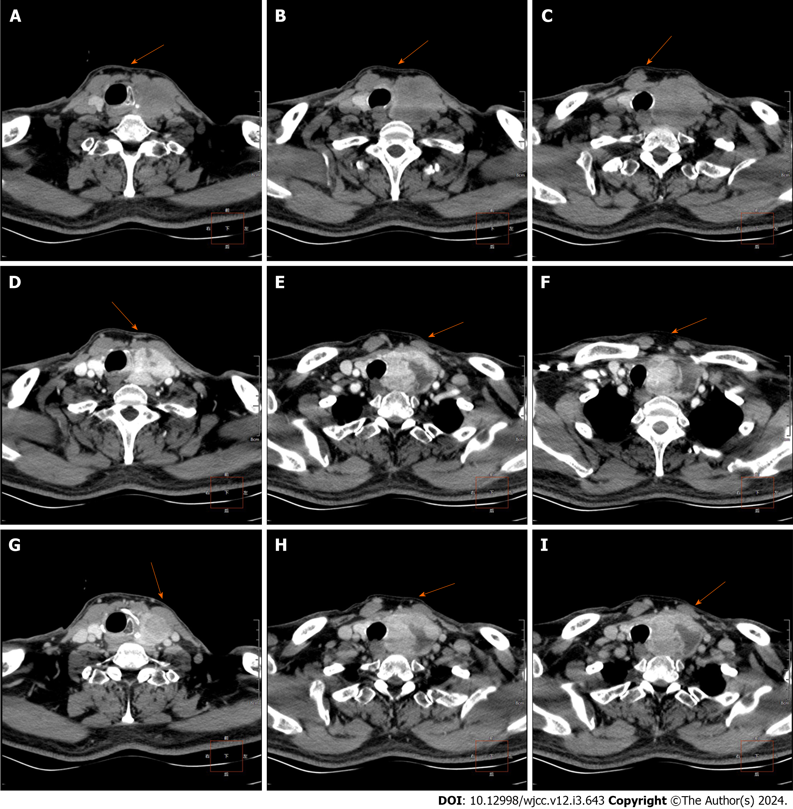
A 77-year-old male patient was admitted to hospital after a physical examination revealed a goiter. No other special clinical symptoms and signs. Before admission, chest enhanced computed tomography (CT) scan showed that the left lobe of the thyroid gland was enlarged and slightly low-density mass was seen, about 49 mm × 43 mm × 68 mm in size, the boundary was not clear, its internal density was uneven, the enhanced scan was significantly uneven and enhanced, local protrusion into the upper mediastinum, compression of adjacent structures, rightward compression of the trachea, and local lumen was slightly narrowed.
A history of hypertension, regular oral antihypertensive medication treatment, and well-controlled blood pressure.
Nothing special.
Nothing special.
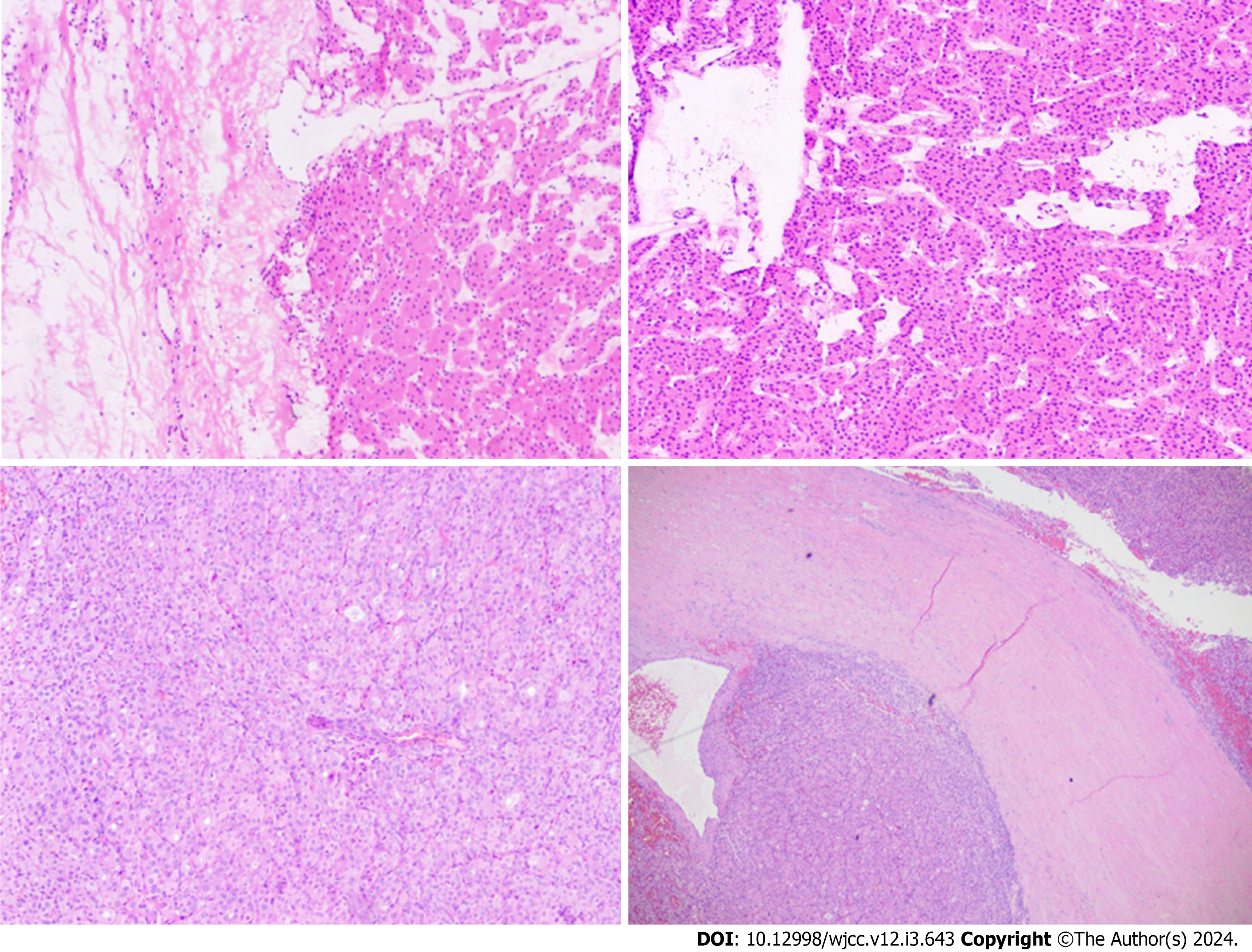
Combined with subimmunohistochemical findings, consistent with follicular thyroid cancer (Left goiter ice stone). Immunohistochemical results: Tg (+), TTF-1 (+), Calcitonin (CT) (I), Ki-67 (+, about 20%), CD34 (-).
Thyroid plain CT scan plus enhanced imaging showed that the volume of the left lobe of the thyroid increased and slightly low-density mass was seen, the size was about 49mm×43mm×68mm, the boundary was not clear, the internal density was not uniform, and the enhanced scan was obviously uneven and enhanced, local protrusion into the upper mediastinum, compression of adjacent structures, rightward compression of the trachea, and local lumen was slightly narrowed. The size, shape and density of the right lobe of the thyroid gland were normal. There was no obvious lymph node shadow in the neck (Figure 1).
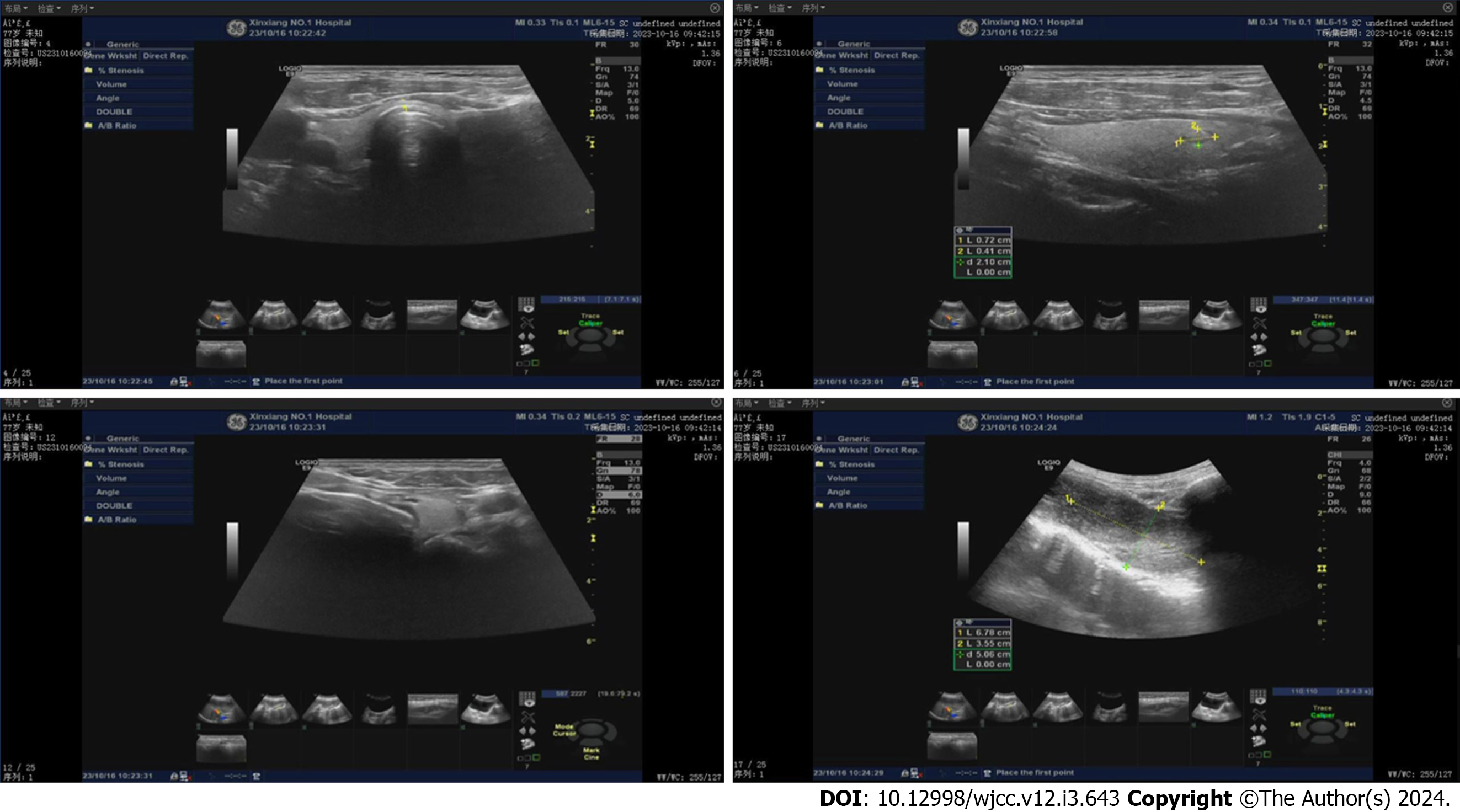
Real-time ultrasound examination of thyroid and elastic imaging showed that the right lobe of thyroid gland was 1.7 cm × 1.4 cm, the left lobe was 5.2 cm × 3.3 cm, and the isthmus was 0.2 cm thick. The thyroid gland tissue echo was uniform, and a solid cystic nodule was found in the right lobe of thyroid gland with a size of 0.7 cm × 0.4 cm, clear boundary and regular shape. CDFI: Visible peripheral blood flow signal, and elasticity score: Multiple solid cystic nodules were observed in the left lobe of the thyroid gland, mainly solid components, with a size of 6.8 cm × 3.6 cm, clear boundary, regular shape, and peripheral blood flow signals (Figure 2).
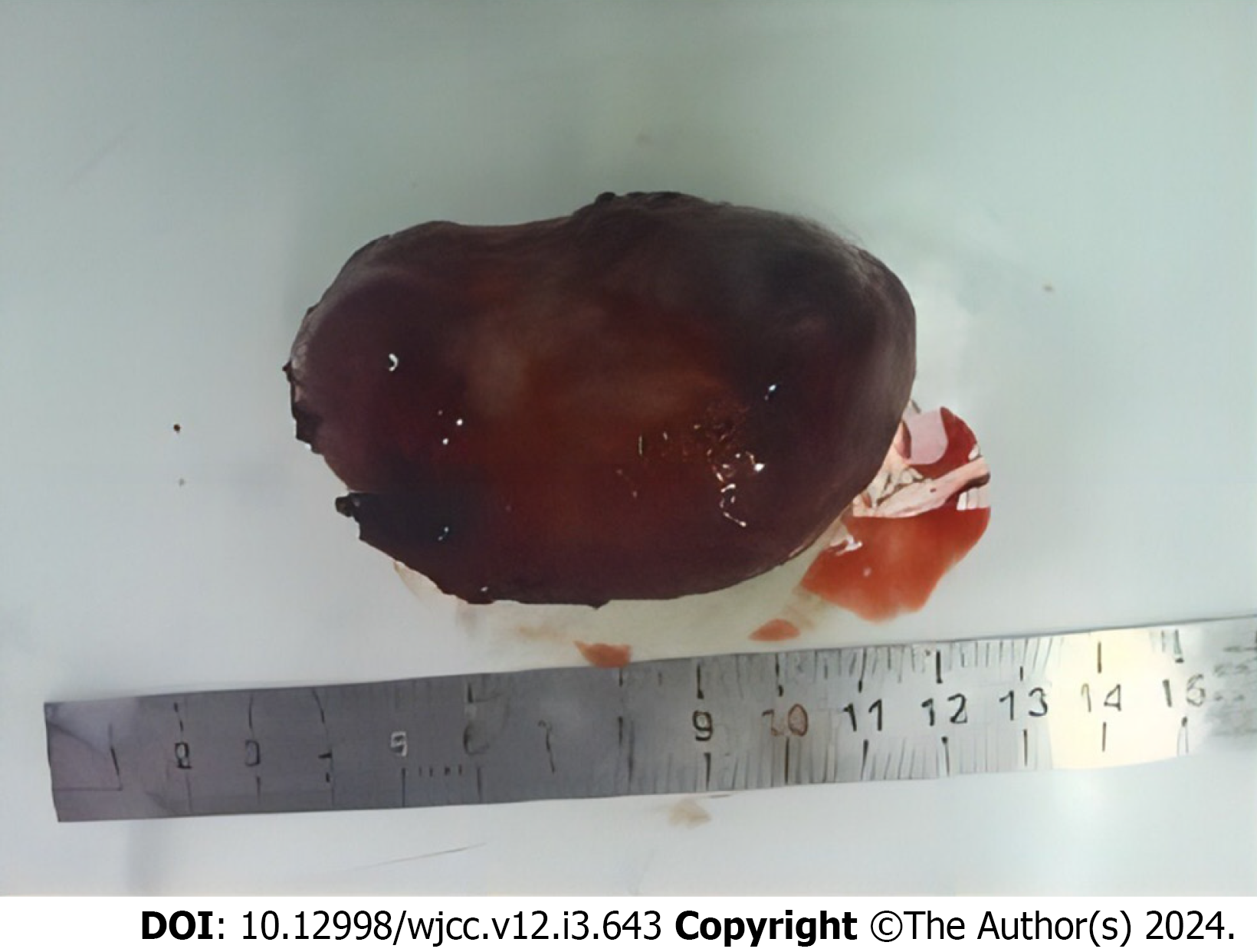
The pathological diagnosis was micro-invasive follicular thyroid cancer (Figures 3 and 4).
We recommend that patients continue to receive treatment after surgery and to do regular follow-up observations.
The patient recovered after operation, and no recurrence was found after 6 mo.
In this project, we studied a large retrosternal goiter in an elderly patient. Giant retrosternal goiter is a specific type of thyroid tumor characterized by the growth of a goiter to the back of the sternum[9-11]. According to the nature and growth rate of the tumor, it can be divided into two types: benign and malignant[12]. Benign giant retrosternal goiter usually grows slowly, but because of its location, it can cause difficulty breathing and swallowing, with a noticeable impact on the patient's quality of life[13]. Malignant giant retrosternal goiter is potentially malignant and requires a more aggressive treatment strategy. The clinical manifestations of giant retrosternal goiter in the elderly usually include dyspnea, dysphagia, hoarseness, and neck mass[14]. These symptoms are similar to other diseases and can easily lead to misdiagnosis or delayed diagnosis. Therefore, clinicians facing these symptoms in patients should be on high alert for the possibility of giant retrosternal goiter and conduct detailed physical examination and imaging evaluation to confirm the diagnosis and develop appropriate treatment[15-17]. The relatively low incidence of giant retrosternal goiter in old age makes the disease even rarer. Due to its rarity, the medical community has limited knowledge and awareness of the disease, which is easily overlooked or misdiagnosed[18]. However, it is precisely because of its potentially serious harm that we believe it is necessary to conduct in-depth research on this rare disease in order to improve diagnostic accuracy and treatment effectiveness[19]. For the treatment of giant retrosternal goiter in the elderly, surgical resection is usually the preferred method. The outcome of surgery depends largely on the nature of the tumor, preoperative preparation, and the overall health of the patient. For benign giant retrosternal goiter, surgery usually results in a significant improvement in the patient's symptoms and has a good long-term prognosis[20]. However, for malignant lesions, further treatment may be required after surgery, such as radiation therapy or chemoradiotherapy, and the prognosis is relatively poor.
The imaging features of giant retrosternal goiter are one of the important diagnostic bases for this disease. Typically, a CT or magnetic resonance imaging examination of the patient's neck will reveal the following features: A large retrosternal goiter is located behind the sternum and usually presents as a uniform mass that takes up space in the upper rib cage and part of the neck[21]. The immunohistochemical features of thyroid follicular carcinoma include thyroid specific markers such as thyroglobulin and thyroid peroxidase, CK19, HMGA2, PAX8, Tg, TTF-1, Ki-67, etc. These markers are often significantly expressed in follicular carcinoma and help in diagnosis, treatment selection and prognosis assessment. Immunohistochemical analysis is very important for the diagnosis and treatment of follicular cancer and provides strong support and guidance. In terms of clinical manifestations, the symptoms of giant retrosternal goiter in the elderly can be diverse, including poor breathing, swallowing discomfort, sore throat, throat swelling, hoarseness and so on[22]. Patients often see a doctor for these symptoms, but because they are not specific, they are easily confused with other neck or chest diseases.
Therefore, a comprehensive consideration of clinical and radiological features is essential for correct diagnosis and treatment planning. For such patients, comprehensive clinical evaluation should be actively conducted to ensure early detection and treatment of giant retrosternal goiter in the elderly, thereby improving the quality of life and prognosis of patients.
Through comprehensive pathological diagnosis and immunohistochemical analysis of a giant retrosternal mass in an elderly patient, we discussed the characteristics of this rare disease. The results showed that the immunohistochemical markers of micro-invasive follicular thyroid cancer had a certain diversity. Among them, CK19, HMGA2, PAX8, Tg, TTF-1 and Ki-67 play a crucial role in pathological diagnosis.
Provenance and peer review: Unsolicited article; Externally peer reviewed.
Peer-review model: Single blind
Specialty type: Medicine, general and internal
Country/Territory of origin: China
Peer-review report’s scientific quality classification
Grade A (Excellent): 0
Grade B (Very good): B
Grade C (Good): C
Grade D (Fair): 0
Grade E (Poor): 0
P-Reviewer: Hasan A, Egypt S-Editor: Yan JP L-Editor: A P-Editor: Zhang YL
| 1. | Nakamura R, Okuda K, Chiba K, Matsui T, Oda R, Tatematsu T, Yokota K, Nakanishi R. A large intrathoracic goiter with tracheal stenosis: Complete resection using a robot-assisted thoracoscopic approach. Thorac Cancer. 2022;13:1874-1877. [RCA] [PubMed] [DOI] [Full Text] [Full Text (PDF)] [Reference Citation Analysis (0)] |
| 2. | Pan Y, Chen C, Yu L, Zhu S, Zheng Y. Airway Management of Retrosternal Goiters in 22 Cases in a Tertiary Referral Center. Ther Clin Risk Manag. 2020;16:1267-1273. [RCA] [PubMed] [DOI] [Full Text] [Full Text (PDF)] [Cited by in Crossref: 1] [Cited by in RCA: 4] [Article Influence: 0.8] [Reference Citation Analysis (0)] |
| 3. | Cankar Dal H. Difficult airway management and emergency tracheostomy in a patient with giant goiter presenting with respiratory arrest: A case report. Exp Ther Med. 2022;24:499. [RCA] [PubMed] [DOI] [Full Text] [Full Text (PDF)] [Reference Citation Analysis (0)] |
| 4. | Figueiredo AA, Simões-Pereira J. Giant retrosternal goitre causing lung atelectasis. Endokrynol Pol. 2022;73:794-795. [RCA] [PubMed] [DOI] [Full Text] [Reference Citation Analysis (0)] |
| 5. | Sun X, Chen C, Zhou R, Chen G, Jiang C, Zhu T. Anesthesia and airway management in a patient with acromegaly and tracheal compression caused by a giant retrosternal goiter: a case report. J Int Med Res. 2021;49:300060521999541. [RCA] [PubMed] [DOI] [Full Text] [Full Text (PDF)] [Reference Citation Analysis (0)] |
| 6. | D'hulst L, Deroose C, Strybol D, Coolen J, Gheysens O. 18F-FDG PET/CT and MRI of a Mediastinal Malignant Granular Cell Tumor With Associated Recurrent Pericarditis. Clin Nucl Med. 2018;43:589-590. [RCA] [PubMed] [DOI] [Full Text] [Cited by in Crossref: 3] [Cited by in RCA: 3] [Article Influence: 0.4] [Reference Citation Analysis (0)] |
| 7. | Khalid MA, Iqbal J, Memon HL, Hanif FM, Butt MOT, Luck NH, Majid Z. Dyspepsia Amongst End Stage Renal Disease Undergoing Hemodialysis: Views from a Large Tertiary Care Center. J Transl Int Med. 2018;6:78-81. [RCA] [PubMed] [DOI] [Full Text] [Full Text (PDF)] [Cited by in Crossref: 6] [Cited by in RCA: 7] [Article Influence: 1.0] [Reference Citation Analysis (0)] |
| 8. | Lampridis S, Lau MC, Mhandu P, Parissis H. Concomitant off-pump coronary artery bypass grafting and total thyroidectomy for a large retrosternal goitre: a case report and review of the literature. J Thorac Dis. 2016;8:E362-E368. [RCA] [PubMed] [DOI] [Full Text] [Cited by in Crossref: 1] [Reference Citation Analysis (0)] |
| 9. | Zuo T, Gao Z, Chen Z, Wen B, Chen B, Zhang Z. Surgical Management of 48 Patients with Retrosternal Goiter and Tracheal Stenosis: A Retrospective Clinical Study from a Single Surgical Center. Med Sci Monit. 2022;28:e936637. [RCA] [PubMed] [DOI] [Full Text] [Full Text (PDF)] [Cited by in RCA: 5] [Reference Citation Analysis (0)] |
| 10. | Battistella E, Pomba L, Sidoti G, Vignotto C, Toniato A. Retrosternal Goitre: Anatomical Aspects and Technical Notes. Medicina (Kaunas). 2022;58. [RCA] [PubMed] [DOI] [Full Text] [Full Text (PDF)] [Reference Citation Analysis (0)] |
| 11. | Wu B, Xie X, Li X, Zhu Q, Zhou C. Large intramural hematoma of the esophagus after endoscopic injection sclerotherapy: A case report. Medicine (Baltimore). 2023;102:e32752. [RCA] [PubMed] [DOI] [Full Text] [Reference Citation Analysis (0)] |
| 12. | Chen Q, Su A, Zou X, Liu F, Gong R, Zhu J, Li Z, Wei T. Clinicopathologic Characteristics and Outcomes of Massive Multinodular Goiter: A Retrospective Cohort Study. Front Endocrinol (Lausanne). 2022;13:850235. [RCA] [PubMed] [DOI] [Full Text] [Full Text (PDF)] [Cited by in Crossref: 1] [Reference Citation Analysis (0)] |
| 13. | Simões AB, D'Avila LH, Silva CK, Valentini DF Jr. A very rare case of an intrathoracic pancreatic pseudocyst. Rev Esp Enferm Dig. 2023;. [RCA] [PubMed] [DOI] [Full Text] [Cited by in Crossref: 1] [Reference Citation Analysis (0)] |
| 14. | Nesti C, Wohlfarth B, Borbély YM, Kaderli RM. Case Report: Modified Thoracoscopic-Assisted Cervical Resection for Retrosternal Goiter. Front Surg. 2021;8:695963. [RCA] [PubMed] [DOI] [Full Text] [Full Text (PDF)] [Cited by in Crossref: 1] [Cited by in RCA: 4] [Article Influence: 1.0] [Reference Citation Analysis (0)] |
| 15. | Moore E, Merali N, Abbassi-Ghadi N. Herpes simplex oesophagitis leading to perforation and mediastinal collection: a case report and review of literature. Ann R Coll Surg Engl. 2023;105:94-96. [RCA] [PubMed] [DOI] [Full Text] [Cited by in Crossref: 1] [Reference Citation Analysis (0)] |
| 16. | Palani P, Lokeshwaran S, Sunil Kumar K, Girish G, Vidya MN, Gupta N. A 33-Year-Old Woman With Breathing Difficulty, Inspiratory Wheeze, and Hemoptysis. Chest. 2023;163:e141-e145. [RCA] [PubMed] [DOI] [Full Text] [Reference Citation Analysis (0)] |
| 17. | Cantinotti M, Giordano R, Marchese P, Franchi E, Viacava C, Pak V, Murzi B, Arcieri L, Poli V, Federici D, Koestenberger M, Assanta N. Retrosternal Clots After Fontan Surgery by Systematic Evaluation With Transthoracic Ultrasound. J Cardiothorac Vasc Anesth. 2020;34:951-955. [RCA] [PubMed] [DOI] [Full Text] [Cited by in Crossref: 1] [Cited by in RCA: 1] [Article Influence: 0.2] [Reference Citation Analysis (0)] |
| 18. | Chen YH, Lin PC, Chen YL, Yiang GT, Wu MY. Point-of-Care Ultrasonography Helped to Rapidly Detect Pneumomediastinum in a Vomiting Female. Medicina (Kaunas). 2023;59. [RCA] [PubMed] [DOI] [Full Text] [Reference Citation Analysis (0)] |
| 19. | Bernaldo-de-Quirós E, Cózar B, López-Esteban R, Clemente M, Gil-Jaurena JM, Pardo C, Pita A, Pérez-Caballero R, Camino M, Gil N, Fernández-Santos ME, Suarez S, Pion M, Martínez-Bonet M, Correa-Rocha R. A Novel GMP Protocol to Produce High-Quality Treg Cells From the Pediatric Thymic Tissue to Be Employed as Cellular Therapy. Front Immunol. 2022;13:893576. [RCA] [PubMed] [DOI] [Full Text] [Full Text (PDF)] [Cited by in Crossref: 1] [Cited by in RCA: 8] [Article Influence: 2.7] [Reference Citation Analysis (0)] |
| 20. | Knobel M. An overview of retrosternal goiter. J Endocrinol Invest. 2021;44:679-691. [RCA] [PubMed] [DOI] [Full Text] [Cited by in Crossref: 12] [Cited by in RCA: 15] [Article Influence: 3.8] [Reference Citation Analysis (0)] |
| 21. | Nagaty M, Shehata MS, Elkady AS, Eid M, Nady M, Youssef A, Henish MIE, Monazea K, Noreldin RI, Nasr M, Fayad S, Abdelwahed MS, Hasan A. An assessment of the role of surgical loupe technique in prevention of postthyroidectomy complications: a comparative prospective study. Ann Med Surg (Lond). 2023;85:446-452. [RCA] [PubMed] [DOI] [Full Text] [Cited by in Crossref: 5] [Cited by in RCA: 7] [Article Influence: 3.5] [Reference Citation Analysis (0)] |
| 22. | Hauch A, Al-Qurayshi Z, Randolph G, Kandil E. Total thyroidectomy is associated with increased risk of complications for low- and high-volume surgeons. Ann Surg Oncol. 2014;21:3844-3852. [RCA] [PubMed] [DOI] [Full Text] [Cited by in Crossref: 217] [Cited by in RCA: 231] [Article Influence: 21.0] [Reference Citation Analysis (0)] |


