Published online Jan 26, 2024. doi: 10.12998/wjcc.v12.i3.495
Peer-review started: November 13, 2023
First decision: November 22, 2023
Revised: December 4, 2023
Accepted: January 2, 2024
Article in press: January 2, 2024
Published online: January 26, 2024
Processing time: 66 Days and 0 Hours
Morgagni hernias are rare anomalies that are easily misdiagnosed or missed.
To summarize the ultrasound (US) imaging characteristics of Morgagni hernias through a comparison of imaging and surgical results.
The records of children with Morgagni hernias who were hospitalized at two hospitals between January 2013 and November 2023 were retrospectively re
Between 2013 and 2023, we observed nine (five male and four female) children with Morgagni hernias. Upper abdominal scanning revealed a widening of the prehepatic space, with an abnormal channel extending from the xiphoid process to the right or left side of the thoracic cavity. The channel had intestinal duct and intestinal gas echoes. Hernia contents were found in the transverse colon (n = 6), the colon and small intestine (n = 2), and the colon and stomach (n = 1). Among the patients, seven had a right-sided lesion, two had a left-sided lesion, and all of them had hernial sacs.
US imaging can accurately determine the location, extent, and content of Morgagni hernias. For suspected Mor
Core Tip: Morgagni hernias are rare and easily misdiagnosed or missed. In this report, retrosternal hernias accidentally discovered by ultrasound. The ultrasonic and clinical characteristics are summarized to provide a simple and effective basis for early diagnosis. Ultrasonic imaging can accurately determine the location, extent, and content of Morgagni hernias. For suspected Morgagni hernias, we recommend performing sonographic screening first.
- Citation: Shi HQ, Chen WJ, Yin Q, Zhang XH. Ultrasound diagnosis of congenital Morgagni hernias: Ten years of experience at two Chinese centers. World J Clin Cases 2024; 12(3): 495-502
- URL: https://www.wjgnet.com/2307-8960/full/v12/i3/495.htm
- DOI: https://dx.doi.org/10.12998/wjcc.v12.i3.495
A Morgagni hernia is an unusual congenital herniation of the abdominal contents through the costochondral triangles of the anterior diaphragm[1]. Few reports on Morgagni hernias in children are available. Morgagni hernias are easily mis
The records of nine patients with Morgagni hernias diagnosed by US imaging and confirmed by surgery at two children’s hospitals between 2013 and 2023 were collected.
US scans were performed by a team of experienced examiners by using Philips EPIQ 7C (Netherlands). The scans were acquired at convex and linear array transducer frequencies of 5-14 MHz. The upper abdomen and chest of each patient in the supine, lateral, or sitting position were scanned to reveal the whole diaphragm and the site in front of the liver and behind the sternum. The positions, contents, morphologies, structures, and connections of the hernias were recorded.
All nine patients underwent contrast gastrointestinal imaging (GI) (TOSHIBA, Japan). Two patients underwent chest computed tomography (CT) scans (PHILIP Brilliance 64 spiral CT-Netherlands). The patients were diagnosed with Morgagni hernias after US imaging, but their GI results did not show any signs of the disease.
The study was approved by the Medical Ethics Committee of Fujian Children’s Hospital and Hunan Children’s Hospital, and waived of Informed Consent from parents. All methods were performed in accordance with the Declaration of Hel
SPSS 20.0 was used for statistical analysis. Counting data are expressed as cases and percentages. The agreement between US, and GI/CT and surgery results was calculated using cross - tabulation.
Nine children (five male and four female) who were diagnosed with Morgagni hernias that were confirmed by surgery were included in this study (Table 1). The ages of the patients ranged from three months to three years and six months. The chief complaints were repeated coughing in five patients, vomiting in two patients, and constipation in one patient. In one patient, beating of the heart could be seen on the chest wall, which is one of the manifestations of Cantrell’s pentagonal syndrome (Table 1).
| Sex | Age | Chief complaint | US | Contrast GI, contrast CT | Surgery |
| Male | 1 yr | Cough | Right/intestinal tract/fourth rib | Right/intestinal tract | Right/colon and small intestine |
| Female | 3 mo 4 d | Constipation | Right/intestinal tract/fifth rib | GI: Negative, no results of CT | Right/transverse colon |
| Female | 1 yr | Cough | Left/intestinal tract/second rib | Left/intestinal tract | Left/transverse colon and omentum |
| Female | 3 yr 6 mo | Cough | Right/intestinal tract/fourth rib | Right/intestinal tract | Right/transverse colon |
| Female | 9 mo | Vomiting | Right/intestinal tract/sixth rib | GI: Negative. CT: Right/intestinal tract | Right/transverse colon and omentum |
| Male | 1 yr 5 mo | Forechest fluctuating mass | Left/intestinal tract/fifth rib | Left/intestinal tract | Left/transverse colon |
| Male | 2 yr | Cough | Right/intestinal tract/fourth rib | Right/intestinal tract | Right/transverse colon |
| Male | 6 mo 16 d | Cough | Right/intestinal tract/fifth rib | Right/intestinal tract | Right/colon and small intestine |
| Male | 4 mo | Vomiting | Right/colon and stomach | Right/colon and stomach | Right/colon and stomach |
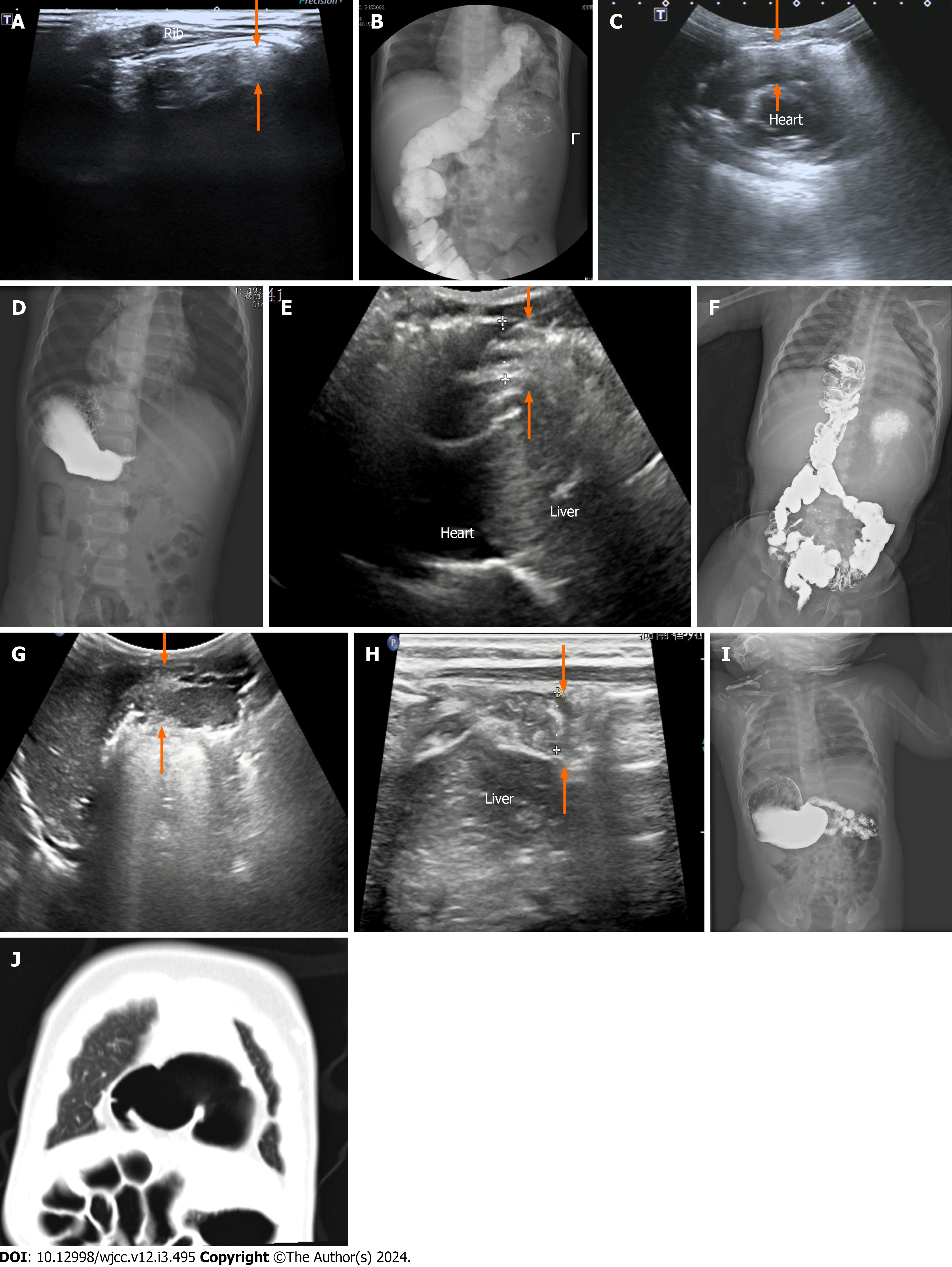
All nine Morgagni hernias were first identified by US: (1) Upper abdominal scanning revealed a widening of the pre
The GI of seven patients showed herniation of the transverse colon or bowl into the thoracic cavity (Figures 1B, D, and F) and herniation of the stomach and colon into the thoracic cavity. Two patients showed no abnormalities (Figure 1I). A CT scan revealed a right Morgagni hernia with an intestinal tube in two patients (Figure 1J).
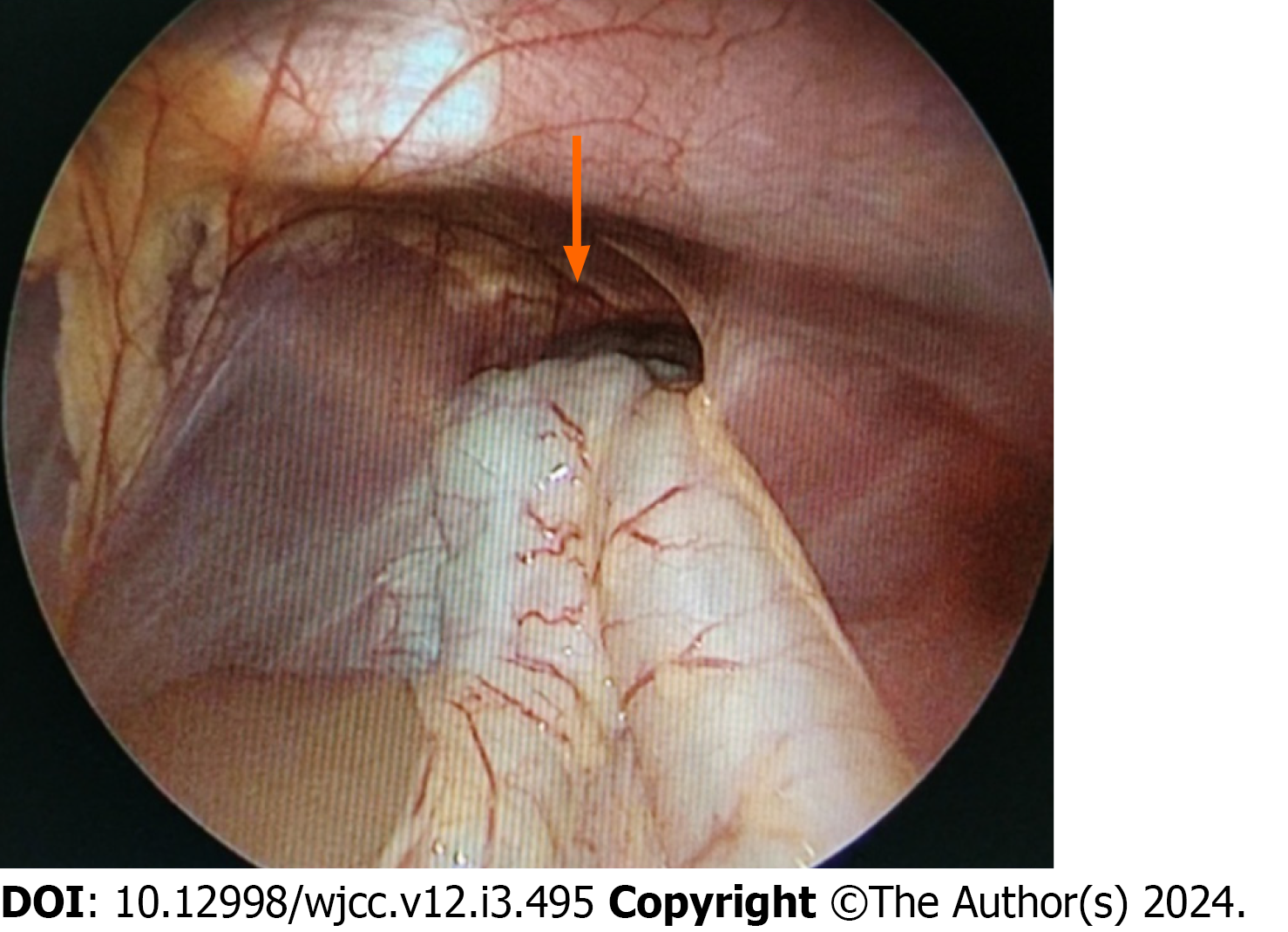
Seven patients underwent laparoscopic surgery, one underwent open diaphragmatic repair, and one (Cantrell’s pen
The consistency between ultrasounic diagnosis and surgical results was 100%, and the consistency between GI diagnosis and surgical results was 77%.
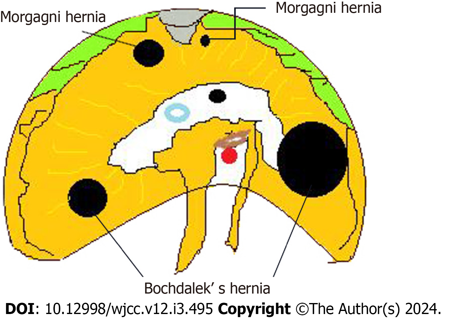
In 1769, the Italian anatomist Giovanni Battista Morgagni described the herniation of abdominal contents through sternochondral triangles via cadaver observation. In 1828, Larrey described a surgical approach to the pericardial sac through the same triangles. Given the early work of Morgagni and Larrey, the right- and left-hand costochondral triangles have been designated as the foramen of Morgagni and the space of Larrey, respectively (Figure 3)[3]. These triangles form when the pars sternalis and a costochondral arch fuse and close around the internal thoracic artery as it becomes the superior epigastric artery. Occasionally, these spaces fail to fully close, allowing the herniation of abdominal contents into the thorax. This type of herniation is referred to as a Morgagni hernia regardless of the laterality[1].
Morgagni hernias are confined to the posterior sternal triangle, so they are different from Bochdalek[4] (mainly in the left posterolateral, with diaphragmatic defect) and hiatal (hiatal and adjacent hiatal holes) hernias. The symptoms of Morgagni hernias are not as typical as those of Bochdalek and hiatal hernias, and the onset of Morgagni hernias is occult and therefore easy to ignore clinically. A review of the literature showed that only a few reports on Morgagni hernias are available. Hosokawa et al[2] performed a meta-analysis of all articles on Morgagni hernias published from 1997 to 2017, with a total of 296 cases. Most of the Morgagni hernias were found accidentally by X-ray or CT examination (240/298 cases), and only seven cases were detected accidentally by US examination[2]. A few studies have summarized the cha
The present study showed the US manifestations of nine children with Morgagni hernias. First, US examination could detect the position of the hernias (on the left or right). Among the nine patients, seven and two patients had hernias on the right and left sides, respectively, indicating that the incidence on the right side was higher than that on the left side. Similarly, a previous report showed that 90% of chest hernias are located on the right side, and 10% are on the left side[2].
Second, US examination could reveal the contents of the hernias. The surgical results showed herniation of the trans
Third, US examination could measure the width of the hernia sac orifice, estimate the position of the hernia contents in the anterior thoracic cavity, determine its relationship with the surrounding anatomy, and provide a basis for further clinical treatment. In the present group, all nine patients had hernia sacs, with an average size of approximately 21-25 cm2. These Morgagni hernias were small and had limited extension, a result that is consistent with those of previous reports[5,6]. Therefore, this type of hernia does not easily compress the lungs or other organs and has mild clinical symptoms or lacks typical manifestations (respiratory distress, vomiting, etc.). In this study, five children presented with repeated cough, and three children presented with vomiting and constipation. All nine children were incidentally diagnosed through US and subjected to GI and CT scanning. Therefore, US examination is an important method for diagnosing Morgagni hernias.
US examination has limitations. Although US can roughly locate the height of the hernia sac, it cannot accurately evaluate the range of the whole hernia sac and has poor spatial resolution. In addition, US depends on the experience of the scanner, who should always keep in mind the US characteristics of Morgagni hernias. Direct signs, such as abnormal channels between the sternum and liver and intestinal tubes and gas entering the chest cavity through these channels, are key. Peristalsis occurs in the anterior chest cavity, and widening of the anterior hepatic space is suggestive of a Morgagni hernia.
Morgagni hernias in children are diagnosed by US detection of the peristalsis of the prehepatic area and intrathoracic intestine and the movement of intestinal contents. This study provides a new, reliable basis for the clinical diagnosis of this type of malformation.
A Morgagni hernia is an unusual congenital herniation. It is easily misdiagnosed or missed because their symptoms are mild and atypical. In the present report, retrosternal hernias accidentally discovered by ultrasound (US) are described, and their ultrasonic manifestations are analyzed. The US and clinical characteristics are summarized to provide a simple and effective basis for early diagnosis.
Through this report, we can understand more about the clinical and ultrasonic characteristics of rare retrosternal hernia diseases. To add much new insightful information to the field.
To summarize the US imaging characteristics of Morgagni hernias through a comparison of imaging and surgical results.
The records of nine patients with Morgagni hernias diagnosed by US imaging and confirmed by surgery at two children’s hospitals between 2013 and 2023 were collected. The clinical symptoms of the case were summarized. The location, contents and size of the hernia sac were recorded by ultrasound. The clinical and ultrasonic characteristics of the hernia were summarized by comparing with gastrointestinal imaging/computed tomography and surgery.
Between 2013 and 2023, we observed nine (five male and four female) children with Morgagni hernias. All nine Morgagni hernias were first identified by US: (1) Upper abdominal scanning revealed a widening of the prehepatic space, with an abnormal channel extending from under the xiphoid process to the right or left side of the thoracic cavity. Two hernias were on the left side, and seven were on the right side. The abdominal intestinal tube and intestinal air echo crossed this area to the chest in all nine cases; and (2) Chest scanning showed echoes of the bowel and stomach. Intestinal peristalsis and intestinal content movement were observed during the scans.
US imaging can accurately determine the location, extent, and content of Morgagni hernias. Direct signs, such as abnormal channels between the sternum and liver and intestinal tubes and gas entering the chest cavity through these channels, are key. Peristalsis occurs in the anterior chest cavity, and widening of the anterior hepatic space is suggestive of a Morgagni hernia.
The research perspective of this study is to analysed the clinical findings, US features, and operative details of children with Morgagni hernias. In the future studies, we will continue to increase the sample size for more in-depth research, and will analyze the postoperative recurrence rate of retrosternal hernia.
First, I would like to give my heartfelt thanks to my academic supervisor Prof. Xue-Hua Zhang for his invaluable instruction and inspiration. Without his previous advice and guidance, this study could not have been completed. Addi
Provenance and peer review: Unsolicited article; Externally peer reviewed.
Peer-review model: Single blind
Specialty type: Medicine, research and experimental
Country/Territory of origin: China
Peer-review report’s scientific quality classification
Grade A (Excellent): 0
Grade B (Very good): B
Grade C (Good): 0
Grade D (Fair): 0
Grade E (Poor): 0
P-Reviewer: Shalaby MN, Egypt S-Editor: Wang JJ L-Editor: A P-Editor: Xu ZH
| 1. | Leshen M, Richardson R. Bilateral Morgagni Hernia: A Unique Presentation of a Rare Pathology. Case Rep Radiol. 2016;2016:7505329. [RCA] [PubMed] [DOI] [Full Text] [Full Text (PDF)] [Cited by in Crossref: 1] [Cited by in RCA: 1] [Article Influence: 0.1] [Reference Citation Analysis (0)] |
| 2. | Hosokawa T, Takahashi H, Tanami Y, Sato Y, Hosokawa M, Kato R, Kawashima H, Oguma E. Usefulness of Ultrasound in Evaluating the Diaphragm in Neonates and Infants With Congenital Diaphragmatic Hernias. J Ultrasound Med. 2019;38:1109-1113. [RCA] [PubMed] [DOI] [Full Text] [Cited by in Crossref: 4] [Cited by in RCA: 6] [Article Influence: 1.0] [Reference Citation Analysis (0)] |
| 3. | Chandrasekharan PK, Rawat M, Madappa R, Rothstein DH, Lakshminrusimha S. Congenital Diaphragmatic hernia - a review. Matern Health Neonatol Perinatol. 2017;3:6. [RCA] [PubMed] [DOI] [Full Text] [Full Text (PDF)] [Cited by in Crossref: 108] [Cited by in RCA: 145] [Article Influence: 18.1] [Reference Citation Analysis (2)] |
| 4. | Zhang XH, Chen WJ, Zhou CG, Lei R, Wang J. [Ultrasound and clinical features of congenital diaphragmatic hernia in 31 children]. Chin J Ultrasound Med. 2019;35:236-238. |
| 5. | Slepov O, Kurinnyi S, Ponomarenko O, Migur M. Congenital retrosternal hernias of Morgagni: Manifestation and treatment in children. Afr J Paediatr Surg. 2016;13:57-62. [RCA] [PubMed] [DOI] [Full Text] [Full Text (PDF)] [Cited by in Crossref: 10] [Cited by in RCA: 10] [Article Influence: 1.1] [Reference Citation Analysis (1)] |
| 6. | Golden J, Barry WE, Jang G, Nguyen N, Bliss D. Pediatric Morgagni diaphragmatic hernia: a descriptive study. Pediatr Surg Int. 2017;33:771-775. [RCA] [PubMed] [DOI] [Full Text] [Cited by in Crossref: 18] [Cited by in RCA: 15] [Article Influence: 1.9] [Reference Citation Analysis (0)] |


