Published online Aug 26, 2024. doi: 10.12998/wjcc.v12.i24.5622
Revised: May 28, 2024
Accepted: June 24, 2024
Published online: August 26, 2024
Processing time: 92 Days and 8.8 Hours
Ileal atresia is a congenital abnormality where there is significant stenosis or complete absence of a portion of the ileum. The overall diagnostic accuracy of prenatal ultrasound in detecting jejunal and ileal atresia is low. We report a case of ileal atresia diagnosed prenatally by ultrasound examination with the “key
We report a case of ileal atresia diagnosed in utero at 31 weeks' of gestation. Pre
The prenatal diagnosis of ileal atresia is difficult. The sonographic features of the “keyboard sign” and “coffee bean sign” are helpful in diagnosing the location of congenital jejunal and ileal atresia.
Core Tip: The prenatal diagnosis of ileal atresia is difficult. The sonographic features of “keyboard sign” and “coffee bean sign” are helpful to diagnose the location of congenital jejunal and ileal atresia.
- Citation: Fei ZH, Zhou QY, Fan L, Yin C. “Keyboard sign” and “coffee bean sign” in the prenatal diagnosis of ileal atresia: A case report. World J Clin Cases 2024; 12(24): 5622-5627
- URL: https://www.wjgnet.com/2307-8960/full/v12/i24/5622.htm
- DOI: https://dx.doi.org/10.12998/wjcc.v12.i24.5622
Small bowel atresia is a congenital obstruction of the lumen of the duodenum, jejunum or ileum. Small bowel atresia is one of the most common causes of congenital bowel obstruction, with an incidence between 0.57 and 6.6 per 10000 Live births[1,2]. The pathophysiology of small bowel atresia has not yet been completely elucidated[3]. The most accredited hypothesis relates to an early mesenteric vascular accident leading to ischemia and obliteration of the intestinal lumen[4].
Although mortality due to small bowel atresia is low, especially in isolated cases, newborns with small bowel atresia are still affected by significant morbidities such as sepsis, short bowel syndrome and a need for prolonged total parenteral nutrition and hospital stay[1,5].
While duodenal atresia is commonly detected prenatally by the presence of the “double-bubble” sign, antenatal de
The detection rate of jejunal and ileal atresia by ultrasound has not improved in the last decade, thus highlighting the lack of standard prenatal ultrasound signs for the diagnosis of jejunal and ileal atresia and the need for further studies to define them[1]. We report a case of ileal atresia diagnosed prenatally by ultrasound examination with the “keyboard sign” and “coffee bean sign”.
A 26-year-old woman gravida 1, para 0, with a slightly wider colon was referred to our prenatal diagnosis center at 31 weeks' of gestation.
At 22 weeks of pregnancy, during the first prenatal ultrasound screening, the intestinal diameter of the pregnant woman was normal. However, at 32 weeks of gestation, a follow-up ultrasound examination revealed an increase in intestinal diameter.
Maternal history included no smoking, alcohol consumption, or any known teratogenic exposure.
Physical examination here was no other medical or surgical history, no family history, and no consanguinity.
The general condition of the woman was good, no edema.
A low risk for Down's screening, Torch test negative.
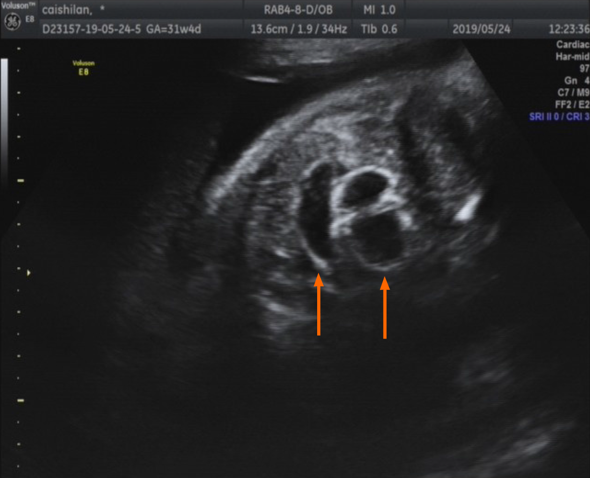
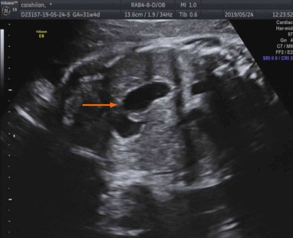
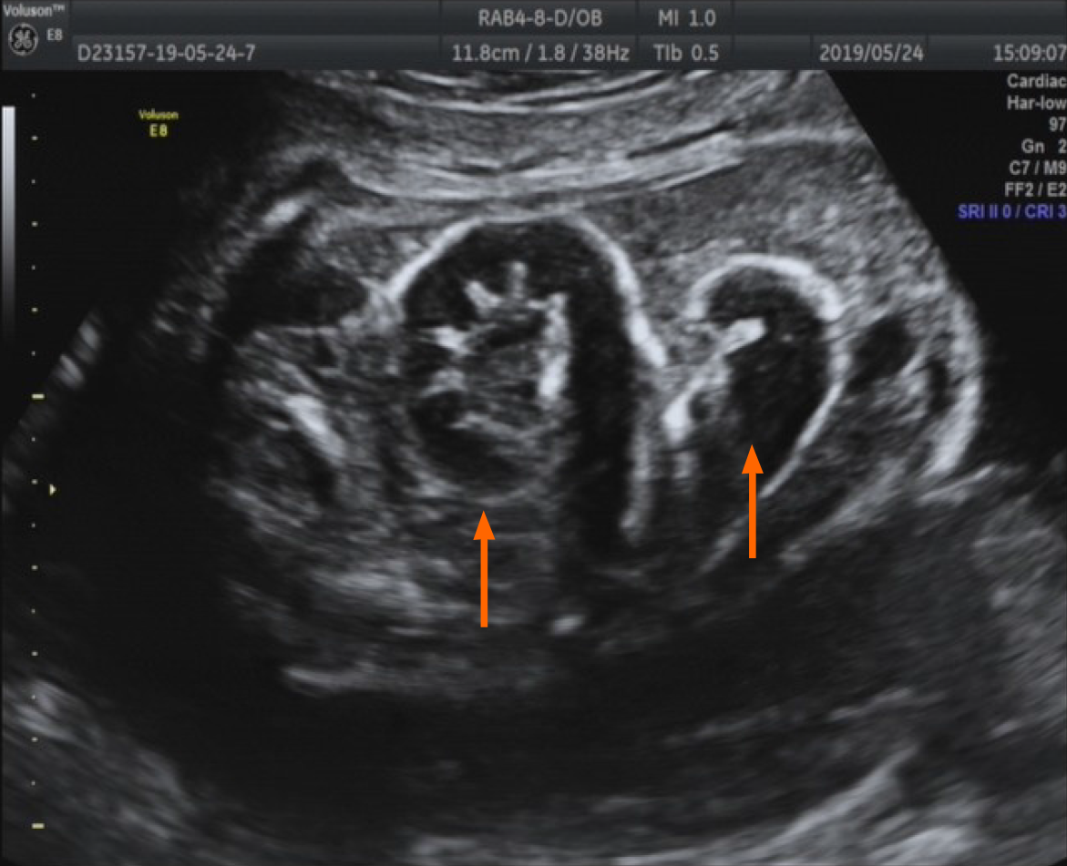
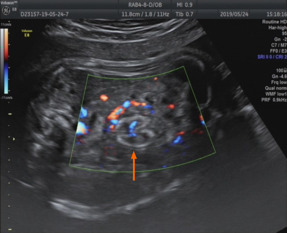
Prenatal sonography showed that the amniotic index was 9.9 cm. There were no abnormalities in the gastric bubble. The duodenum was not dilated. In the middle abdomen of the fetus, there were two rows of intestines arranged in an ‘S’ shape in parallel (Figure 1), with an inner diameter of 1.2 cm and good sound transmission. The echo of the intestinal wall was similar to that of bone. Continuous scanning of the cross-section showed that there was a ‘C’ shape of intestine in the middle and lower abdomen, with an inner diameter of 1.4 cm. The sound inside was Intestinal turbidity. The echo of the intestinal wall was normal. Scanning upward from the anus revealed that the rectum was approximately 0.9 cm wide, the sigmoid colon was 0.7 cm, and the empty structure of the remaining colon was unclear. After 30 minutes, a second examination showed that the size of the gastric bubble was normal, and the duodenum did not dilate. Only a row of intestinal canal was seen horizontally arranged in the middle abdomen (Figure 2). The inner diameter of the intestine canal was 1.1 cm, and the sound transmission was good. Three hours later, re-examination showed that there were two rows of intestines arranged in an “S” shape in the middle abdomen. The inner diameters were 1.7 cm and 1.6 cm, res
The intestinal tract was significantly dilated, and the intestinal wall resembled piano keys. It showed a typical “key
Type II ileal atresia.
After thorough genetic eugenics consultation, the woman chose an induced abortion and agreed to an autopsy.
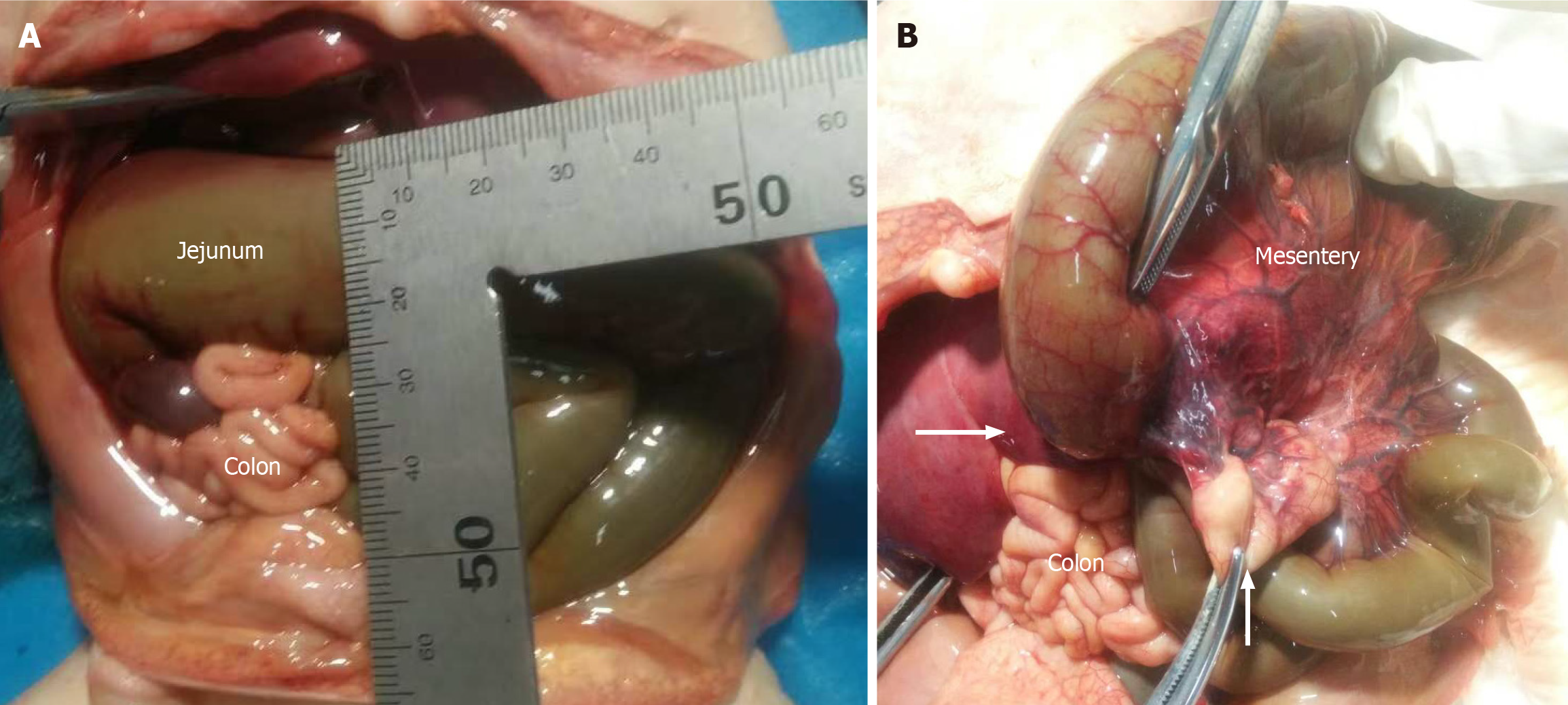
The woman was counseled accordingly and she elected to have the pregnancy terminated. Autopsy examination con
Small bowel atresia can occur anywhere throughout the small intestine, with approximately 54% in the jejunum and 43% in the ileum[3]. Small bowel atresia can be classified into 4 types. Type I atresia is caused by a luminal diaphragm with a continuing outer muscular layer. In type II, the two blind ends of the bowel are attached by a fibrous cord. Type III atresia is complete separation of the bowel ends. Our case had type II ileal atresia[3,9,10]. The accuracy of prenatal ultrasound examination in detecting jejunal and ileal atresia is reported in the literature to be highly variable[11]. However, we accurately judged our case of ileal atresia based on the ultrasound image features of the “keyboard sign” and “coffee bean sign”.
In 1979, scholars found that expansion of the jejunum would show the “keyboard sign”[12]. The discovery of the “keyboard sign” requires significant clinical experience[13]. In recent years, there have been few reports on the “keyboard sign”, as sonographic demonstration of this sign is difficult. In this case, the typical “keyboard sign” appeared for only 5 min, as the shape and position of the intestine were constantly changing. The dilated intestine arranged laterally in the middle abdomen was easily mistaken for the transverse colon. In addition, the “C” shaped dilated bowel in the lower abdomen was easily mistaken for the ascending or descending colon. On the contrary, anatomical results often showed that the expanded jejunum and ileum occupied the whole abdominal cavity. The adjacent colon was small and curly. In order to reduce misdiagnosis, we cannot just identify the intestinal canal by location during ultrasound examination. Instead, the digestive tract should be scanned in sequence.
The position and shape of small intestine distention were changeable due to peristalsis. The dilated small intestine often fills the whole abdominal cavity, which makes the empty colon difficult to display. The distended colon was located in the periphery without obvious peristalsis. The position of the hepatic flexure and the splenic flexure were relatively fixed. The sigmoid colon in the oblique coronal section of the lower abdomen was S-shaped.
Although there was no obvious boundary between the jejunum and ileum, there were differences in the structure of the intestinal wall. The annular plica of jejunum mucosa was greater and higher, with more and higher villi. The villi gradually decreased to the distal end and became longitudinal plica up to the end of the ileum[14]. The structural dif
In our case, there were two different characteristics of the “keyboard sign”. The reasons for this are as follows: The “keyboard sign” in the proximal part of the intestine was regular, while that in the distal part was rare and irregular. This was considered to be caused by different positions of the intestine. The “keyboard sign” in the proximal part was due to jejunum expansion, and that in the distal part was located at the jejunum ileum junction or formed by ileum expansion. The ultrasonographic features of the “keyboard sign” only appeared for 5 min, which was considered to be related to pressure of the intestinal fluid. Only when a large amount of intestinal fluid accumulates to form a certain pressure can the “keyboard sign” be formed[12,13]. When the pressure decreased after peristalsis, the “keyboard sign” disappeared or failed to form.
In our patient, there was a strong echo due to the “coffee bean sign” located under the “keyboard sign”. The echo was similar to that of bone. We considered that this was due to the decrease in fluid content in the wall of the intestine and cystic fibrosis of the intestinal wall. The presence of the “coffee bean sign” was caused by intestinal atresia. The intestine was discontinuous and distorted. Therefore, according to the strong echo “coffee bean sign”, we considered that this was the closed ileum. Ileal atresia was confirmed after delivery.
Most cases of intestinal obstruction are not apparent until the third trimester and the sonographic features are not unique. It is necessary to pay attention to the characteristics of the intestine, especially the typical ultrasound image characteristics. The sonographic features of the “keyboard sign” and “coffee bean sign” are helpful in diagnosing the location of congenital jejunal and ileal atresia.
We thank Dr. Nie D and Dr. Li JC for proofreading the manuscript, and Professor Zhou QC from the Second Xiangya Hospital, Central South University for the guidance on this project.
| 1. | Yin C, Tong L, Ma M, Tan X, Luo G, Fei Z, Nie D. The application of prenatal ultrasound in the diagnosis of congenital duodenal obstruction. BMC Pregnancy Childbirth. 2020;20:387. [RCA] [PubMed] [DOI] [Full Text] [Full Text (PDF)] [Cited by in Crossref: 1] [Cited by in RCA: 4] [Article Influence: 0.8] [Reference Citation Analysis (0)] |
| 2. | John R, D'Antonio F, Khalil A, Bradley S, Giuliani S. Diagnostic Accuracy of Prenatal Ultrasound in Identifying Jejunal and Ileal Atresia. Fetal Diagn Ther. 2015;38:142-146. [RCA] [PubMed] [DOI] [Full Text] [Cited by in Crossref: 23] [Cited by in RCA: 23] [Article Influence: 2.3] [Reference Citation Analysis (0)] |
| 3. | Yin C, Tong L, Nie D, Fei Z, Tan X, Ma M. Significance of the 'line sign' in the diagnosis of congenital imperforate anus on prenatal ultrasound. BMC Pediatr. 2022;22:15. [RCA] [PubMed] [DOI] [Full Text] [Full Text (PDF)] [Reference Citation Analysis (0)] |
| 4. | Heij HA, Moorman-Voestermans CG, Vos A. Atresia of jejunum and ileum: is it the same disease? J Pediatr Surg. 1990;25:635-637. [RCA] [PubMed] [DOI] [Full Text] [Cited by in Crossref: 21] [Cited by in RCA: 19] [Article Influence: 0.5] [Reference Citation Analysis (0)] |
| 5. | Pediatric Gastrointestinal Disease. Pathophysiology, Diagnosis, Management, vol. 1 (2nd edn), Walker WA, Dyrie PR, Hamilton JR, Walker-Smith JA, Watkins JB (eds). Mosby: London, 1996. |
| 6. | 6 Iacobelli BD, Zaccara A, Spirydakis I, Giorlandino C, Capolupo I, Nahom A, Bagolan P. Prenatal counselling of small bowel atresia: watch the fluid! Prenat Diagn. 2006;26:214-217. [RCA] [PubMed] [DOI] [Full Text] [Cited by in Crossref: 9] [Cited by in RCA: 9] [Article Influence: 0.5] [Reference Citation Analysis (0)] |
| 7. | Lu J, Law KM, Lyu GR, Chen BH, Yang GZ, Chen QH, Leung TY. Sonographic 'barber-pole' sign in fetal jejunoileal obstruction is suggestive of apple-peel atresia. Ultrasound Obstet Gynecol. 2022;60:580-581. [RCA] [PubMed] [DOI] [Full Text] [Reference Citation Analysis (0)] |
| 8. | Yu W, Ailu C, Bing W. Sonographic diagnosis of fetal intestinal volvulus with ileal atresia: a case report. J Clin Ultrasound. 2013;41:255-257. [RCA] [PubMed] [DOI] [Full Text] [Cited by in Crossref: 10] [Cited by in RCA: 14] [Article Influence: 1.2] [Reference Citation Analysis (0)] |
| 9. | Grosfeld JL, O'Neill JA, Coran AG. Pediatric surgery. 6th ed. Philadelphia: Mosby, 2009: 1269-1288. |
| 10. | Cho FN, Yang TL, Kan YY, Sung PK. Prenatal sonographic findings in a fetus with congenital isolated ileal atresia. J Chin Med Assoc. 2004;67:366-368. [PubMed] |
| 11. | Matsuzawa N, Yamamoto Y, Kido Y, Itakura A. EP10.13: Investigation of the obstructive site and prenatal ultrasound features in fetal small bowel atresia. Ultrasound in Obstet Gyne. 2023;62:155-156. [DOI] [Full Text] |
| 12. | Bedi DG, Fagan CJ, Nocera RM. Sonographic diagnosis of bowel obstruction presenting with fluid-filled loops of bowel. J Clin Ultrasound. 1985;13:23-30. [PubMed] [DOI] [Full Text] |
| 13. | Fleischer AC, Dowling AD, Weinstein ML, James AE Jr. Sonographic patterns of distended, fluid-filled bowel. Radiology. 1979;133:681-685. [RCA] [PubMed] [DOI] [Full Text] [Cited by in Crossref: 53] [Cited by in RCA: 38] [Article Influence: 0.8] [Reference Citation Analysis (0)] |
| 14. | Liu XZ, Ding WL. Systematic anatomy. People's Medical Publishing House, 2018. |


