Published online Aug 6, 2024. doi: 10.12998/wjcc.v12.i22.5276
Revised: June 7, 2024
Accepted: June 18, 2024
Published online: August 6, 2024
Processing time: 53 Days and 21.8 Hours
Crochetage sign is a specific electrocardiographic manifestation of ostium secundum atrial septal defects (ASDs), which is associated with the severity of the left-to-right shunt. Herein, we reported a case of selective his bundle pacing (S-HBP) that eliminated crochetage sign in a patient with ostium secundum ASD.
A 77-year-old man was admitted with a 2-year history of chest tightness and shortness of breath. Transthoracic echocardiography revealed an ostium secun
S-HBP eliminated crochetage sign on electrocardiogram. Crochetage sign may be a manifestation of a conduction system disorder.
Core Tip: Crochetage sign is a characteristic electrocardiographic manifestation seen in patients with ostium secundum atrial septal defects. We reported the case of a patient with secundum atrial septal defect, atrial fibrillation, and second-degree atrioventricular block who received selective his bundle pacing (S-HBP) treatment. Subsequently, crochetage sign was eliminated by S-HBP. Crochetage sign may indicate a conduction system disease.
- Citation: Mu YG, Liu KS. Selective his bundle pacing eliminates crochetage sign: A case report. World J Clin Cases 2024; 12(22): 5276-5282
- URL: https://www.wjgnet.com/2307-8960/full/v12/i22/5276.htm
- DOI: https://dx.doi.org/10.12998/wjcc.v12.i22.5276
With an incidence of 6%-10% after birth, atrial septal defect (ASD) is one of the most prevalent congenital heart diseases, with secundum ASD being the most common[1]. Because ASD's clinical presentation may be inconspicuous, optimal diagnostic methods remain challenging[2]. Several electrocardiogram (ECG) findings are thought to be sensitive for the diagnosis of ASD; hence, the ECG can be used as a helpful ASD screening tool. However, echocardiography remains the primary diagnostic tool for ASD[3].
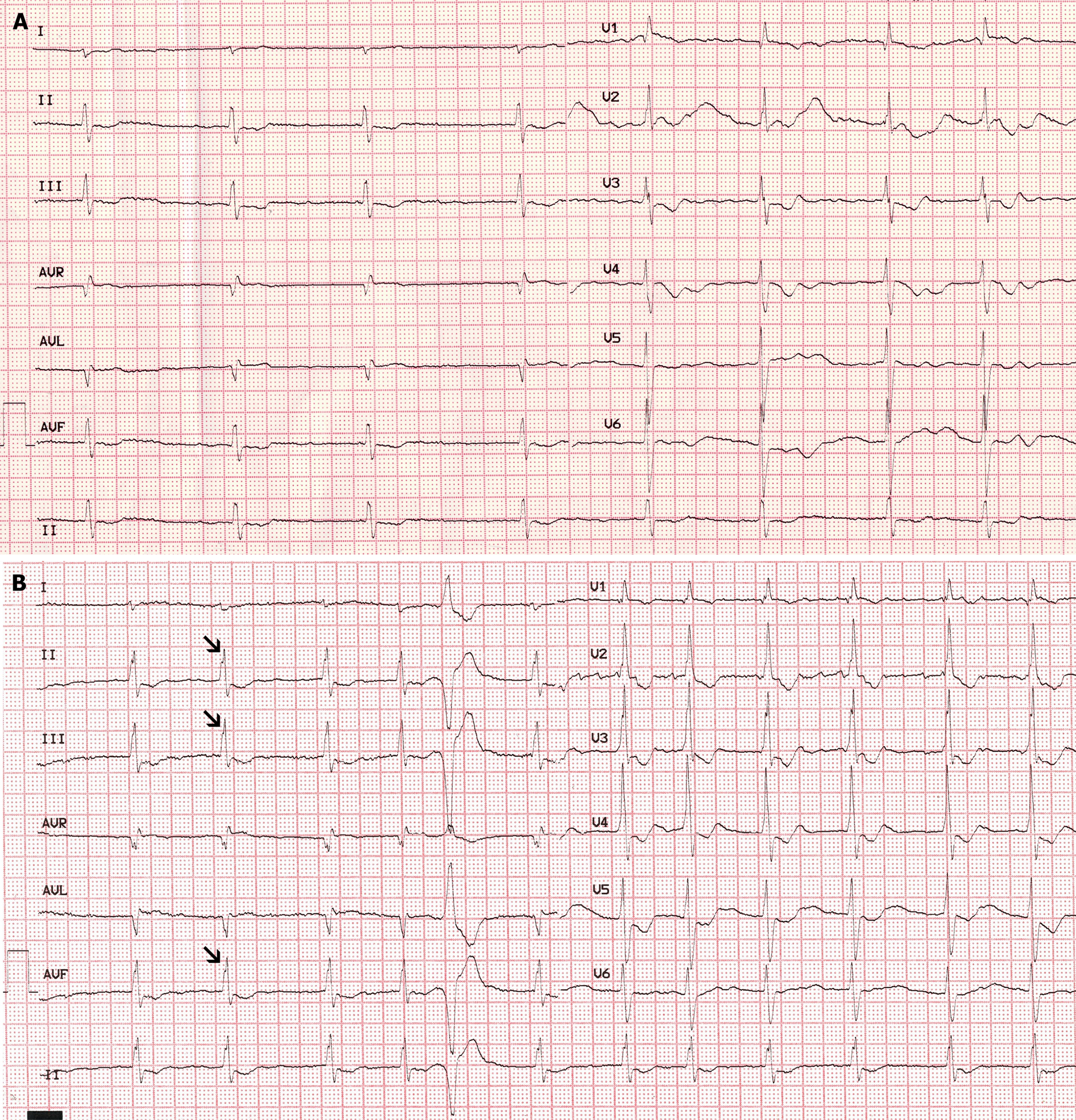
One ECG finding associated with ASD is crochetage sign, which is a specific sign of ASD in the ostium secundum. Crochetage sign, similar to a crochet needle, is characterized by fragmented QRS complexes with a notch in the ascending portion or at the peak of the R-wave, which are typically observed in limb leads II, III, and aVF[4]. For secundum ASD, the sensitivity and specificity of this sign are higher when more inferior leads are involved. When crochetage pattern is seen in all three inferior leads, the specificity of this sign ranges from 92% to 100%[3]. The right bundle branch block (RBBB) on electrocardiography can further improve the specificity and sensitivity of crochetage sign[5]. Therefore, the appearance of crochetage sign on ECG is an important indicator of ASD, aiding in its early diagnosis. Previous studies showed that 35.1% of patients with ASD who underwent surgical closure experienced a disappearance of crochetage sign on their ECGs[6]. However, the disappearance of crochetage sign after cardiac pacing has not yet been reported. Herein, we report the case of a man with secundum ASD, atrial fibrillation, and a second-degree atrioventricular (AV) block. After receiving selective his bundle pacing (S-HBP) therapy, the crochetage sign on his ECG disappeared.
A 77-year-old man presented to our hospital complaining of chest tightness and shortness of breath for 2 years.
The patient complained of chest tightness and shortness of breath for 2 years. Symptoms appeared with activity, gradually resolved with rest, and worsened over the past 2 days, prompting the patient to seek treatment at our hospital.
The patient had no history of hypertension, type 2 diabetes mellitus, or coronary heart disease.
The patient denied smoking, any significant drinking history, or any family disease history.
Vital signs were as follows: Body temperature, 36.7 ºC; Blood pressure, 122/76 mmHg; Heart rate, 61 beats per min; and Respiratory rate, 18 breaths per min. Both lungs produced coarse respiratory sounds and wet rales. The heart rate was irregular, cardiac sounds were decreased, and no murmur was detected. No cyanosis or lower limb edema was observed.
The brain natriuretic peptide level was 622 pg/mL (reference range: 0-80 pg/mL), and the serum potassium level was 3.04 mmol/L (reference range: 3.2-5.3 mmol/L). Routine blood testing revealed normal liver, kidney, thyroid, and blood coagulation functions. Electrocardiography suggested atrial fibrillation with a prolonged relative risk interval; a notch near the apex of the R-wave in leads II, III, and aVF; and incomplete RBBB (Figure 1).
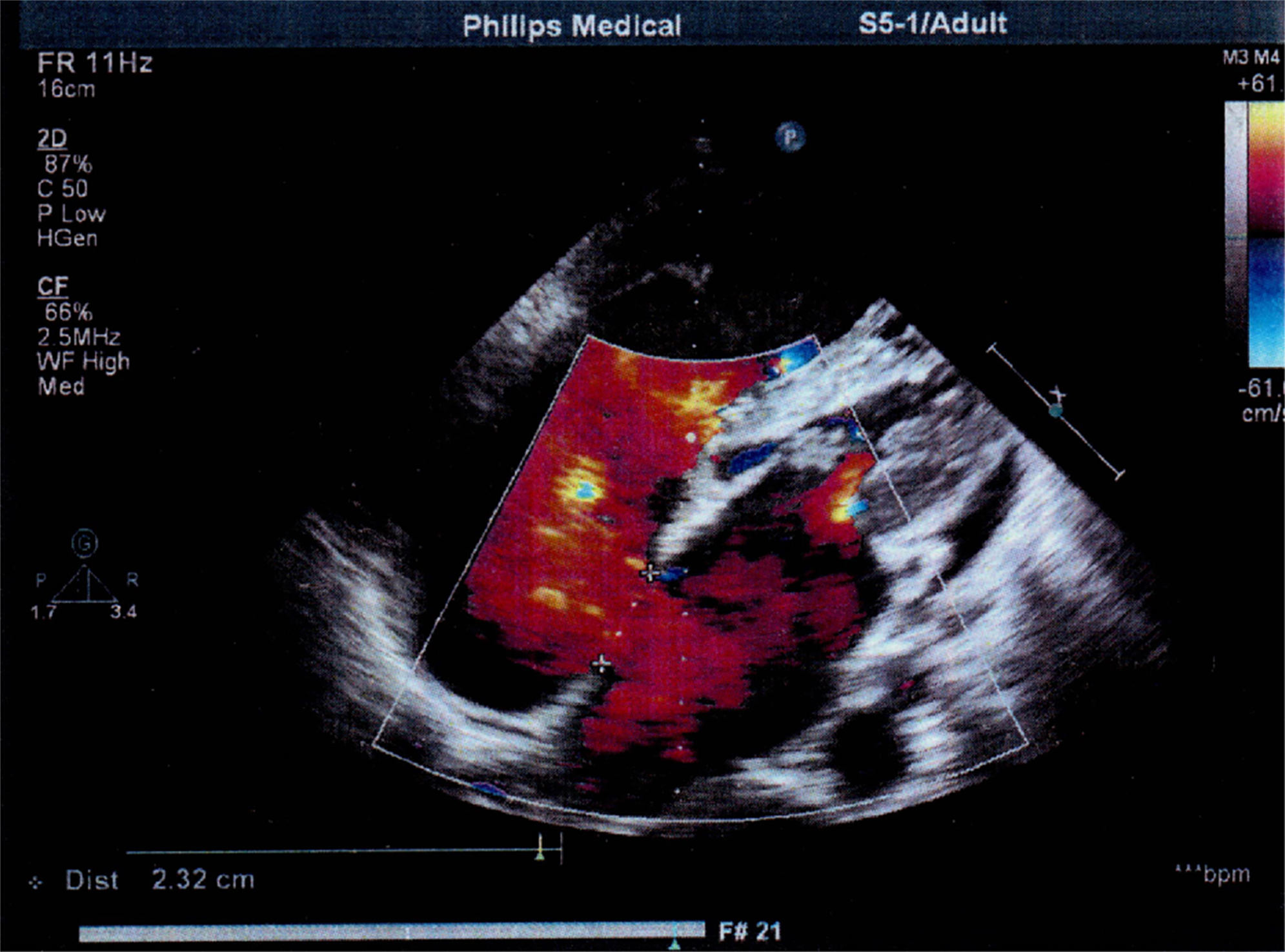
Transthoracic echocardiography (TTE) revealed a secundum ASD (Figure 2), severe tricuspid regurgitation, and mild mitral regurgitation. The left atrial diameter was 54 mm, left ventricular diameter was 33 mm, right atrial diameter was 66 mm, right ventricular (RV) diameter was 56 mm, and left ventricular ejection fraction was 75%.
The final diagnosis was a secundum ASD, atrial fibrillation with second-degree AV block (AVB), and heart failure.
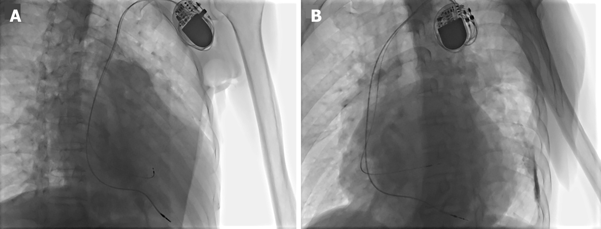
Considering the adverse effects of chronic RV pacing, we decided to perform HBP. A 3830 SelectSecure lead (Medtronic Inc., Minneapolis, MN, United States) was implanted in the his-bundle region (Figure 3). Pacing parameters were acceptable, with a capture threshold of 0.8 V/0.4 ms, sensitivity of 7 mV, and bipolar impedance at 570 Ω. A 5076-58 cm lead was implanted in the RV apex as a backup. Finally, a dual-chamber pacemaker (Medtronic Sensia SEDRL1; Medtronic Inc., Minneapolis, MN, United States) was implanted subcutaneously, with a 3830 lead connected to the atrial port and a 5076–58 cm lead connected to the ventricular port.
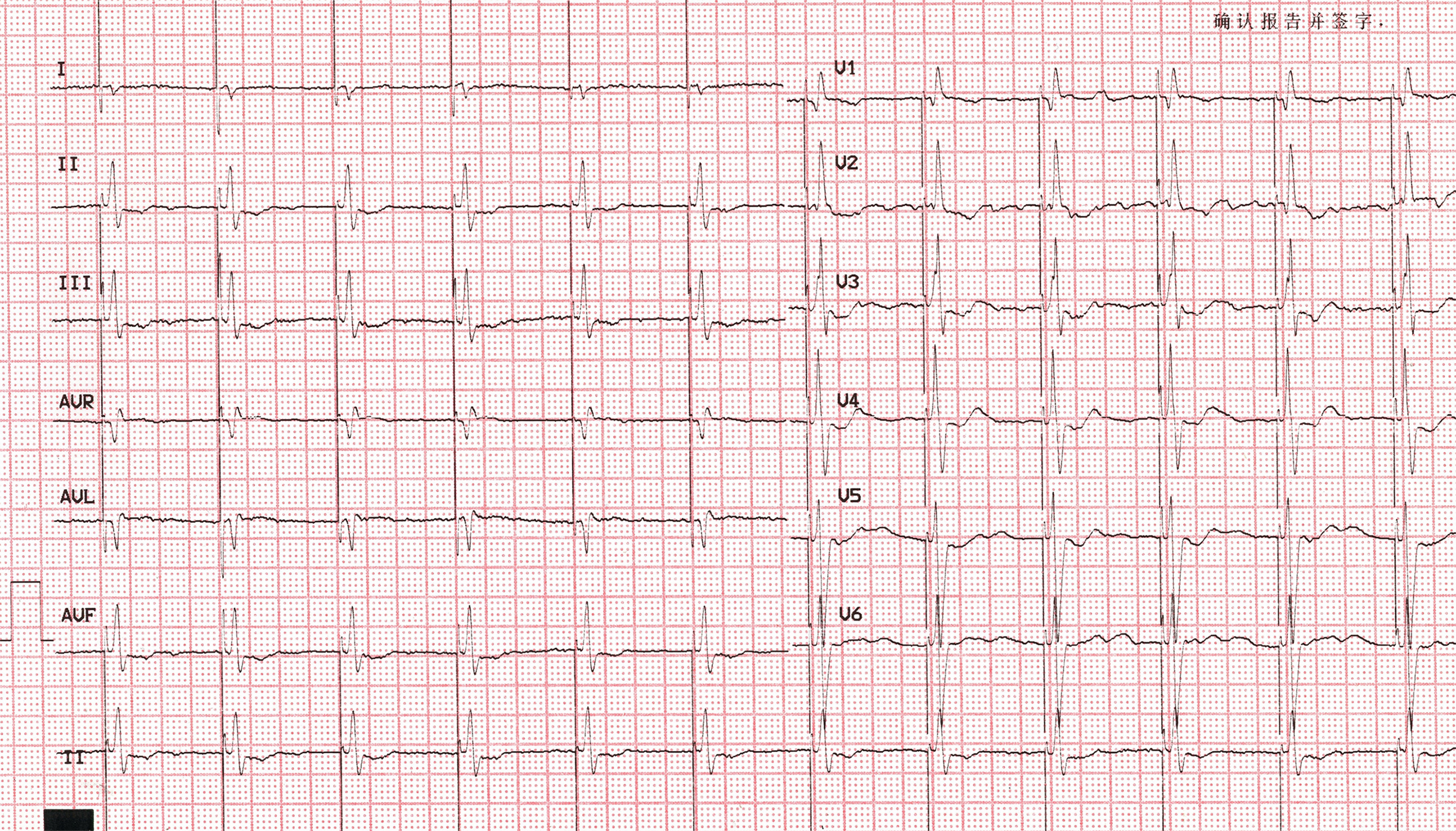
After the procedure, the crochetage sign disappeared during HBP on electrocardiography (Figure 4). The patient received regular follow-up at the outpatient clinic. Pacemaker programming indicated normal functioning of both sensing and pacing.
ASDs are the most common heart defects in adults[7]. ASDs can cause heart failure, arrhythmic problems, and paradoxical embolism-related morbidity or death[2]. Many individuals with ASDs develop first-degree AVB, which is mostly caused by a prolonged intra-atrial conduction time[8]. Over time, a left-to-right shunt may cause cellular damage, decreased reservoir function, myocardial cell hypertrophy, and RV dilatation. Stretching myocardial fibers might alter their conductive properties, which could ultimately result in bradyarrhythmia or atrial tachyarrhythmia[9]. Some ASDs are hereditary and linked to cardiac conduction abnormalities such as bundle branch block or AVB[10]. ASD in patients with AVB may be related to a genetic mutation in myocardial transcription factors, such as NKX2.5, GATA4, and TBX5j, which are crucial for the development of the AV conduction system[8,10]. These include Holt–Oram syndrome, characterized by ASDs and conduction abnormalities such as sinus node dysfunction, AVB, atrial fibrillation, and RBBB[10].
Patients diagnosed with ostium-primum ASDs more commonly have higher-grade AV nodal conduction anomalies. AV nodal conduction disorders are uncommon in other forms of ASD[8]. The incidence of heart block in patients with ASD is unclear, and ASD with high-grade AV block is relatively uncommon[11]. Our patient had a second-degree AVB and atrial fibrillation, which are rare. His bundle pacing was then performed. The postoperative ECG revealed an isoelectric interval in all 12 leads, with the pacing spike to QRS onset equaling the native HV interval (35–55 ms). The QRS morphology was identical to that of the intrinsic rhythm, confirming S-HBP.
Crochetage sign refers to the presence of a notch in the initial 80 ms of the QRS complex in leads II, III and aVF, which resembles the shape of a crochet needle[12]. This sign is most frequently observed in individuals with ASD and can also be observed in other disorders such as patent foramen ovale (PFO), pulmonary embolism, and RV hypertrophy[11]. Recent research has revealed that, crochetage sign combined with RBBB and TTE had good performance for PFO prediction, with an accuracy, sensitivity, and specificity of 0.712, 0.801, and 0.439, respectively[13]. In the initial published research on crochetage, 73% of patients with ASD showed this pattern[14]. Crochetage sign is considered a characteristic presentation of secundum ASDs. When the pattern is seen in all three inferior leads, the specificity of this sign ranges from 92% to 100%[3]. The presence of a RBBB on the ECG considerably increases the specificity and sensitivity of crochetage sign for the diagnosis of ASD[3]. It has also been associated with the severity of the left-to-right shunt, and patients with ASD sizes of ≥ 5 mm showed a higher incidence of crochetage sign[6]. Because of its high sensitivity and specificity in diagnosing ASD, crochetage sign is a useful diagnostic tool[12]. Moreover, a retrospective clinical study demonstrated that the presence of crochetage sign was an independent predictor for late atrial arrhythmia among patients with secundum ASD who received transcatheter closure[15].
The underlying electrophysiological mechanism of crochetage sign is not fully understood, but many researchers believe that it is related to delayed RV depolarization due to RV volume overload[15]. Crochetage sign disappears in approximately 50% of patients after successful correction of ASD using percutaneous or surgical procedures[3,6]. Interestingly, in our case, crochetage sign disappeared on the ECG after S-HBP; however, the patient had not undergone atrial septal closure. We concluded that crochetage sign may be a manifestation of conduction system impairment. Based on the theory of longitudinal dissociation in the his bundle, conduction impairment may originate within the proximal his bundle because the pacing exceeded the injury level, and conduction through the His-Purkinje system was recovered, thus eliminating crochetage sign[16].
The exact mechanism underlying the development of crochetage sign has not yet been elucidated. Some patients with ASD closure experience crochetage signs that disappear from their ECG. Here, we report the case of a patient with ASD whose crochetage sign disappeared after selective his bundle pacing. Subsequently, we deduced that crochetage sign could be a manifestation of a cardiac conduction system injury. Future research is needed to explore the mechanism behind crochetage sign.
| 1. | Martin SS, Shapiro EP, Mukherjee M. Atrial septal defects - clinical manifestations, echo assessment, and intervention. Clin Med Insights Cardiol. 2014;8:93-98. [RCA] [PubMed] [DOI] [Full Text] [Full Text (PDF)] [Cited by in Crossref: 9] [Cited by in RCA: 31] [Article Influence: 3.1] [Reference Citation Analysis (0)] |
| 2. | Bayar N, Arslan Ş, Köklü E, Cagirci G, Cay S, Erkal Z, Ayoglu RU, Küçükseymen S. The importance of electrocardiographic findings in the diagnosis of atrial septal defect. Kardiol Pol. 2015;73:331-336. [RCA] [PubMed] [DOI] [Full Text] [Cited by in Crossref: 6] [Cited by in RCA: 7] [Article Influence: 0.7] [Reference Citation Analysis (0)] |
| 3. | Heller J, Hagège AA, Besse B, Desnos M, Marie FN, Guerot C. "Crochetage" (notch) on R wave in inferior limb leads: a new independent electrocardiographic sign of atrial septal defect. J Am Coll Cardiol. 1996;27:877-882. [RCA] [PubMed] [DOI] [Full Text] [Cited by in Crossref: 49] [Cited by in RCA: 50] [Article Influence: 1.7] [Reference Citation Analysis (0)] |
| 4. | Zeman J, Kochiashvili A, Naik R, Muco E, Kim AS. Crochet Leads the Way. JACC Case Rep. 2023;13:101814. [RCA] [PubMed] [DOI] [Full Text] [Reference Citation Analysis (0)] |
| 5. | Singh H, Pannu AK, Dahiya N, Suri V, Bhalla A, Kumari S. 'Crochetage' sign of atrial septal defect. QJM. 2020;113:133-134. [RCA] [PubMed] [DOI] [Full Text] [Reference Citation Analysis (0)] |
| 6. | Shen L, Liu J, Li JK, Xu M, Yuan L, Zhang GQ, Wang JY, Huang YJ. The Significance of Crochetage on the R wave of an Electrocardiogram for the Early Diagnosis of Pediatric Secundum Atrial Septal Defect. Pediatr Cardiol. 2018;39:1031-1035. [RCA] [PubMed] [DOI] [Full Text] [Cited by in Crossref: 2] [Cited by in RCA: 2] [Article Influence: 0.3] [Reference Citation Analysis (0)] |
| 7. | Webb G, Gatzoulis MA. Atrial septal defects in the adult: recent progress and overview. Circulation. 2006;114:1645-1653. [RCA] [PubMed] [DOI] [Full Text] [Cited by in Crossref: 341] [Cited by in RCA: 329] [Article Influence: 17.3] [Reference Citation Analysis (0)] |
| 8. | Williams MR, Perry JC. Arrhythmias and conduction disorders associated with atrial septal defects. J Thorac Dis. 2018;10:S2940-S2944. [RCA] [PubMed] [DOI] [Full Text] [Cited by in Crossref: 14] [Cited by in RCA: 23] [Article Influence: 3.3] [Reference Citation Analysis (0)] |
| 9. | Albæk DHR, Udholm S, Ovesen AL, Karunanithi Z, Nyboe C, Hjortdal VE. Pacemaker and conduction disturbances in patients with atrial septal defect. Cardiol Young. 2020;30:980-985. [RCA] [PubMed] [DOI] [Full Text] [Cited by in Crossref: 3] [Cited by in RCA: 6] [Article Influence: 1.2] [Reference Citation Analysis (0)] |
| 10. | Aoki H, Horie M. Electrical disorders in atrial septal defect: genetics and heritability. J Thorac Dis. 2018;10:S2848-S2853. [RCA] [PubMed] [DOI] [Full Text] [Cited by in Crossref: 2] [Cited by in RCA: 2] [Article Influence: 0.3] [Reference Citation Analysis (0)] |
| 11. | Fernandez Hazim C, Shaban M, Cordero D, Urena Neme AP, Rodriguez Guerra MA. Crochetage, the Forgotten Electrocardiographic Sign. Cureus. 2023;15:e46498. [RCA] [PubMed] [DOI] [Full Text] [Reference Citation Analysis (0)] |
| 12. | Sarma A. Crochetage Sign: An Invaluable Independent ECG Sign in Detecting ASD. Indian J Crit Care Med. 2021;25:234-235. [RCA] [PubMed] [DOI] [Full Text] [Full Text (PDF)] [Cited by in Crossref: 1] [Cited by in RCA: 1] [Article Influence: 0.3] [Reference Citation Analysis (0)] |
| 13. | Jin P, Jiao P, Feng J, Shi L, Ma L. The predictive value of abnormal electrocardiogram for patent foramen ovale: A retrospective study. Clin Cardiol. 2023;46:1504-1510. [RCA] [PubMed] [DOI] [Full Text] [Reference Citation Analysis (0)] |
| 14. | Toscano Barboza E, Brandenburg RO, Swan HJ. Atrial septal defect; the electrocardiogram and its hemodynamic correlation in 100 proved cases. Am J Cardiol. 1958;2:698-713. [RCA] [PubMed] [DOI] [Full Text] [Cited by in Crossref: 35] [Cited by in RCA: 30] [Article Influence: 0.4] [Reference Citation Analysis (0)] |
| 15. | Celik M, Yilmaz Y, Kup A, Karagoz A, Kahyaoglu M, Cakmak EO, Celik FB, Sengor BG, Guner A, Izci S, Kilicgedik A, Candan O, Kahveci G, Gecmen C, Kaymaz C. Crochetage sign may predict late atrial arrhythmias in patients with secundum atrial septal defect undergoing transcatheter closure. J Electrocardiol. 2021;67:158-165. [RCA] [PubMed] [DOI] [Full Text] [Cited by in Crossref: 1] [Cited by in RCA: 4] [Article Influence: 1.0] [Reference Citation Analysis (0)] |
| 16. | Teng AE, Massoud L, Ajijola OA. Physiological mechanisms of QRS narrowing in bundle branch block patients undergoing permanent His bundle pacing. J Electrocardiol. 2016;49:644-648. [RCA] [PubMed] [DOI] [Full Text] [Cited by in Crossref: 20] [Cited by in RCA: 27] [Article Influence: 3.0] [Reference Citation Analysis (0)] |


