Published online Aug 6, 2024. doi: 10.12998/wjcc.v12.i22.5253
Revised: May 23, 2024
Accepted: June 4, 2024
Published online: August 6, 2024
Processing time: 72 Days and 21.4 Hours
Diaphragmatic paralysis is typically associated with phrenic nerve injury. Neonatal diaphragmatic paralysis diagnosis is easily missed because its manifestations are variable and usually nonspecific.
We report a 39-week-old newborn delivered via vaginal forceps who presented with tachypnea but without showing other birth-trauma-related manifestations. The infant was initially diagnosed with pneumonia. However, the newborn still exhibited tachypnea despite effective antibiotic treatment. Chest radiography revealed right diaphragmatic elevation. M-mode ultrasonography revealed decreased movement of the right diaphragm. The infant was subsequently diagnosed with diaphragmatic paralysis. After 4 weeks, tachypnea improved. Upon re-examination using M-mode ultrasonography, the difference in bilateral diaphragmatic muscle movement was smaller than before.
Appropriate use of M-mode ultrasound to quantify diaphragmatic excursions could facilitate timely diagnosis and provide objective evaluation.
Core Tip: Diaphragmatic paralysis is typically caused by phrenic nerve injury in the context of birth-related trauma or cardiothoracic surgery. Diagnosis of diaphragmatic paralysis is easily missed because its signs are usually nonspecific. The infant in this case had a lung infection but without showing other manifestations due to birth-related trauma. The infant exhibited tachypnea despite effective antibiotic treatment. Chest radiography revealed an elevated right hemidiaphragm. Diagnosis of diaphragmatic paralysis was confirmed by ultrasonography, which revealed decreased motion of the right diaphragm. Appropriate use of M-mode ultrasound to quantify diaphragmatic excursions could facilitate timely diagnosis and provide objective evaluation.
- Citation: Zeng Y, Luo P, Zhao DR, Wang FY, Song B. Neonatal tachypnea caused by diaphragmatic paralysis: A case report. World J Clin Cases 2024; 12(22): 5253-5257
- URL: https://www.wjgnet.com/2307-8960/full/v12/i22/5253.htm
- DOI: https://dx.doi.org/10.12998/wjcc.v12.i22.5253
Diaphragmatic paralysis is typically caused by phrenic nerve injury in the context of birth-related trauma or cardiothoracic surgery[1]. Neonatal diaphragmatic paralysis is usually unilateral, commonly involving the right side[2]. Diaphragmatic paralysis diagnosis is easily missed because its manifestations are variable and usually nonspecific. In this report, we describe neonatal diaphragmatic paralysis and its diagnosis using M-mode ultrasonography.
A 39-week-old newborn was admitted to the hospital for tachypnea.
The newborn, delivered via vaginal forceps, presented with tachypnea but without showing other manifestations due to birth trauma.
The newborn had no remarkable past illness.
There were no notable medical issues in the newborn’s family.
The infant exhibited tachypnea, with a respiratory rate of 70 breaths/minutes. The findings of the nervous system examination were unremarkable, without noting other manifestations due to birth trauma.
Laboratory test results showed an increased infection index.
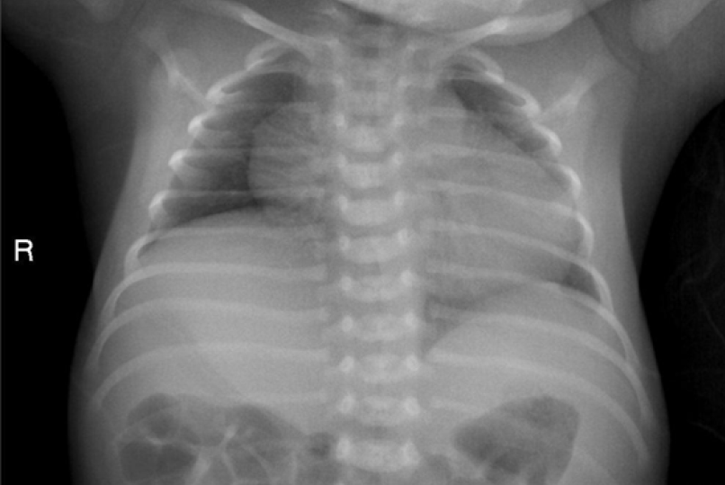
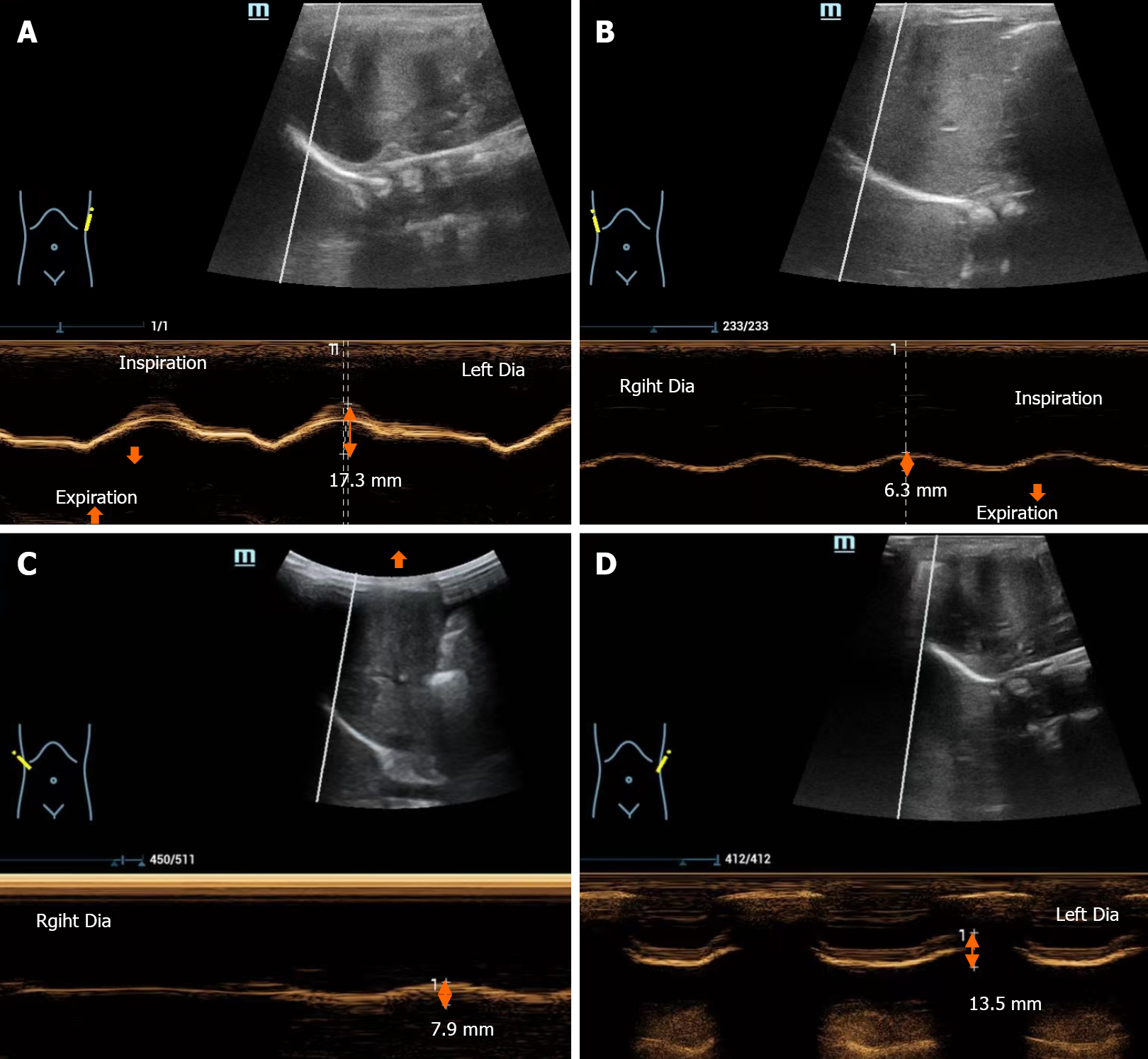
Chest radiography revealed right diaphragmatic elevation and increased patch density in the upper field of the left lung (Figure 1). M-mode ultrasonography revealed normal movement of the left diaphragm (Figure 2A) and decreased movement of the right diaphragm in the supine position (Figure 2B).
The infant was ultimately diagnosed with pneumonia and diaphragmatic paralysis.
The infant was administered a course of antibiotic treatment. One week later, re-examination by chest radiography showed no infection. Since the levels of other laboratory infection indicators were also normal, antibiotic administration was stopped. Due to a respiration rate of 70 breaths/minutes, the infant was initiated on nasal high-flow oxygen therapy at a flow rate of 8 L/minutes. However, after 1 week, the infant continued to exhibit tachypnea. Thus, breathing support was continued for a further 1 week.
Four weeks later, there was improvement in tachypnea. Upon reassessment using M-mode ultrasonography, the difference in bilateral diaphragmatic muscle movement was noted to be smaller compared with that before (Figure 2C and D).
Diaphragmatic paralysis is typically caused by phrenic nerve injury sustained in the context of birth-related trauma or cardiothoracic surgery[1]. Given weak intercostal muscles and increased mediastinal mobility in infants and young children, adequate ventilation is almost totally dependent on diaphragmatic function initially[3]. Consequently, diaphragmatic paralysis affects breathing more severely in children even with unilateral paralysis. Consistent with this, infants tolerate diaphragmatic paralysis much less effectively than do older children[4,5]. Unfortunately, the diagnosis of diaphragmatic paralysis is easily missed because its manifestations are variable and usually nonspecific. Clinical manifestations include unexplained difficulty with mechanical ventilation, asymmetric breathing patterns, abnormal upper abdominal movements, as well as recurrent pneumonia and/or tachypnea.
Neonatal diaphragmatic paralysis is usually unilateral, commonly involving the right side[2].
| Classification | Definition |
| Normal motion | The amplitude of diaphragmatic excursion is > 4 mm, and the difference of amplitude between the hemidiaphragms is < 50% |
| Decreased motion | The amplitude of diaphragmatic excursion is ≤ 4 mm and the difference of amplitude is > 50% |
| Absent motion | Absent motion is considered by a flat line trace |
| Paradoxical motion | Paradoxical motion is considered when diaphragm moves away from the transducer in inspiration |
Diaphragmatic paralysis in this case was caused by phrenic nerve injury caused by birth-related trauma. The infant in this case had a lung infection but without showing other manifestations due to birth-related trauma, such as brachial plexus palsy. The infant was initially diagnosed with pneumonia and treated accordingly. However, since the infant still exhibited tachypnea despite effective antibiotic treatment and chest radiography revealed an elevated right hemidiaphragm, diaphragmatic paralysis was suspected. The diagnosis was confirmed by ultrasonography, which revealed decreased motion of the right diaphragm.
Neonatal diaphragmatic paralysis can be easily misdiagnosed. Appropriate use of M-mode ultrasonography to quantify diaphragmatic excursions could facilitate a timely diagnosis and provide an objective evaluation.
| 1. | Rizeq YK, Many BT, Vacek JC, Reiter AJ, Raval MV, Abdullah F, Goldstein SD. Diaphragmatic paralysis after phrenic nerve injury in newborns. J Pediatr Surg. 2020;55:240-244. [RCA] [PubMed] [DOI] [Full Text] [Cited by in Crossref: 4] [Cited by in RCA: 11] [Article Influence: 2.2] [Reference Citation Analysis (0)] |
| 2. | Volpe JJ. Injuries of Extracranial, Cranial, Intracranial, Spinal Cord, and Peripheral Nervous System Structures. Volpe's Neurology of the Newborn (Sixth Edition). Elsevier, 2018: 1093.e5-1123.e5. [DOI] [Full Text] |
| 3. | Mok Q, Ross-Russell R, Mulvey D, Green M, Shinebourne EA. Phrenic nerve injury in infants and children undergoing cardiac surgery. Br Heart J. 1991;65:287-292. [RCA] [PubMed] [DOI] [Full Text] [Cited by in Crossref: 47] [Cited by in RCA: 46] [Article Influence: 1.4] [Reference Citation Analysis (0)] |
| 4. | Mickell JJ, Oh KS, Siewers RD, Galvis AG, Fricker FJ, Mathews RA. Clinical implications of postoperative unilateral phrenic nerve paralysis. J Thorac Cardiovasc Surg. 1978;76:297-304. [RCA] [PubMed] [DOI] [Full Text] [Cited by in Crossref: 80] [Cited by in RCA: 69] [Article Influence: 1.5] [Reference Citation Analysis (0)] |
| 5. | Smith CD, Sade RM, Crawford FA, Othersen HB. Diaphragmatic paralysis and eventration in infants. J Thorac Cardiovasc Surg. 1986;91:490-497. [RCA] [PubMed] [DOI] [Full Text] [Cited by in Crossref: 30] [Cited by in RCA: 13] [Article Influence: 0.3] [Reference Citation Analysis (0)] |
| 6. | McCauley RG, Labib KB. Diaphragmatic paralysis evaluated by phrenic nerve stimulation during fluoroscopy or real-time ultrasound. Radiology. 1984;153:33-36. [RCA] [PubMed] [DOI] [Full Text] [Cited by in Crossref: 32] [Cited by in RCA: 30] [Article Influence: 0.7] [Reference Citation Analysis (0)] |
| 7. | Alexander C. Diaphragm movements and the diagnosis of diaphragmatic paralysis. Clin Radiol. 1966;17:79-83. [RCA] [PubMed] [DOI] [Full Text] [Cited by in Crossref: 130] [Cited by in RCA: 118] [Article Influence: 2.0] [Reference Citation Analysis (0)] |
| 8. | Boussuges A, Rives S, Finance J, Brégeon F. Assessment of diaphragmatic function by ultrasonography: Current approach and perspectives. World J Clin Cases. 2020;8:2408-2424. [RCA] [PubMed] [DOI] [Full Text] [Full Text (PDF)] [Cited by in CrossRef: 53] [Cited by in RCA: 95] [Article Influence: 19.0] [Reference Citation Analysis (3)] |
| 9. | Sehgal A, Fernando S, Ditchfield M. M-Mode Imaging of the Diaphragm in Phrenic Nerve Palsy Due to Birth Trauma. J Pediatr. 2022;246:281-282. [RCA] [PubMed] [DOI] [Full Text] [Cited by in RCA: 2] [Reference Citation Analysis (0)] |


