Published online Jun 26, 2024. doi: 10.12998/wjcc.v12.i18.3529
Revised: February 7, 2024
Accepted: May 9, 2024
Published online: June 26, 2024
Processing time: 239 Days and 2.9 Hours
Leiomyomas (LMs) are mesenchymal tumors that arise from smooth muscle cells. LMs most commonly arise in organs with an abundance of smooth muscle such as the uterus and gastrointestinal tract. Conversely, LMs are rarely detected in the head and neck region. In this study, we report a rare case of laryngeal LM (LLM) and summarized the clinical characteristics of reported LLMs to help clinicians better understand this rare disease and improve its diagnosis, treatment, and postoperative course.
A 49-year-old man was admitted to our ENT outpatient clinic with a chief com
Surgical resection is the preferred treatment for LLMs, its early diagnosis and differential diagnosis have important clinical significance.
Core Tip: We present a rare case of laryngeal leiomyoma (LLM) admitted to our institution, while also providing a comprehensive review and synthesis of the clinical characteristics associated with reported LLMs. Our aim is to enhance clinicians' comprehension of this uncommon disease, thereby facilitating improved diagnosis, treatment strategies, and postoperative patient recovery.
- Citation: Wu Y, Li JM, Zhang TJ, Wang X. Laryngeal leiomyoma: A case report and review of literature. World J Clin Cases 2024; 12(18): 3529-3533
- URL: https://www.wjgnet.com/2307-8960/full/v12/i18/3529.htm
- DOI: https://dx.doi.org/10.12998/wjcc.v12.i18.3529
Leiomyomas (LMs) are mesenchymal tumors that originate from smooth muscle cells. LMs most commonly arise in organs with an abundance of smooth muscle such as the uterus and gastrointestinal tract. Conversely, LMs are rarely detected in the head and neck region, and the incidence of oral LM is only 0.065%, accounting for 0.42% of all soft tissue tumors in the oral cavity. To the best of our knowledge, no more than 50 cases have been reported to date. In this study, we report a rare case of laryngeal LM (LLM) in a patient admitted to our center. We also reviewed and summarized the clinical characteristics of reported LLMs to help clinicians gain a better understanding of this uncommon disease and improve the diagnosis, treatment, and postoperative course.
A 49-year-old man was admitted to our ENT outpatient clinic with a chief complaint of pharynx discomfort persisting for 2 months.
During his clinical course, he had no fever, cough, sore throat, or difficulties in swallowing or breathing.
He had no previous medical history.
He was a non-smoker without a remarkable medical history or family history of head and neck tumors.
Through indirect laryngoscopy, revealed a solitary, pink mass at the tubercle of epiglottis.
Biopsy was performed via laryngoscopy, and the pathological examination revealed spindle cell proliferation, which is typical of smooth muscle-derived tumors. The tissues were submitted for pathological examination, revealing polypoid tumor tissue at the base. Hematoxylin and eosin staining demonstrated that the mass was composed of myogenic, indistinct shape tumor cells surrounding the submucosal small salivary glands without sign of necrosis or mitoses. Positive immunohistochemical staining for desmin and smooth-muscle actin indicated a smooth muscle origin (Figure 1).
Laryngoscopy performed under topical anesthesia revealed a solitary, pink mass at the tubercle of epiglottis.
LLM.
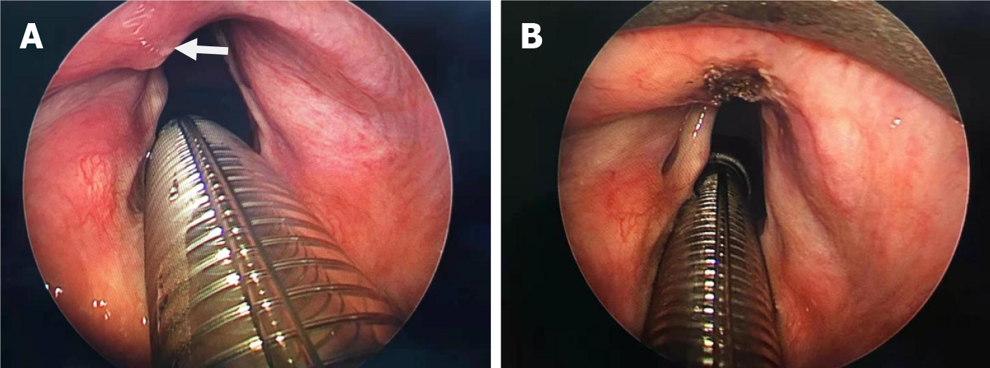
Surgery via laryngeal endoscopy was performed under general anesthesia, and the lesion was excised easily (Figure 2). No other interventions were given during treatment.
After surgery, the patient condition was stable, and he was discharged 2 d after surgery (Figure 3). During the 1-year postoperative period, the patient condition remained stable without evidence of recurrence. The patient provided informed consent for publication of his case.
LMs are benign tumors originating from the smooth muscle components of organs. They mostly arise in the uterus, small intestine, and esophagus. By contrast, LMs rarely occur in the head and neck region because of the paucity of smooth muscle[1]. Three different types of LM are recognized: Common LM, vascular LM (angiomyoma), and epithelioid LM. Although smooth muscle is rarely present in laryngeal tissues, all three types of LM have been found in the larynx[2]. Histopathologically, angiomyoma consists of many variant blood vessels. Whirlpools of smooth muscle fibers can be observed around the vascular endothelium. However, they lack mitotic and well-differentiated smooth muscle bundles, and they are sometimes accompanied by mucoid lesions[3]. Epithelioid LMs manifest as tumor cell masses with round or oval nucleus, with or without vacuoles, and clear cell changes in the rich eosinophilic cytoplasm[4]. Common LMs are characterized by intertwined spindle cells, cigar-shaped nuclei, eosinophilic cytoplasm, and positivity for smooth-muscle actin or desmin[1]. The findings of the current case are consistent with the pathological diagnosis of common LMs. LMs rarely arise in the larynx. We searched the PubMed and Geen Medical databases using LLM as a keyword and analyzed the search results (Table 1). To our knowledge, including this case, only 12 studies reported relevant patients[1-3,5-13]. The patients included 11 men and 5 women aged 7-75 years old, and half of the patients were aged 40–70 years (median age, 48 years). Apparently, a male dominance (11/16) was observed as shown in Table1. LMs that occur in the upper aerodigestive tract can be distributed in the nose, pharynx, and trachea. The most commonly reported sites of laryngeal LMs were the supraglottic area (7/16), the glottal area (5/16) and the subglottic area (4/16). The most common symptom of LMs in the glottic region was hoarseness. Most subglottic LMs originate from the posterior wall and the walls of small blood vessels[5], and most patients were admitted to the emergency department because of dyspnea. Dysphagia and foreign body sensation in the throat were also typical symptoms[3,6]. The current case involved a supraglottic LM. In addition, it is believed that although the malignant transformation of laryngeal LMs has not been reported, benign LMs must be distinguished from leiomyosarcoma, and the mitotic rate is the most reliable standard indicating the malignancy of LMs[2].
| Ref. | Age | History | Gender | Symptom | Side and location | Treatment | Follow-up |
| Nagaraju et al[1] | 18 | 3 wk | Female | Hoarseness | Left vocal cord | Microlaryngoscopy | Recurrence |
| McKiernan et al[2] | 28 | 6 months | Female | Hoarseness | The right vocal fold | Microlaryngoscopy | No recurrence |
| Xu et al[3] | 53 | Not mentioned | Male | Hoarseness | Left vocal cord | Microlaryngoscopy | No recurrence |
| Xu et al[3] | 47 | 6 yr | Male | Foreign body sensation | Right aryepiglottic fold | Tracheotomy and lateral neck approach | No recurrence |
| Xu et al[3] | 62 | Not mentioned | Male | Globus feeling | Lingual surface of the epiglottis | Tracheotomy and lateral neck approach | No recurrence |
| Kaya et al[5] | 11 | Suddenly | Female | Respiratory distress | Subglottic lateral wall of the larynx | Microlaryngoscopy | Recurrence |
| Karatay and Pars et al[6] | 40 | Not mentioned | Female | Difficulty in swallowing | Right aryepiglottic fold | Indirect laryngoscopy | No recurrence |
| Karma et al[7] | 71 | 6 months | Female | Respiratory distress | Left lateral subglottic wall | Tracheotomy and thyrotomy route | No recurrence |
| Gallagher and Sinacori[8] | 56 | 2 yr | Male | Hoarseness | Right false vocal cord | Microlaryngoscopy | No recurrence |
| Neivert and Royer[9] | 10 | 7 months | Male | Hoarseness | Vestibule of the larynx | Tracheotomy and laryngoscopy | No recurrence |
| Neivert and Royer[9] | 67 | Not mentioned | Male | Not mentioned | The left ventricle | Indirect laryngoscopy | No recurrence |
| Neivert and Royer[9] | 75 | 16 yr | Male | Respiratory distress | Vestibule of the larynx | Tracheotomy and thyrotomy route | No recurrence |
| Matsumoto et al[10] | 53 | 2 months | Male | Hoarseness | The left vocal cord | Microlaryngoscopy | No recurrence |
| Chang et al[11] | 7 | 2 wk | Male | Respiratory distress | Below the right vocal cord | Microlaryngoscopy | No recurrence |
| Lawal et al[12] | 7 | Suddenly | Male | Respiratory distress | Right false cord and subglottic area | Microlaryngoscopy | No recurrence |
| Ebert et al[13] | 65 | Several years | Male | Not mentioned | Left vocal cord | Laryngectomy | No recurrence |
To date, the treatment of LLM has not been standardized because of its rarity, and thus treatment decisions are often made empirically from case to case. In these 16 case reports, the most recommended treatment was complete surgical resection via endoscopy (11 cases) or external surgery (5 cases). The method of surgery depends on the location of the tumor. Because of the large mass, one reported patient with laryngeal LM required emergency tracheal intubation at the time of diagnosis. If the tumor seriously affects breathing, tracheotomy should be performed first, and then the tumor can be removed through a neck approach[7]. In addition, if videolaryngoscopy reveals a smooth surface and no new orga
As an extremely rare tumor, surgical resection remains the preferred treatment for LLM. Although the recurrence of LM after resection is rare, it is possible. Therefore, early diagnosis and differentiation of LM from leiomyosarcoma have important clinical significance.
| 1. | Nagaraju G, Suresh S, Raju K, Azeem Mohiyuddin S, Shuaib M. Recurrent laryngeal leiomyoma. J Laryngol Voice. 2012;2:95. [DOI] [Full Text] |
| 2. | McKiernan DC, Watters GW. Smooth muscle tumours of the larynx. J Laryngol Otol. 1995;109:77-79. [RCA] [PubMed] [DOI] [Full Text] [Cited by in Crossref: 25] [Cited by in RCA: 22] [Article Influence: 0.7] [Reference Citation Analysis (0)] |
| 3. | Xu Y, Zhou S, Wang S. Vascular leiomyoma of the larynx: a rare entity. Three case reports and literature review. ORL J Otorhinolaryngol Relat Spec. 2008;70:264-267. [RCA] [PubMed] [DOI] [Full Text] [Cited by in Crossref: 17] [Cited by in RCA: 8] [Article Influence: 0.5] [Reference Citation Analysis (0)] |
| 4. | Hellquist HB, Hellqvist H, Vejlens L, Lindholm CE. Epithelioid leiomyoma of the larynx. Histopathology. 1994;24:155-159. [RCA] [PubMed] [DOI] [Full Text] [Cited by in Crossref: 14] [Cited by in RCA: 14] [Article Influence: 0.5] [Reference Citation Analysis (0)] |
| 5. | Kaya S, Saydam L, Ruacan S. Laryngeal leiomyoma. Int J Pediatr Otorhinolaryngol. 1990;19:285-288. [RCA] [PubMed] [DOI] [Full Text] [Cited by in Crossref: 11] [Cited by in RCA: 10] [Article Influence: 0.3] [Reference Citation Analysis (0)] |
| 6. | Karatay S, Pars B. Leiomyoma laryngis. AMA Arch Otolaryngol. 1959;69:224-226. [RCA] [PubMed] [DOI] [Full Text] [Cited by in Crossref: 2] [Cited by in RCA: 2] [Article Influence: 0.0] [Reference Citation Analysis (0)] |
| 7. | Karma P, Hyrynkangas K, Räsänen O. Laryngeal leiomyoma. J Laryngol Otol. 1978;92:411-415. [RCA] [PubMed] [DOI] [Full Text] [Cited by in Crossref: 8] [Cited by in RCA: 8] [Article Influence: 0.2] [Reference Citation Analysis (0)] |
| 8. | Gallagher TQ, Sinacori JT. Laryngeal leiomyoma. Ear Nose Throat J. 2010;89:346-347. [RCA] [PubMed] [DOI] [Full Text] [Cited by in Crossref: 26] [Cited by in RCA: 17] [Article Influence: 0.7] [Reference Citation Analysis (0)] |
| 9. | Neivert H, Royer L. Leiomyoma of the larynx. Arch Otolaryngol (1925). 1946;44:214-218. [RCA] [PubMed] [DOI] [Full Text] [Cited by in Crossref: 8] [Cited by in RCA: 6] [Article Influence: 0.1] [Reference Citation Analysis (0)] |
| 10. | Matsumoto T, Nishiya M, Ichikawa G, Fujii H. Leiomyoma with atypical cells (atypical leiomyoma) in the larynx. Histopathology. 1999;34:532-536. [RCA] [PubMed] [DOI] [Full Text] [Cited by in Crossref: 7] [Cited by in RCA: 7] [Article Influence: 0.3] [Reference Citation Analysis (0)] |
| 11. | Chang YC, Lin CD, Chiang IP, Cheng YK, Tsai MH. Subglottic leiomyoma: report of a case. J Formos Med Assoc. 2002;101:795-797. [PubMed] |
| 12. | Lawal B, Jaafar R, Mohamad I, Mohamad H, Ab Hamid SS, Hassan NFHN, Zawawi N. Multiple laryngeal leiomyoma as a cause of acute upper airway obstruction. Egyptian J Ear Nose Throat Allied Sci. 2014;15:251-253. [DOI] [Full Text] |
| 13. | Ebert W, Scholz HJ. Leiomyomas of the larynx. Zentralbl Allg Pathol. 1979;123:580-583. [PubMed] |


