Published online Jun 16, 2024. doi: 10.12998/wjcc.v12.i17.3221
Revised: March 18, 2024
Accepted: April 16, 2024
Published online: June 16, 2024
Processing time: 105 Days and 9.5 Hours
An intrauterine device (IUD) is a contraceptive device placed in the uterine cavity and is a common contraceptive method for Chinese women. However, an IUD may cause complications due to placement time, intrauterine pressure and other factors. Ectopic IUDs are among the most serious complications. Ectopic IUDs are common in the myometrium and periuterine organs, and there are few reports of ectopic IUDs in the urinary bladder, especially in the anterior wall.
A 52-year-old woman was hospitalized due to a urinary bladder foreign body found via abdominal ultrasound and computed tomography (CT) examination. The patient had a 2-year history of recurrent abdominal distension and lower ab
Abdominal ultrasound and CT are effective methods for detecting ectopic IUDs. The IUD is located in the urinary bladder and requires early surgical treatment. The choice of surgical method is determined by comprehensively considering the depth of the IUD in the bladder muscle layer, the situation of complicated cal
Core Tip: There are few reports of ectopic intrauterine devices (IUDs) in the anterior wall of urinary bladder. A 52-year-old woman was diagnosed an ectopic IUD in the anterior wall of urinary bladder via abdominal ultrasound and computed tomography examination. The IUD and oval calculus were successfully and completely removed using a laparoscope. The choice of surgical method is determined by comprehensively considering the position of the IUD in the urinary bladder, the size of the calculus, and the situation of intravesical inflammation.
- Citation: Liu SX, Dong XY. Using laparoscope to remove an ectopic intrauterine device in the anterior wall of urinary bladder: A case report. World J Clin Cases 2024; 12(17): 3221-3225
- URL: https://www.wjgnet.com/2307-8960/full/v12/i17/3221.htm
- DOI: https://dx.doi.org/10.12998/wjcc.v12.i17.3221
An intrauterine device (IUD) is a contraceptive device placed in the uterine cavity and is a commonly used contraceptive method for women of childbearing age[1,2]. An IUD is usually made of materials such as stainless steel, plastic, and si
A 52-year-old woman was hospitalized for a urinary bladder foreign body found via abdominal ultrasound and com
The patient has a mild tenderness in the lower abdomen, accompanied by frequent urination, urgency and dysuria.
The patient had a 2-year history of recurrent abdominal distension and lower abdominal pain, accompanied by frequent urination, urgency, dysuria and other discomfort. In the past 2 years, the patient has been diagnosed with urinary tract infection many times in the outpatient department and treated with quinolones and cephalosporins, but the treatment efficacy is poor. The patient said that she had an implanted IUD in the uterine cavity 20 years prior.
The patient had no previous or family history of similar illnesses.
Physical examination after admission revealed no percussion pain in the bilateral renal areas, no tenderness in the ureter areas, and mild tenderness in the lower abdomen.
C-reactive protein and blood cell analysis revealed no abnormalities. Urine tests revealed the following results: blood +1 (normal range Neg), leukocyte esterase +1 (normal range Neg), specific gravity > 1.030 (normal range 1.010-1.025), red blood cell = 50/μL (normal range 0.00-17.00/μL), and white blood cell = 50/μL (normal range 0.00-28.00/μL).
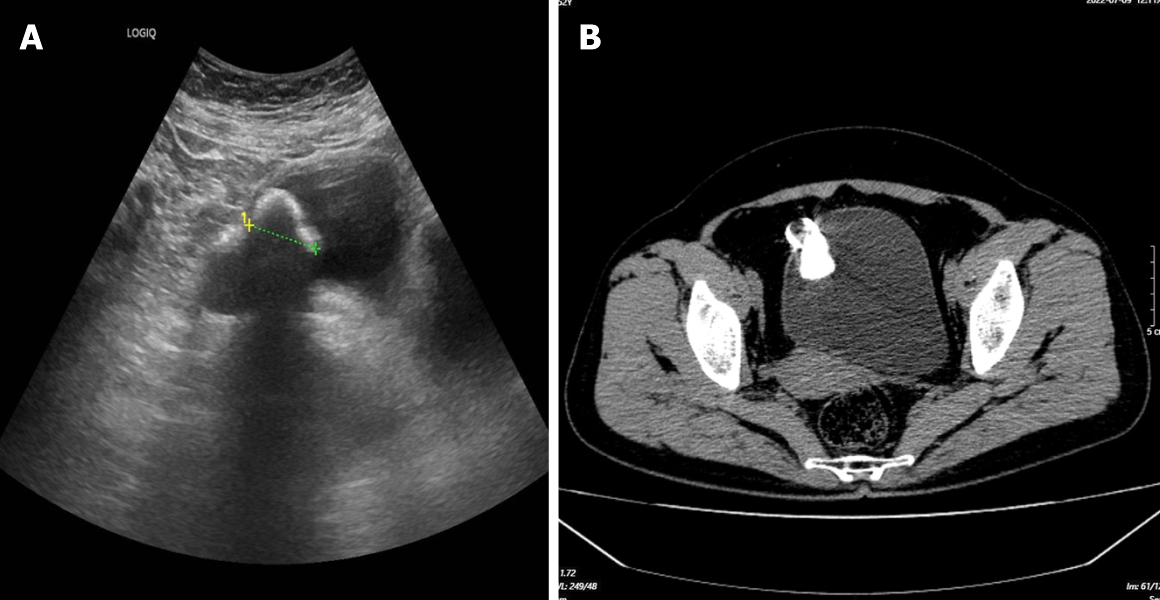
Bladder ultrasound and pelvic CT revealed a ring-shaped foreign body in the anterior wall of urinary bladder (Figure 1), an ectopic IUD be considered.
Considering the patient’s history and laboratory and imaging findings, the patient was diagnosed with ectopic IUD in urinary bladder.
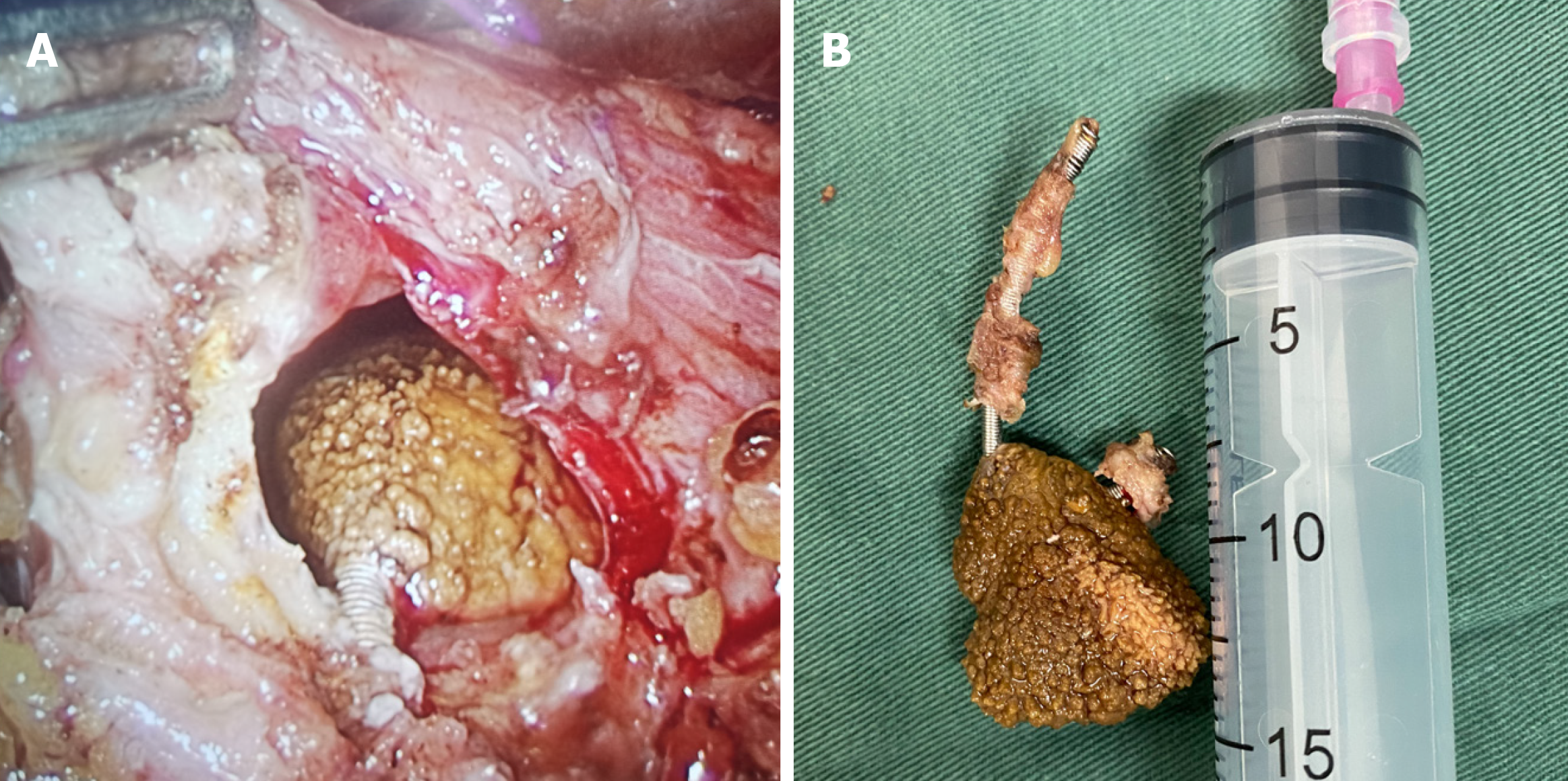
The surgery is carried out under a general anesthesia. Laparoscopic surgery was performed after the intestinal tract and fallopian tube adhesions were separated, an annular IUD embedded into the muscularis was found in the right anterior wall of urinary bladder, and an oval calculus with a diameter of approximately 2 cm was attached to the surface of the bladder cavity (Figure 2A). The IUD and calculus were successfully and completely removed, and the bladder wall was sutured to the full thickness (Figure 2B).
A urinary indwelling catheter was used after the operation, and antibiotics were administered to prevent infection. The patient was discharged with a urinary catheter after 3 d of operation. The patient returned to the hospital 2 wk later for removal of the urinary catheter. The patient urinated normally without vaginal bleeding, abdominal pain or other dis
There are many reasons for ectopic IUD: (1) Due to an unskilled operator, the position and size of the uterus are not accurately judged, and the direction of the probe or placer into the uterine cavity is wrong; alternatively, the operator is too rough and fails to pay attention when encountering resistance at the bending region and the probe is forced through, resulting in uterine wall damage, perforation and ectopic IUD[7]; (2) When placing an IUD on a woman with a scarred uterus, the scar tissue is easily damaged due to the lack of elasticity of connective tissue at the incision in the uterus, and carelessness can cause ectopic IUD. In addition, when women enter the perimenopausal period, the uterine cavity begins to shrink, which can cause the IUD to deform and embed into the myometrium; the longer the menopausal period is, the more serious the uterine atrophy and the greater the incidence of IUD incarceration[7]; and (3) The size and shape of the IUD do not match the shape of the uterine cavity.
The incidence of IUD penetrating the uterine wall is less than 1/1000, and there are few reports indicating IUD in the urinary bladder, especially in the anterior wall[8]. The location of the IUD in the urinary bladder may be associated with the chronic effect of the IUD in the uterus, which can lead to the formation of an inflammatory reaction, through which the IUD penetrates the uterine wall and contacts the bladder wall to cause inflammatory adhesion, resulting in the erosion and rupture of the bladder wall and the entry of the IUD into the urinary bladder. In addition, this difference may also be related to the intense exercise of patients after IUD implantation. The ectopic IUD in this patient was caused by chronic inflammation, and the IUD was located in the anterior wall of the urinary bladder. Calculus had formed on the surface due to chronic inflammation.
The diagnosis of ectopic IUD is relatively simple. Transvaginal and abdominal ultrasound is an effective method for identifying ectopic IUD[9]. Abdominal X-ray imaging is very effective for diagnosing metal IUDs, but it is unable to lo
When it is confirmed that the IUD is located in the urinary bladder, surgery should be performed as soon as possible. At present, there are cystoscopy, hysteroscopy combined with cystoscopy, laparoscopy and open surgery. The choice of surgical method is determined by comprehensively considering the depth of the IUD in the bladder muscle layer, the si
In conclusion, we reported a case of an ectopic IUD in the anterior wall of urinary bladder that was documented 20 years after IUD insertion. Abdominal ultrasound and CT are effective methods for detecting ectopic IUDs. The IUD is located in the urinary bladder and requires early surgical treatment. At present, there are cystoscopy, hysteroscopy combined with cystoscopy, laparoscopy and open surgery. The choice of surgical method is determined by comprehensively considering the position of the IUD in the urinary bladder, the size of the calculus, and the situation of intravesical inflammation.
| 1. | Averbach SH, Ermias Y, Jeng G, Curtis KM, Whiteman MK, Berry-Bibee E, Jamieson DJ, Marchbanks PA, Tepper NK, Jatlaoui TC. Expulsion of intrauterine devices after postpartum placement by timing of placement, delivery type, and intrauterine device type: a systematic review and meta-analysis. Am J Obstet Gynecol. 2020;223:177-188. [RCA] [PubMed] [DOI] [Full Text] [Cited by in Crossref: 23] [Cited by in RCA: 49] [Article Influence: 9.8] [Reference Citation Analysis (0)] |
| 2. | Bahamondes MV, Bahamondes L. Intrauterine device use is safe among nulligravidas and adolescent girls. Acta Obstet Gynecol Scand. 2021;100:641-648. [RCA] [PubMed] [DOI] [Full Text] [Cited by in Crossref: 5] [Cited by in RCA: 11] [Article Influence: 2.8] [Reference Citation Analysis (0)] |
| 3. | Beltz AM, Demidenko MI, Chaku N, Klump KL, Joseph JE. Intrauterine Device Use: A New Frontier for Behavioral Neuroendocrinology. Front Endocrinol (Lausanne). 2022;13:853714. [RCA] [PubMed] [DOI] [Full Text] [Full Text (PDF)] [Cited by in Crossref: 5] [Cited by in RCA: 8] [Article Influence: 2.7] [Reference Citation Analysis (0)] |
| 4. | Levin G, Dior UP, Gilad R, Benshushan A, Shushan A, Rottenstreich A. Pelvic inflammatory disease among users and non-users of an intrauterine device. J Obstet Gynaecol. 2021;41:118-123. [RCA] [PubMed] [DOI] [Full Text] [Cited by in Crossref: 6] [Cited by in RCA: 2] [Article Influence: 0.4] [Reference Citation Analysis (0)] |
| 5. | Berzenji L, Hosten L, Van Schil P. The lost intrauterine device. Eur J Cardiothorac Surg. 2021;60:1477. [RCA] [PubMed] [DOI] [Full Text] [Reference Citation Analysis (0)] |
| 6. | Wang L, Wang H, Yuan M, Lu Q. Ileal bladder fistula following ectopic intrauterine contraceptive device. Asian J Surg. 2020;43:1117-1118. [RCA] [PubMed] [DOI] [Full Text] [Reference Citation Analysis (0)] |
| 7. | Long S, Colson L. Intrauterine Device Insertion and Removal. Prim Care. 2021;48:531-544. [RCA] [PubMed] [DOI] [Full Text] [Cited by in Crossref: 1] [Cited by in RCA: 8] [Article Influence: 2.0] [Reference Citation Analysis (0)] |
| 8. | Yu HT, Chen Y, Xie YP, Gan TB, Gou X. Ectopic intrauterine device in the bladder causing cystolithiasis: A case report. World J Clin Cases. 2022;10:3194-3199. [RCA] [PubMed] [DOI] [Full Text] [Full Text (PDF)] [Cited by in RCA: 4] [Reference Citation Analysis (1)] |
| 9. | Simó Alari F, Gutierrez I. Intrauterine device extraction through laparoscopic hysterotomy. Eur J Contracept Reprod Health Care. 2021;26:261-263. [RCA] [PubMed] [DOI] [Full Text] [Cited by in Crossref: 1] [Cited by in RCA: 1] [Article Influence: 0.3] [Reference Citation Analysis (0)] |
| 10. | Liu G, Li F, Ao M, Huang G. Intrauterine devices migrated into the bladder: two case reports and literature review. BMC Womens Health. 2021;21:301. [RCA] [PubMed] [DOI] [Full Text] [Full Text (PDF)] [Cited by in RCA: 19] [Reference Citation Analysis (0)] |


