Editorial Open Access
Copyright ©The Author(s) 2024. Published by Baishideng Publishing Group Inc. All rights reserved.
World J Clin Cases. Jun 16, 2024; 12(17): 2921-2924
Published online Jun 16, 2024. doi: 10.12998/wjcc.v12.i17.2921
Revolutionizing disease diagnosis and management: Open-access magnetic resonance imaging datasets a challenge for artificial intelligence driven liver iron quantification
Jaber H Jaradat, Faculty of Medicine, Mutah University, Al-Karak 61101, Jordan
Abdulqadir J Nashwan, Department of Nursing, Hamad Medical Corporation, Doha 3050, Qatar
ORCID number: Jaber H Jaradat (0000-0002-6488-4664); Abdulqadir J Nashwan (0000-0003-4845-4119).
Author contributions: Jaradat JH and Nashwan AJ contributed to the manuscript's conceptualization, writing, editing, and literature review. All authors have read and approved the final manuscript.
Conflict-of-interest statement: All the authors report no relevant conflicts of interest for this article.
Open-Access: This article is an open-access article that was selected by an in-house editor and fully peer-reviewed by external reviewers. It is distributed in accordance with the Creative Commons Attribution NonCommercial (CC BY-NC 4.0) license, which permits others to distribute, remix, adapt, build upon this work non-commercially, and license their derivative works on different terms, provided the original work is properly cited and the use is non-commercial. See: https://creativecommons.org/Licenses/by-nc/4.0/
Corresponding author: Abdulqadir J Nashwan, MSc, Research Scientist, Department of Nursing, Hamad Medical Corporation, Rayyan Road, Doha 3050, Qatar. anashwan@hamad.qa
Received: February 26, 2024
Revised: April 4, 2024
Accepted: April 18, 2024
Published online: June 16, 2024
Processing time: 98 Days and 22.4 Hours

Abstract

Artificial intelligence (AI), particularly machine learning (ML) and deep learning (DL) techniques, such as convolutional neural networks (CNNs), have emerged as transformative technologies with vast potential in healthcare. Body iron load is usually assessed using slightly invasive blood tests (serum ferritin, serum iron, and serum transferrin). Serum ferritin is widely used to assess body iron and drive medical management; however, it is an acute phase reactant protein offering wrong interpretation in the setting of inflammation and distressed patients. Magnetic resonance imaging is a non-invasive technique that can be used to assess liver iron. The ML and DL algorithms can be used to enhance the detection of minor changes. However, a lack of open-access datasets may delay the advancement of medical research in this field. In this letter, we highlight the importance of standardized datasets for advancing AI and CNNs in medical imaging. Despite the current limitations, embracing AI and CNNs holds promise in revolutionizing disease diagnosis and treatment.

Key Words: Liver diseases; Magnetic resonance imaging; Iron quantification; Machine learning; Deep learning

Core Tip: This editorial emphasizes the revolutionary impact of artificial intelligence (AI), particularly machine learning and deep learning techniques like convolutional neural networks (CNNs), in healthcare. Highlighting the limitations of traditional, slightly invasive blood tests for assessing body iron load, it advocates for magnetic resonance imaging 's non-invasive advantages. The editorial underscores the critical role of AI and CNNs in improving disease diagnosis and treatment, especially through the precise detection of minor changes in liver iron levels. However, it points out the significant hurdle of lacking open-access datasets, which hampers medical research progress. The call for standardized datasets is a crucial step towards leveraging AI in medical imaging, promising to transform patient care and management.


INTRODUCTION

Artificial intelligence, the simulation of human intelligence by machines, is an emerging transformative technology in various fields, particularly healthcare. Machine learning (ML) and deep learning (DL) are two major classes of artificial intelligence (AI) that have revolutionized data analysis and decision-making. Convolutional neural networks (CNNs) are a subclass of DL, in which they are designed to learn spatial hierarchies of features adaptively and automatically. In healthcare, the availability of large amounts of data of all types, such as text, audio, images, and videos, makes the application of AI in this field promising and effective. CNNs are increasingly used to interpret medical images, including magnetic resonance imaging (MRI) scans, with remarkable accuracy and efficiency[1].

The growing capabilities of AI and CNNs offer immense potential for reducing human error and capturing minor and trivial differences that are difficult for a well-trained expert radiologist to capture and improve patient care, diagnosis, and treatment across various medical specialties[1].

Despite their increasing capabilities, it is essential to acknowledge the current limitations of AI in healthcare, such as interpretability, data privacy concerns, and the need for large and diverse datasets to train algorithms[1]. In medicine, large datasets are not an obstacle or concern because of the tremendous amount of patient data available and accessible to institutions. However, the challenge lies in curating and preparing the data and making it accessible to researchers (open access).

IRON QUANTIFICATION

Iron body quantification and measurement are usually performed via slightly invasive methods, such as routine blood tests (serum ferritin, serum iron, and transferrin saturation)[2]. Serum ferritin is an acute phase reactant protein, elevated in inflammation and distressed patients, therefore, it might give wrong impression about body iron[2,3]. Noninvasive procedures are increasingly utilized for quantifying and assessing body iron load, and MRI is the best noninvasive examination of the liver for quantifying iron in iron-overloaded patients and helps in the early detection of liver fibrosis, liver cirrhosis, and liver cancer induced by iron overload[4]. MRI is a major diagnostic imaging modality that has been applied and leveraged for the diagnosis and detection of various diseases. It is characterized by high structural specificity and many different windows, each of which is used to visualize different structural differences, some of which are computer reconstructions[4]. However, routine blood tests are widely used owing to their cost-effectiveness compared to MRI, which has low sensitivity and specificity compared to MRI in sick patients[2]. MRI remains a valuable tool for disease diagnosis and research because of its safety and noninvasive nature. However, its high cost and accessibility in some regions may make it the second choice in areas with limited resources[5].

MRI BASED DISEASE DETECTION

Evidence drawn from the literature on the use of AI for iron quantification and the detection of various liver diseases is insufficient. However, current literature shows promising results regarding iron quantification using MRI, and various models and algorithms have been used[3]. Furthermore, MRI has several windows, each focusing on certain body structures; however, it is still uncertain which MRI window is the most accurate for training ML and DL algorithms[5]. Compared to AI and ML methods, CNNs offer advantages in terms of accuracy, efficiency, and automation, significantly enhancing the diagnostic process and improving patient outcomes[1].

CALL FOR OPEN-ACESS LIVER MRI DATASETS

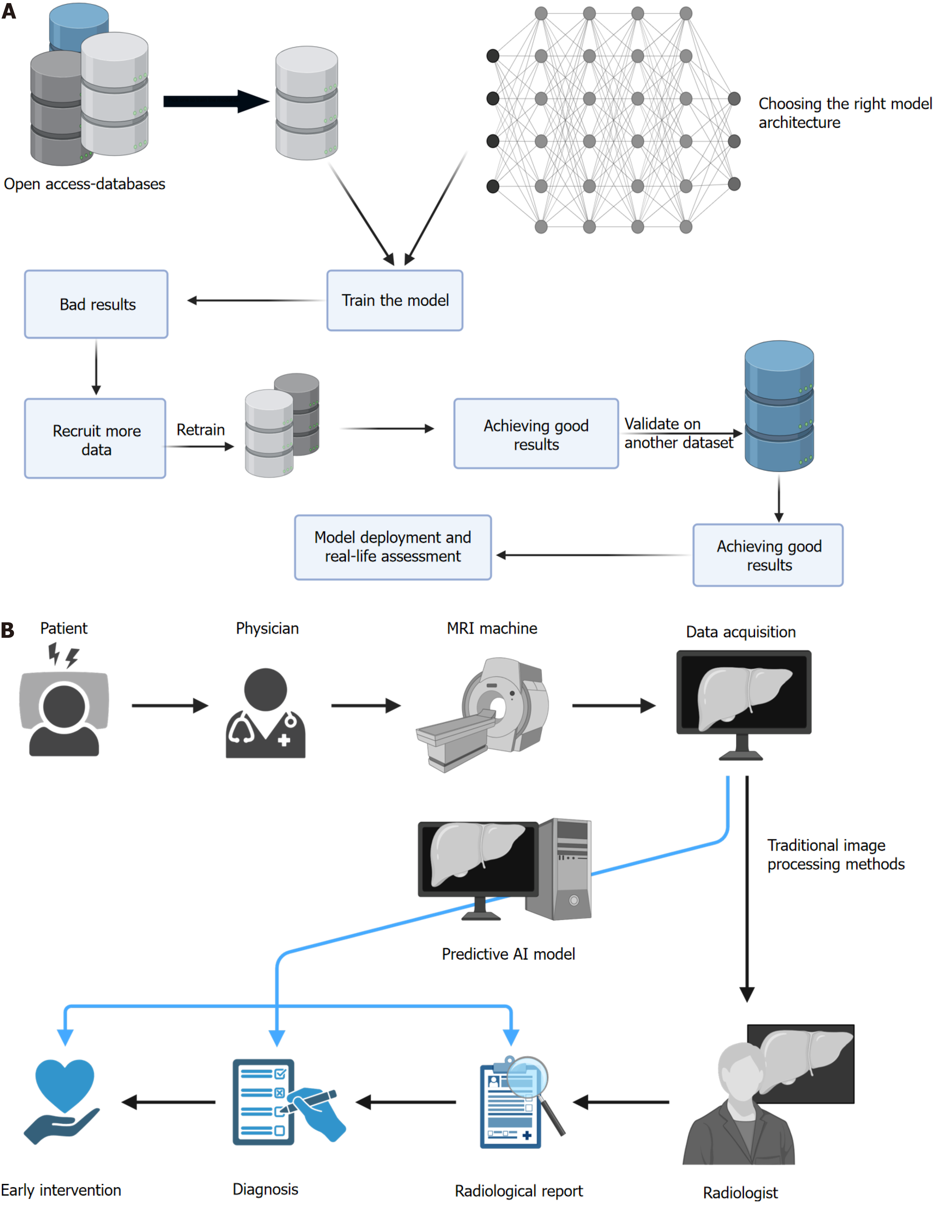
To harness the full potential of CNNs and various ML algorithms in iron quantification and disease diagnosis, there is a demand for the creation and public release of standardized liver MRI datasets (see Figure 1). These datasets will not only accelerate research, but also improve diagnosis and ultimately benefit patients with iron overload disorders. By advocating the development and sharing of such datasets, we can advance healthcare and patient care, while maximizing the potential of AI and CNNs in medical imaging. Moreover, the potential of CNNs and ML algorithms in assessing body iron load extends beyond the liver, with opportunities to apply similar techniques to other organs, such as the heart, for iron quantification.

Figure 1
Figure 1 Illustrates the general benefits of utilizing open-access datasets. A: Initially, we had multiple datasets (three datasets in total); however, using the first dataset alone proved to be insufficient for training a satisfactory model, therefore, we incorporated another dataset into the training data. Following this, we evaluated the model on an external dataset to confirm the efficacy of the model, yielding a good result. The model is then transferred to the deployment stage; B: Clinical testing of the model and if proven to be effective, it is started to be tested on a larger scale, and lastly used in the computer-aided-diagnosis. MRI: Magnetic resonance imaging; AI: Artificial intelligence. This figure was created with BioRender.com.

Ferritin may provide false assessments of body iron levels in distressed patients. Well-documented large-scale datasets covering various MRI windows. Large-scale studies exploring various AI algorithms for iron quantification using MRI are required. Machine learning and deep learning have revolutionized disease diagnosis and treatment; therefore, they must be trained on large-scale multi-country, well-curated datasets.

CONCLUSION

In conclusion, the integration of AI and CNNs in healthcare, particularly medical imaging, presents a significant opportunity to improve patient care and advance medical research. While a major challenge of such technologies is dataset availability and accessibility, open-access liver MRI datasets for iron quantification are lacking. Therefore, we advocate the creation of large, multi-country datasets to harness the full potential of these technologies in disease diagnosis, treatment, and patient outcomes.

Footnotes

Provenance and peer review: Invited article; Externally peer reviewed.

Peer-review model: Single blind

Specialty type: Medicine, research and experimental

Country of origin: Qatar

Peer-review report’s classification

Scientific Quality: Grade B

Novelty: Grade B

Creativity or Innovation: Grade B

Scientific Significance: Grade B

P-Reviewer: Wang TJ, China S-Editor: Liu H L-Editor: A P-Editor: Xu ZH

References
1.  Yamashita R, Nishio M, Do RKG, Togashi K. Convolutional neural networks: an overview and application in radiology. Insights Imaging. 2018;9:611-629.  [PubMed]  [DOI]  [Cited in This Article: ]  [Cited by in Crossref: 1202]  [Cited by in F6Publishing: 1067]  [Article Influence: 177.8]  [Reference Citation Analysis (0)]
2.  Hernando D, Levin YS, Sirlin CB, Reeder SB. Quantification of liver iron with MRI: state of the art and remaining challenges. J Magn Reson Imaging. 2014;40:1003-1021.  [PubMed]  [DOI]  [Cited in This Article: ]  [Cited by in Crossref: 163]  [Cited by in F6Publishing: 193]  [Article Influence: 19.3]  [Reference Citation Analysis (0)]
3.  Nashwan AJ, Alkhawaldeh IM, Shaheen N, Albalkhi I, Serag I, Sarhan K, Abujaber AA, Abd-Alrazaq A, Yassin MA. Using artificial intelligence to improve body iron quantification: A scoping review. Blood Rev. 2023;62:101133.  [PubMed]  [DOI]  [Cited in This Article: ]  [Cited by in Crossref: 1]  [Reference Citation Analysis (0)]
4.  Feng Q, Yi J, Li T, Liang B, Xu F, Peng P. Narrative review of magnetic resonance imaging in quantifying liver iron load. Front Med (Lausanne). 2024;11:1321513.  [PubMed]  [DOI]  [Cited in This Article: ]  [Reference Citation Analysis (0)]
5.  Tziomalos K, Perifanis V. Liver iron content determination by magnetic resonance imaging. World J Gastroenterol. 2010;16:1587-1597.  [PubMed]  [DOI]  [Cited in This Article: ]  [Cited by in CrossRef: 33]  [Cited by in F6Publishing: 30]  [Article Influence: 2.1]  [Reference Citation Analysis (0)]