Published online Jun 6, 2024. doi: 10.12998/wjcc.v12.i16.2831
Revised: February 15, 2024
Accepted: April 9, 2024
Published online: June 6, 2024
Processing time: 192 Days and 23.3 Hours
Computed tomography (CT) small bowel three-dimensional (3D) reconstruction is a powerful tool for the diagnosis of small bowel disease and can clearly show the intestinal lumen and wall as well as the outside structure of the wall. The hori
Bleeding caused by small intestinal polyps is often difficult to diagnose in clinical practice. This study reports a 29-year-old male patient who was admitted to the hospital with black stool and abdominal pain for 3 months. Using the combination of CT-3D reconstruction and capsule endoscopy, the condition was diagnosed correctly, and the polyps were removed using single-balloon enteroscopy-endoscopic retrograde cholangiopancreatography without postoperative complications.
The role of CT-3D in gastrointestinal diseases was confirmed. CT-3D can assist in the diagnosis and treatment of gastrointestinal diseases in combination with capsule endoscopy and small intestinal microscopy.
Core Tip: The computed tomography three-dimensional reconstruction can assist the capsule endoscopy-assisted single-balloon enteroscopy of the small bowel for the diagnosis and treatment of difficult-to-diagnose small bowel polyps.
- Citation: Zhang SH, Fan MW, Chen Y, Hu YB, Liu CX. Computed tomography three-dimensional reconstruction in the diagnosis of bleeding small intestinal polyps: A case report. World J Clin Cases 2024; 12(16): 2831-2836
- URL: https://www.wjgnet.com/2307-8960/full/v12/i16/2831.htm
- DOI: https://dx.doi.org/10.12998/wjcc.v12.i16.2831
We report a 29-year-old male patient who was diagnosed giant polyp of small intestine through the computed tomo
The study presents the case of a 29-year-old male, who was admitted to Binzhou Medical University Hospital, Binzhou, Shandong, China with abdominal pain and black stools for 3 months. The abdominal pain was particularly concentrated around the umbilical cord and presented as paroxysmal colic. The pain could be slightly relieved after defecation; however, the patient also reported abdominal distension and fatigue.
The patient has a history of long-term smoking but no history of alcoholism, coffee or strong tea consumption, steroidal anti-inflammatory drug use, abdominal trauma or operation.
The patient had a history of recurrent diarrhea from the past 10 years ago but no history of infectious diseases, such as tuberculosis, viral hepatitis, or contact.
The patient denied a family history of a similar disease. The patient had a history of long-term smoking but no history of alcoholism, coffee or strong tea consumption, steroidal anti-inflammatory drug use, abdominal trauma, or operation.
The patient received treatment at a local hospital three months ago. His blood routine results showed a red blood cell (RBC) level of 3.01 × 10¹²/L and an Hb level of 86 g/L. Using gastroscopy and colonoscopy procedures at the local hospital, only chronic non-atrophic gastritis and terminal ileitis were identified. However, these findings did not provide a clear explanation for the gastrointestinal bleeding. The patient was discharged following conservative treatment. However, after discharge, the patient continued to experience dark red stools, accompanied by abdominal pain, distension, fatigue, and even fainted once.
Consequently, the patient sought treatment at the gastroenterology department of our hospital. The results of the patient's blood test showed RBC level of 4.6 × 10¹²/L, Hb level of 121 g/L, mean cell volume level of 80 fL, mean corpuscular hemoglobin level of 26 pg, mean cellular hemoglobin concentration level of 328 g/L, reticulocyte (RET) level of 0.026, and RET percentage of 0.6%. The patient lost approximately 3 kg weight within 3 months and appeared emaciated.
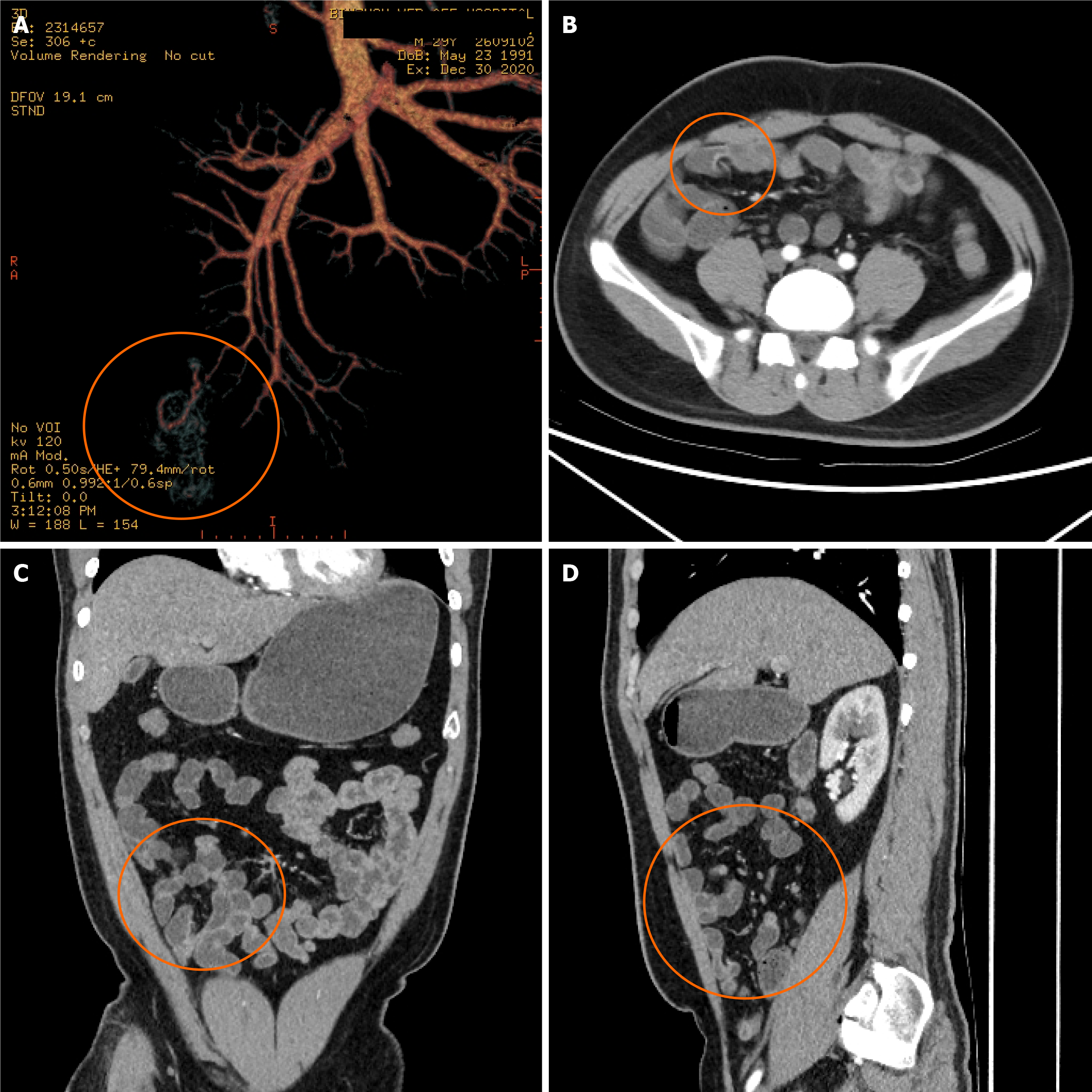
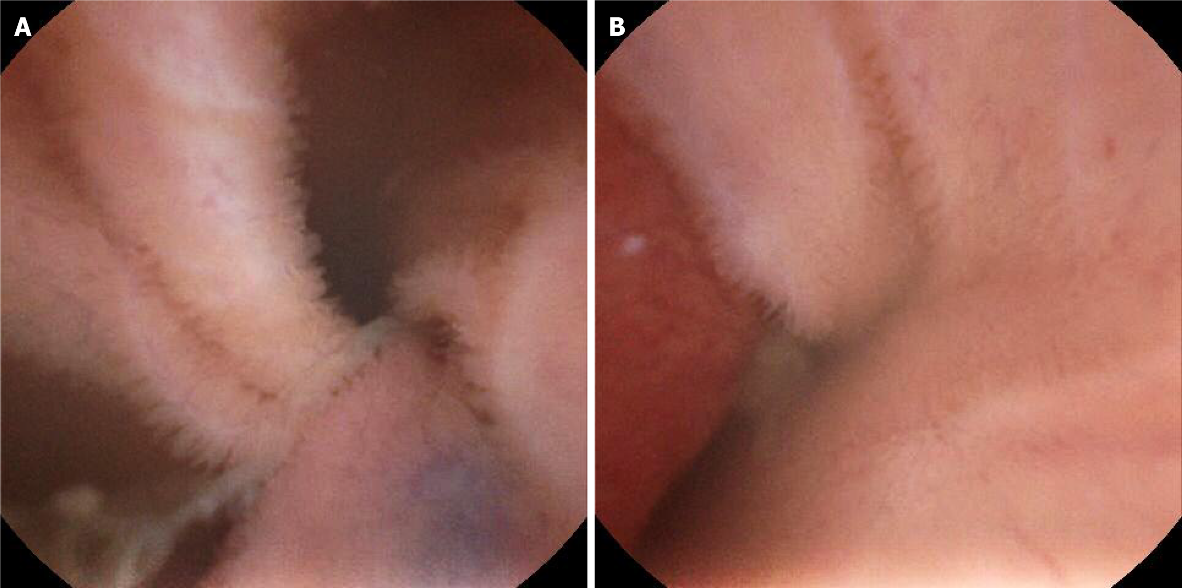
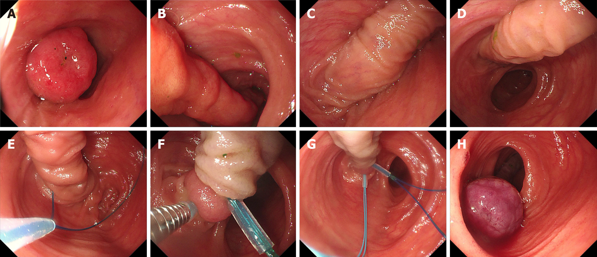
The location of the lesion was suspected to be in the small intestine, and the possible causes included small intestinal stromal tumors and congenital small intestinal vascular malformation. We recommend surgical exploration or enteroscopy for further diagnosis. However, the patient declined surgical exploration and enteroscopy due to concerns about the invasiveness of the procedures. Therefore, we initially conducted a CT angiography of the small intestine to assess the patient's condition. The 3D reconstruction of the small intestine revealed malformations, changes in the ileal wall and mesenteric vessels, formation of the space-occupying lesions, and a large polyp with significant blood vessels passing through the ileum (Figure 1). The patient's small intestine was examined using a magnetron capsule endoscope to observe the shape of the tumor and evaluate its location. The results indicated that the capsule endoscope was in the ileum for approximately 4 h and 51 min. A large protuberant lesion with hyperemia and swelling was observed (Figure 2). Based on the results of small intestinal CT-3D reconstruction and magnetron capsule endoscopy, it was concluded that the gastrointestinal bleeding was caused by a tumor located at the end of the ileum. The patient’s condition was explained to him and his family, and he agreed to undergo enteroscopy and endoscopic treatment of the tumor. After careful consideration, transoral SBE was used to examine the jejunum and upper ileum, which showed no obvious abnormalities. Subsequently, trans-anal SBE was used to identify a large, pedicled tumor measuring approximately 2 cm × 2 cm with erosion at the top (Figure 3A-D).
Based on the patient's symptoms, signs, and gastroenteroscopy results at the local hospital, we believe the patient is experiencing gastrointestinal bleeding.
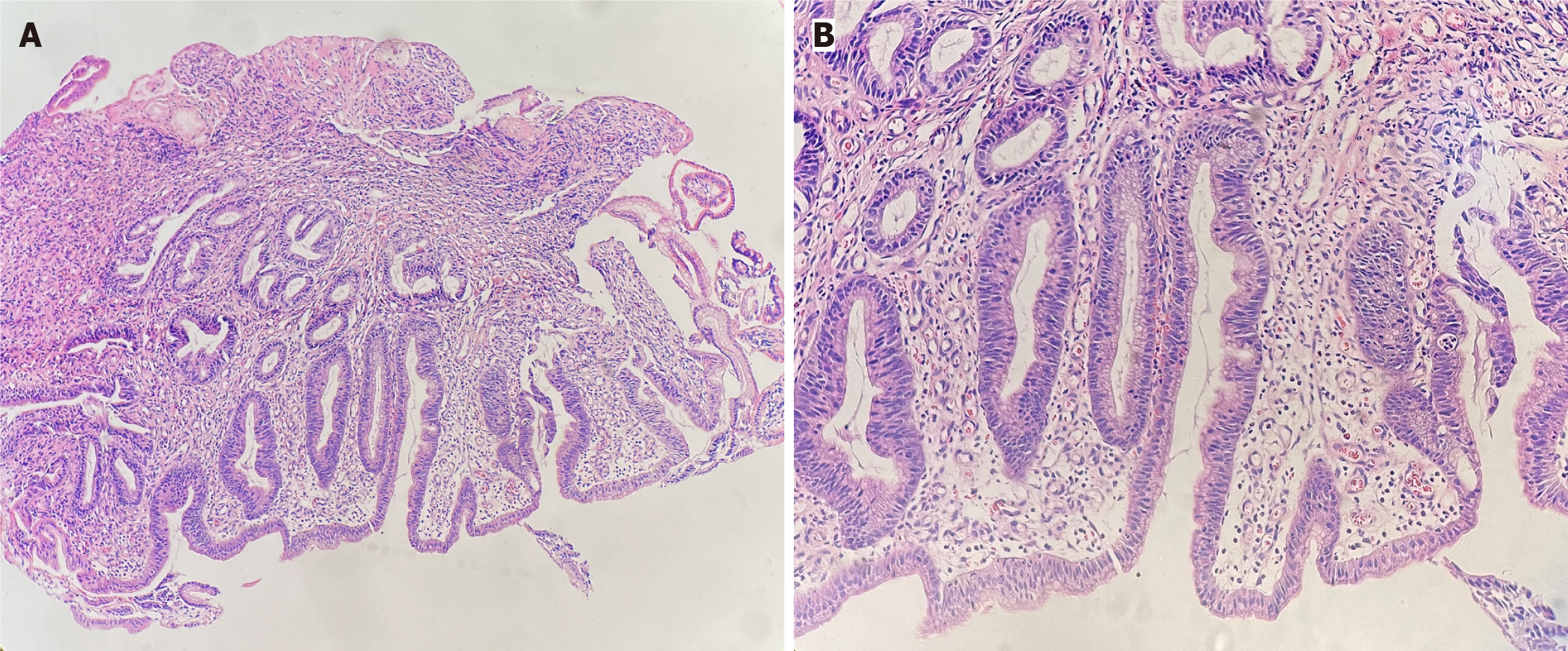
Combined with the CT results of the small intestine, the tumor might cause significant bleeding and perforation by passing through the large blood vessels of ileal polyps and directly through the endoscopic mucosal exfoliation. Therefore, after careful consideration, we chose to perform nylon trap ligation. Once the tumor was completely exposed, two nylon rings were placed at the root of the tumor to make the head of the polyp purplish blue (Figure 3E-H). The tissue biopsy confirmed that the tumor was an adenomatous polyp with low-grade intraepithelial neoplasia, high-grade inflammatory cell infiltration, and local granulation tissue hyperplasia (Figure 4).
The patient did not experience hematochezia after the operation, and his abdominal pain was less severe as compared to that at the time of admission. A week later, the patient underwent a blood routine examination, which showed an Hb level of 143 g/L, indicating effective control of gastrointestinal bleeding. One year later, the patient was readmitted to the hospital for re-examination. The blood routine showed a hemoglobin level of 148 g/L. During the trans-anal enteroscopy, circular scar-like changes were found in the ileocecal valve at 30 cm. This suggested that small intestinal polyps might undergo necrosis and exfoliation after ligation with a nylon ring.
Small intestinal bleeding is a common cause of gastrointestinal bleeding; however, its diagnosis is difficult due to the special shape of the small intestine and the nature of the bleeding lesions[1]. Small bowel polyps are common types of intestinal lesions, which cause small bowel bleeding. The direct resection of the polyp using endoscopic submucosal dissection has been commonly used for the treatment of small intestinal polyps[2]. The traditional small bowel microscopy does not allow for the diagnosis of small bowel bleeding. However, the capsule endoscopy allows for a non-invasive and safe examination of the entire small bowel but requires imaging technology to assist in the examination[3]. The continuous advancements in CT-3D reconstruction technology have led to more accurate and efficient localization of gastrointestinal lesions and can better display the spatial morphology of the small intestine, thereby aiding in the diagnosis of small intestinal diseases[4]. The combination of CT-3D reconstruction with capsule endoscopy can better diagnose small intestinal lesions[5].
Small intestine diseases typically have an insidious onset, and the patients' clinical symptoms are often atypical. Due to the curvature of the small bowel, the intestinal tubes overlap each other, resulting in deep and irregular lesions that are easy to miss and misdiagnose. Therefore, diagnosing small intestine diseases can be challenging. Currently, gastroscopy and colonoscopy are unable to reach the gastrointestinal tract, and capsule endoscopy is uncontrolled and cannot repeatedly observe the lesions in multiple directions; moreover, small intestine microscopy is difficult to operate. Posting the operators' time and energy can be risky and has drawbacks[6]. CT-3D is a non-invasive imaging examination that is highly valuable for diagnosing intestinal tumors, gastrointestinal bleeding, recurrent diarrhea, and other conditions. It helps clinicians locate lesions and choose appropriate endoscopic treatment modalities, thereby reducing the need for multiple endoscopic examinations, alleviating patient pain, and lowering the risk and economic burden of operations.
In conclusion, this study presents a case of a giant terminal ileal polyp, which was diagnosed and treated using capsule endoscopy combined with CT-3D reconstruction and nylon snare ligation under enteroscopy due to bleeding. This study proved that the capsule endoscopy and CT-3D reconstruction of the small intestine might provide valuable clues for the diagnosis and treatment of small intestinal diseases.
Provenance and peer review: Unsolicited article; Externally peer reviewed.
Peer-review model: Single blind
Specialty type: Medicine, General and Internal
Country/Territory of origin: China
Peer-review report’s classification
Scientific Quality: Grade C, Grade C, Grade D
Novelty: Grade B, Grade C, Grade C
Creativity or Innovation: Grade B, Grade C, Grade C
Scientific Significance: Grade B, Grade C, Grade C
P-Reviewer: Bernardes A, Portugal; Chen CH, United States S-Editor: Che XX L-Editor: A P-Editor: Zhao S
| 1. | Lewis BS. Small intestinal bleeding. Gastroenterol Clin North Am. 2000;29:67-95, vi. [PubMed] |
| 2. | Otani K, Watanabe T, Shimada S, Hosomi S, Nagami Y, Tanaka F, Kamata N, Taira K, Yamagami H, Tanigawa T, Shiba M, Fujiwara Y. Clinical Utility of Capsule Endoscopy and Double-Balloon Enteroscopy in the Management of Obscure Gastrointestinal Bleeding. Digestion. 2018;97:52-58. [RCA] [PubMed] [DOI] [Full Text] [Cited by in Crossref: 44] [Cited by in RCA: 39] [Article Influence: 5.6] [Reference Citation Analysis (0)] |
| 3. | Lao IW, Wang J. Adenomatoid tumor of the small intestine: the first case report and review of the literature. Int J Surg Pathol. 2014;22:727-730. [RCA] [PubMed] [DOI] [Full Text] [Cited by in Crossref: 3] [Cited by in RCA: 3] [Article Influence: 0.3] [Reference Citation Analysis (0)] |
| 4. | Xue P, Chen X, Chen S, Shi Y. The Value of CT 3D Reconstruction in the Classification and Nursing Effect Evaluation of Ankle Fracture. J Healthc Eng. 2021;2021:9596518. [RCA] [PubMed] [DOI] [Full Text] [Full Text (PDF)] [Cited by in RCA: 3] [Reference Citation Analysis (0)] |
| 5. | Khan AH, Sohag MHA, Vedaei SS, Mohebbian MR, Wahid KA. Automatic Detection of Intestinal Bleeding using an Optical Sensor for Wireless Capsule Endoscopy. Annu Int Conf IEEE Eng Med Biol Soc. 2020;2020:4345-4348. [RCA] [PubMed] [DOI] [Full Text] [Cited by in Crossref: 2] [Cited by in RCA: 1] [Article Influence: 0.2] [Reference Citation Analysis (0)] |
| 6. | Wei Y, Liu F. [Research status and progress in polyp size measurement under endoscopy]. Zhonghua Xiaohua Neijing Zazhi. 2023;40:234-237. [DOI] [Full Text] |


