Published online Feb 6, 2023. doi: 10.12998/wjcc.v11.i4.918
Peer-review started: September 27, 2022
First decision: December 13, 2022
Revised: December 13, 2022
Accepted: January 12, 2023
Article in press: January 12, 2023
Published online: February 6, 2023
Processing time: 131 Days and 19.4 Hours
Although fluorodeoxyglucose-positron emission tomography/computed tomography (FDG-PET/CT) is widely used for diagnosis and follow-up of large sized vessel vasculitis, it is still not widely used for small to medium sized vessel vasculitis.
This is the case of a 68-year-old male who presented at the emergency department complaining of fever, myalgia, and bilateral leg pain of over two weeks duration, with elevated levels of C-reactive protein. He was subsequently admitted and despite the absence of clinically significant findings, the patient continued to exhibit recurrent fever. A fever of unknown origin workup, which included imaging studies using FDG-PET/CT, revealed vasculitis involving small to medium-sized vessels of both lower extremities, demonstrated by linear hypermetabolism throughout the leg muscles. The patient was treated with methylprednisolone and methotrexate after diagnosis leading to the gradual resolution of the patient’s symptoms. Three weeks later, a follow-up FDG-PET/CT was performed. Previously hypermetabolic vessels were markedly improved.
Our case report demonstrated that FDG-PET/CT has tremendous potential to detect medium-sized vessel inflammation; it can also play a crucial role in prognosticating outcomes and monitoring therapeutic efficacy.
Core Tip: The fluorodeoxyglucose-positron emission tomography/computed tomography can be an option to diagnose small to medium-sized vessel vasculitis and follow-up to assess on the extent and improvement of inflammation in patients with polyarteritis nodosa.
- Citation: Kang JH, Kim J. Polyarteritis nodosa presenting as leg pain with resolution of positron emission tomography-images: A case report. World J Clin Cases 2023; 11(4): 918-921
- URL: https://www.wjgnet.com/2307-8960/full/v11/i4/918.htm
- DOI: https://dx.doi.org/10.12998/wjcc.v11.i4.918
Polyarteritis nodosa (PAN) is characterized by systemic necrotizing vasculitis which can involve medium-sized vessels. This vasculitis is usually difficult to diagnose, thus, imaging study including fluorodeoxyglucose-positron emission tomography/ computed tomography (FDG-PET/CT) would be a possible role to identify this disease.
A 68-year-old male visited to emergency department complaining fever, myalgia, and both leg pain during more than two weeks.
Although he was treated with administered ceftriaxone and metronidazole in other hospital for a week.
There was no specific past illness.
There was no specific personal and family history.
However, his C-reactive protein (CRP) was still high (37.71 mg/dL) and complaining symptoms such as fever, both leg pain was still remained. Therefore, he was admitted to our hospital for further assessment.
His blood, urine culture findings were all negative. And his serologic results such as hepatitis viral marker, rheumatoid factor, anti-cyclic citrullinated peptide antibody, antineutrophil cytoplasmic antibody and anti-nuclear antibody were negative. Because he complained daily fever after admission, fever of unknown origin work up was needed.
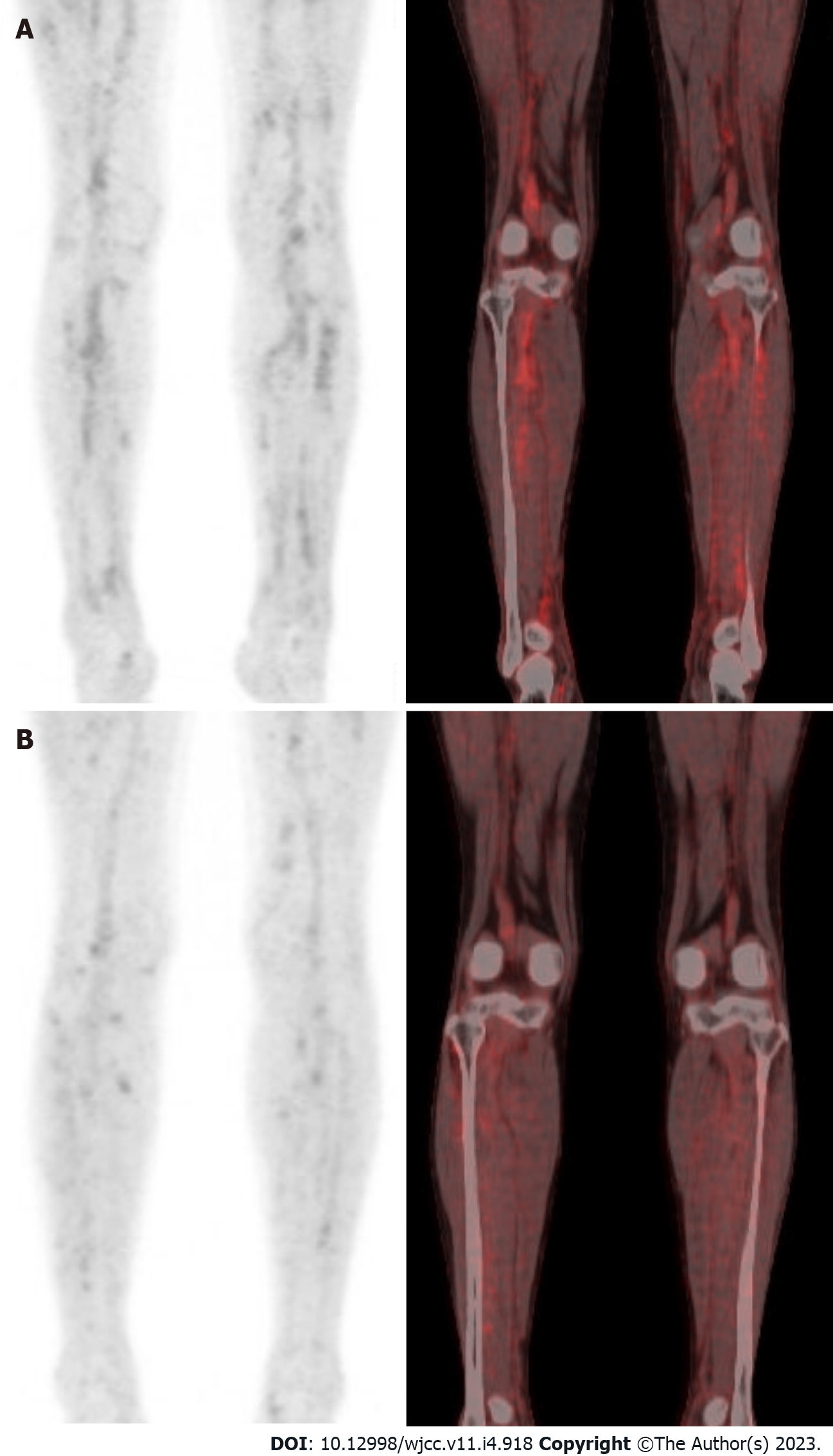
There were no clinically significant findings without two small hemangiomas in the liver on contrast enhanced computed tomography in whole body including neck, chest, and abdomen-pelvic cavity. FDG-PET/CT on day 5 showed vasculitis involving small to medium vessels of both lower extremities by showing somewhat linear hypermetabolism through the muscles (Figure 1).
Finally, he was diagnosed PAN according to criteria by showing satisfied with unexplained more than 4 kg of weight loss, myalgia, new onset more than 90 mmHg of diastolic blood pressure and elevated of blood urea nitrogen > 40mg/dL according to the American College of Rheumatology proposed classification criteria for PAN in 1990[1].
The patient was treated with more than 1mg/kg dosage of methylprednisolone intravenously and immunosuppressants. The patient was treated with high dosage of prednisolone, and methotrexate after diagnosis.
Then, his symptoms were resolved, and his CRP level was 1.19 mg/dL. After 3 wk later, he was performed FDG-PET/CT again to identify his vasculitis state. As a result, previous hypermetabolism of vessels were markedly improved. After resolution of his symptoms, the patient was tapered glucocorticoids and methotrexate and maintained improved status in outpatient clinic.
It is already known that FDG-PET/CT has new diagnostic tool to detect large vessel vasculitis, with its high sensitivity for vessel inflammation[2]. And FDG-PET/CT was shown possibility as a promising prognostic marker by identification of patients having risk of vascular complications. In addition, prior report suggests that FDG-PET/CT can be a role of showing therapeutic efficacy[3].
This patient’s finding indicates that FDG-PET/CT can be an option to diagnose small to medium vessels vasculitis and follow-up to evaluated on the extent and improvement of vessel inflammation in patients with PAN to show therapeutic effects.
Provenance and peer review: Unsolicited article; Externally peer reviewed.
Peer-review model: Single blind
Specialty type: Medicine, research and experimental
Country/Territory of origin: South Korea
Peer-review report’s scientific quality classification
Grade A (Excellent): 0
Grade B (Very good): 0
Grade C (Good): C
Grade D (Fair): 0
Grade E (Poor): 0
P-Reviewer: Wang T, China S-Editor: Liu GL L-Editor: A P-Editor: Liu GL
| 1. | Lightfoot RW Jr, Michel BA, Bloch DA, Hunder GG, Zvaifler NJ, McShane DJ, Arend WP, Calabrese LH, Leavitt RY, Lie JT. The American College of Rheumatology 1990 criteria for the classification of polyarteritis nodosa. Arthritis Rheum. 1990;33:1088-1093. [RCA] [PubMed] [DOI] [Full Text] [Cited by in Crossref: 729] [Cited by in RCA: 666] [Article Influence: 19.0] [Reference Citation Analysis (0)] |
| 2. | Slart RHJA; Writing group; Reviewer group; Members of EANM Cardiovascular; Members of EANM Infection & Inflammation; Members of Committees, SNMMI Cardiovascular; Members of Council, PET Interest Group; Members of ASNC; EANM Committee Coordinator. FDG-PET/CT(A) imaging in large vessel vasculitis and polymyalgia rheumatica: joint procedural recommendation of the EANM, SNMMI, and the PET Interest Group (PIG), and endorsed by the ASNC. Eur J Nucl Med Mol Imaging. 2018;45:1250-1269. [RCA] [PubMed] [DOI] [Full Text] [Full Text (PDF)] [Cited by in Crossref: 213] [Cited by in RCA: 339] [Article Influence: 48.4] [Reference Citation Analysis (0)] |
| 3. | Pelletier-Galarneau M, Ruddy TD. PET/CT for Diagnosis and Management of Large-Vessel Vasculitis. Curr Cardiol Rep. 2019;21:34. [RCA] [PubMed] [DOI] [Full Text] [Cited by in Crossref: 30] [Cited by in RCA: 28] [Article Influence: 4.7] [Reference Citation Analysis (0)] |


