Copyright
©The Author(s) 2023.
World J Clin Cases. Dec 16, 2023; 11(35): 8416-8424
Published online Dec 16, 2023. doi: 10.12998/wjcc.v11.i35.8416
Published online Dec 16, 2023. doi: 10.12998/wjcc.v11.i35.8416
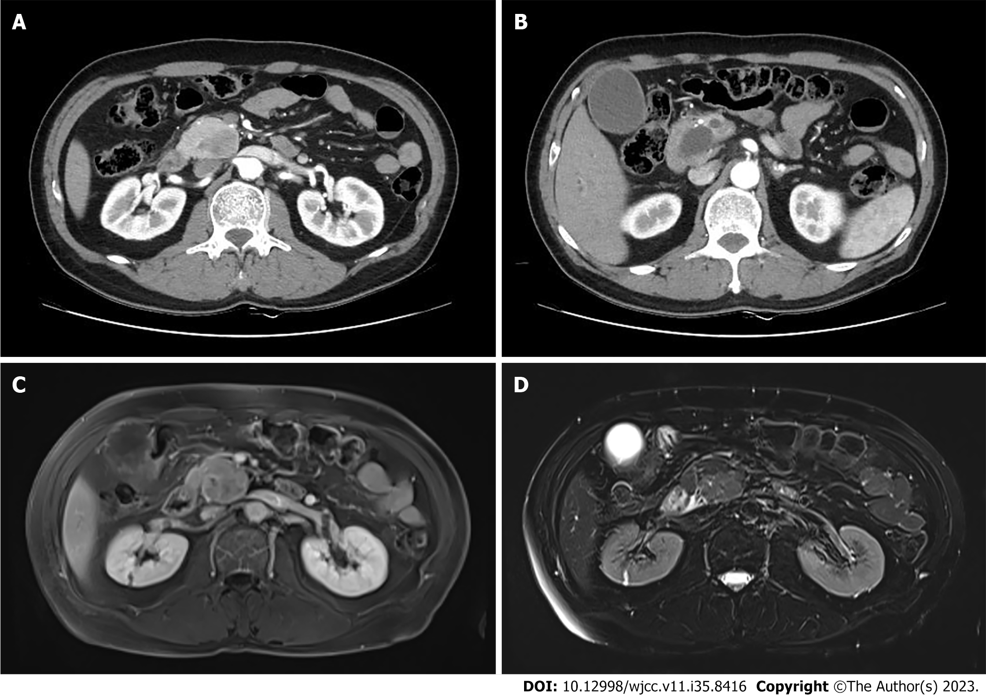
Figure 2 Radiologic findings.
A: Abdominal computed tomography (CT) showed a 3.5 cm sized mass lesion on the pancreas head; B: The CT scan shows a double duct sign, the dilatation of both the pancreatic duct and bile duct, due to the compression of the mass lesion; C: On magnetic resonance imaging (MRI), T1-weighted imaging showed low signal intensity compared to the surrounding pancreas parenchyma; D: On MRI, T2-weighted imaging showed iso-signal intensity.
- Citation: Yi K, Lee J, Kim DU. Metastatic pancreatic solitary fibrous tumor: A case report. World J Clin Cases 2023; 11(35): 8416-8424
- URL: https://www.wjgnet.com/2307-8960/full/v11/i35/8416.htm
- DOI: https://dx.doi.org/10.12998/wjcc.v11.i35.8416


