Published online Nov 6, 2023. doi: 10.12998/wjcc.v11.i31.7706
Peer-review started: September 5, 2023
First decision: September 26, 2023
Revised: October 2, 2023
Accepted: October 27, 2023
Article in press: October 27, 2023
Published online: November 6, 2023
Processing time: 61 Days and 18.1 Hours
Malignant small round cell tumor (MSRCT) metastasis to the common bile duct associated with recurrent biliary hemorrhage is extremely rare. Thus far, there have been no reports of metastatic small round cell tumors of the common bile duct.
Herein, we report the case of a 77-year-old female patient with an MSRCT in the common bile duct. The patient was admitted to hospital due to gastrointestinal hemorrhage and abdominal pain. We found a neoplasm in the common bile duct with active bleeding through a spyglass. We performed biopsy through the spy
MSRCT is a group of tumors with similar cell morphology and diffuse histological structure. Complete tumor resection results in improved survival in patients with MSRCT. Roux-en-Y cholangiojejunostomy was performed. After excision of the common bile duct tumor, the patient felt that the abdominal pain improved and hemorrhage disappeared. The patient underwent routine fecal examination one month after surgery, indicating a negative fecal occult blood test. On May 22, 2023, the patient was reexamined by abdominal computed tomography, and no abdominal space occupying lesions or abdominal lymphadenopathy was found.
Core Tip: Malignant small round cell tumor (MSRCT) in the common bile duct has not been previously documented. Here we present the case of an MSRCT in the common bile duct. A 77-year-old female patient was admitted to hospital due to gastrointestinal hemorrhage and abdominal pain. We found a neoplasm in the common bile duct with active bleeding through a spyglass. We performed biopsy through the spyglass and placed a metal stent to stop bleeding. The pathological result suggested that it was an MSRCT metastasized from the back to the common bile duct.
- Citation: Jin YL, Ruan YJ, Lu GR. Biliary hemorrhage caused by a malignant small round cell tumor in the common bile duct: A case report. World J Clin Cases 2023; 11(31): 7706-7711
- URL: https://www.wjgnet.com/2307-8960/full/v11/i31/7706.htm
- DOI: https://dx.doi.org/10.12998/wjcc.v11.i31.7706
Malignant small round cell tumor (MSRCT) metastasis to the common bile duct associated with recurrent biliary hemorrhage is extremely rare. Thus far, there have been no reports of metastatic small round cell tumors of the common bile duct. We herein report the case of a metastatic MSRCT in the common bile duct.
A 77-year-old Chinese woman presented to the gastroenterology clinic with a complaint of gastrointestinal hemorrhage and abdominal pain for 1 mo.
Symptoms started 1 mo before presentation with gastrointestinal hemorrhage and abdominal pain. The patient felt severe abdominal pain, no radiation-induced pain, and no nausea and vomiting. The patient defecated hemorrhage once a day. The symptoms of abdominal pain were slightly eased about 20 min after defecation.
Nine years ago, this patient underwent the resection of MSRCT in the left shoulder, back, and left supraclavicle. No chemotherapy was taken after operation. Recently, the tumors in the left shoulder, back, and left supraclavicle me
The patient denied any family history of malignant tumours.
On the physical examination, the vital signs were as follows: Body temperature, 36.6 ℃; blood pressure, 97/59 mmHg; heart rate, 84 beats per min; respiratory rate, 16 breaths per min. The abdomen was flat, without tenderness or rebound pain.
The laboratory examinations showed that the patient's hemoglobin level decreased to 58 g/L. Fecal occult blood test was positive. Levels of serum tumour markers were normal. There were no abnormalities in liver and kidney function.
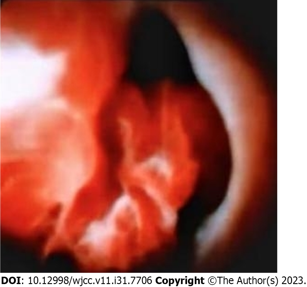
Abdominal computed tomography (CT) and gastrointestinal endoscopy showed no abnormalities. We performed endoscopic retrograde cholangiopancreatography on the patient. The gastroscope showed that the gastroduodenal mucosa was smooth and pale. The main nipple was found to be changed after endoscopic sphincterotomy without blood stains. The nipple was incised with a knife, and dark red liquid was discharged (Figure 1A). On duodenoscopy, the scope was successfully passed through the esophagus, stomach and duodenal bulb, and reached the descending part, and the main nipple was found and was intubated successfully. The amount of contrast agent used was 20 mL. X-ray showed that the common bile duct had developed without expansion, and the maximum diameter was approximately 0.8 cm. It was filled with filling defects of different sizes. The intrahepatic bile duct was partially developed without expansion. There were irregular filling defects in the middle and lower segments of the common bile duct, which could be moved. The bright red liquid was discharged. The cystic pancreatic duct was not developed. The biliary tract was cleared through the zebra guide wire airy catheter, and the bleeding clots and necrotic tissues were removed. Then, the storage guide wire was inserted into the biliary full posterior membrane metal stent (Figure 1B). After release, the stent was in a good position. Before inserting stent, the spyglass was performed. After insertion of the spyglass and after repeated flushing, new organisms could be seen in the middle of the common bile duct, with a rough surface, necrosis of the surface, active bleeding and local lumen stenosis (Figure 2). Spy-Bite was performed. Subsequently, the pathological result suggested that it was an MSRCT.
The pathological result suggested that it was an MSRCT with some cytoplasm and difficult-to-see karyokinesis (Figure 3). Tumors of epithelial cell and neuroendocrine origin were excluded by immunohistochemistry. The pathological examination of bile duct space-occupying lesions showed that they were positive for desmin, vimentin, and CD68 and negative for myogenin, CAM5.2, and S-100. This patient underwent resection of MSRCT in the left shoulder, back and left supraclavicle 9 years previously. No chemotherapy was taken after the operation. Recently, the tumors in the left shoulder, back and left supraclavicle metastasized to the common bile duct. The pathological result suggested MSRCT. The cell morphology and immunohistochemical results were basically consistent with the pathological results of the left shoulder, back and left supraclavicle tumors in 2014. Combined with the medical history, the patient was diagnosed with metastasis of the MSRCT to the common bile duct. Later, we found using fluorescence in situ hybridization that the SS18 gene break test was negative, ruling out the diagnosis of synovial sarcoma (Figure 4).
Roux-en-Y cholangiojejunostomy and loosening of intestinal conglutination were performed. After opening the abdominal cavity, we found expansion of the middle and upper segment of the common bile duct, with a diameter of about 1.3 cm, and a palpable mass in the common bile duct about 2 cm below the confluence of the left and right hepatic ducts, with medium texture and no enlarged lymph nodes around it. Then, we cut the bile duct and discovered a circular yellow and white cauliflower-like neoplasm in the common bile duct. We removed the bile duct lesions. On the premise of clear distal patency of the common bile duct, the common bile duct was transected to prepare for the establishment of a new biliary-intestinal channel. We closed the distal end of the common bile duct, and temporarily clamped the proximal end of the common bile duct with non-destructive forceps to prevent bile from flowing into the abdominal cavity. Then, we cut off the upper segment of the jejunum, lift the transverse colon down along its mesangium, and revealed the duodenal jejunal flexure. We kept the first jejunal artery on the jejunal mesentery, cut off the second jejunal artery, and separated, cut, and ligated the jejunal mesentery, so that the distal jejunum has enough freedom and there is no tension after the above-mentioned choledochojejunostomy. The free distal jejunum was sutured and closed, and then we lifted it to the porta hepatis through the colon for anastomosis. The distal jejunum was lifted 60 cm and anastomosed with the proximal jejunum side to side. A small opening was cut on the distal jejunum lifted from the transverse mesocolon fissure on the side of the opposite edge of the mesentery with the sutured stump. The direction was parallel to the long axis of the intestinal tube, and the size was corresponding to the repaired bile duct orifice. Finally, the abdominal cavity was closed after a drainage tube was placed.
After excision of the common bile duct tumor, the patient felt that the abdominal pain improved and hemorrhage disappeared. The patient underwent routine fecal examination one month after surgery, indicating a negative fecal occult blood test. On May 22, 2023, the patient was reexamined by abdominal CT, and no abdominal space occupying lesions or abdominal lymphadenopathy was found.
MSRCT is an extremely rare malignant tumor that has been clearly defined over the last 10 years. It is a group of tumors with similar cell morphology and diffuse histological structure. Complete tumor resection results in improved survival in patients with MSRCT. Chemotherapy and radiotherapy correlate with improved patient outcomes. Multimodal therapy may improve survival in patients with MSRCT[1]. Generally, MSRCTs include Ewing's sarcoma, peripheral neuroectodermal tumor, rhabdomyosarcoma, synovial sarcoma, non-Hodgkin's lymphoma, retinoblastoma, neuroblastoma, hepatoblastoma, and nephroblastoma or Wilms’ tumor[2]. Regarding the thoracic cavity, cases involving the pleura[3-5], heart[6], colon[7], or lungs[8] have been described. To date, no relevant study has reported MSRCTs of the biliary tract. This is the first case report worldwide of MSRCT metastasized from the left shoulder, back, and left supraclavicle to the common bile duct. In this case report, we used a SpyGlass to detect a neoplasm in the patient’s common bile duct and obtained the tissue for pathological diagnosis. The findings showed that SpyGlass had an irreplaceable important role in the diagnosis of bleeding in the bile duct. Furthermore, the general morphology of MSRCT in the common bile duct was visually displayed using the SpyGlass. For biliary bleeding, in addition to considering common causes (such as trauma and biliary infection), we should also consider whether biliary tumors are present. We need to carefully check for the possibility of MSRCT metastasizing to the biliary tract, especially for patients with a history of MSRCT in the past. This study recognized an extremely rare metastasis of MSRCT in the common bile duct and provided new insight into the difficult diagnosis of biliary bleeding in clinical practice in the future.
MSRCT in the common bile duct has not been previously documented. This report presents the case of an MSRCT in the common bile duct. A 77-year-old female patient was admitted to hospital due to gastrointestinal hemorrhage and abdominal pain. We found a neoplasm in the common bile duct with active bleeding through a SpyGlass. We performed biopsy through the SpyGlass and placed a metal stent to stop bleeding. The pathological result suggested that it was an MSRCT metastasized from the back to the common bile duct.
Provenance and peer review: Unsolicited article; Externally peer reviewed.
Peer-review model: Single blind
Specialty type: Medicine, research and experimental
Country/Territory of origin: China
Peer-review report’s scientific quality classification
Grade A (Excellent): 0
Grade B (Very good): 0
Grade C (Good): C
Grade D (Fair): D
Grade E (Poor): 0
P-Reviewer: Bhardwaj R; Rabago LR, Spain S-Editor: Qu XL L-Editor: Wang TQ P-Editor: Xu ZH
| 1. | Xing PY, Shi YK, Feng FY, Qin Y, Liu P. [Clinical characteristics and treatment of desmoplastic small round cell tumor]. Zhonghua Zhong Liu Za Zhi. 2010;32:139-142. [PubMed] |
| 2. | Donner LR. Cytogenetics and molecular biology of small round-cell tumors and related neoplasms. Current status. Cancer Genet Cytogenet. 1991;54:1-10. [RCA] [PubMed] [DOI] [Full Text] [Cited by in Crossref: 30] [Cited by in RCA: 33] [Article Influence: 1.0] [Reference Citation Analysis (0)] |
| 3. | Parkash V, Gerald WL, Parma A, Miettinen M, Rosai J. Desmoplastic small round cell tumor of the pleura. Am J Surg Pathol. 1995;19:659-665. [RCA] [PubMed] [DOI] [Full Text] [Cited by in Crossref: 126] [Cited by in RCA: 112] [Article Influence: 3.7] [Reference Citation Analysis (0)] |
| 4. | Sápi Z, Szentirmay Z, Orosz Z. Desmoplastic small round cell tumour of the pleura: a case report with further cytogenetic and ultrastructural evidence of 'mesothelioblastemic' origin. Eur J Surg Oncol. 1999;25:633-634. [RCA] [PubMed] [DOI] [Full Text] [Cited by in Crossref: 14] [Cited by in RCA: 15] [Article Influence: 0.6] [Reference Citation Analysis (0)] |
| 5. | Ouarssani A, Atoini F, Ait Lhou F, Rguibi Idrissi M. Desmoplastic small round cell tumor of the pleura. Thorac Cancer. 2011;2:117-119. [RCA] [PubMed] [DOI] [Full Text] [Cited by in Crossref: 3] [Cited by in RCA: 3] [Article Influence: 0.4] [Reference Citation Analysis (0)] |
| 6. | George S, Vaideeswar P, Pandit S, Kane S, Khandeparkar J. Malignant small round cell tumor of the heart: a diagnostic dilemma. Cardiovasc Pathol. 2007;16:56-58. [RCA] [PubMed] [DOI] [Full Text] [Cited by in Crossref: 4] [Cited by in RCA: 6] [Article Influence: 0.3] [Reference Citation Analysis (0)] |
| 7. | Huang J, Sha L, Zhang H, Tang X, Zhang X. Desmoplastic small round cell tumor in transverse colon: report of a rare case. Int Surg. 2015;100:809-813. [RCA] [PubMed] [DOI] [Full Text] [Cited by in Crossref: 5] [Cited by in RCA: 8] [Article Influence: 0.9] [Reference Citation Analysis (0)] |
| 8. | Afif H, Benouhoud N, Aichane A, Trombati N, Bouayad Z. [Lung metastases of a small round cell desmoplastic tumor]. Rev Pneumol Clin. 2006;62:53-55. [RCA] [PubMed] [DOI] [Full Text] [Cited by in Crossref: 1] [Reference Citation Analysis (0)] |


