Published online Oct 26, 2023. doi: 10.12998/wjcc.v11.i30.7502
Peer-review started: September 17, 2023
First decision: September 28, 2023
Revised: October 8, 2023
Accepted: October 11, 2023
Article in press: October 11, 2023
Published online: October 26, 2023
Processing time: 37 Days and 14.5 Hours
For the treatment of distal clavicle fractures, each treatment method has its own advantages and disadvantages, and there is no optimal surgical solution.
Based on this, we report 2 cases of distal clavicle fractures treated utilizing an anterior inferior plate with a single screw placed in the distal, in anticipation of providing a better surgical approach to distal clavicle fracture treatment. Two patients were admitted to the hospital after trauma with a diagnosis of distal clavicle fracture, and were admitted to the hospital for internal fixation of clavicle fracture by incision and reduction, with good postoperative functional recovery.
With solid postoperative fixation and satisfactory prognostic functional recovery, this technique has been shown to be simple, easy to perform and effective.
Core Tip: Distal clavicle fractures account for about 12%-21% of all clavicular fractures and about 25% of distal clavicle fractures are unstable. For conservative treatment of distal clavicle fractures, complications including high incidence of nonunion, abnormal appearance and dysfunction are likely to ensue. Thus, surgical treatment is recommended by scholars. So far, none of the treatment methods has been proven to be the best. Here, we report 2 cases of distal clavicular fractures successfully treated by anteroinferior plating with a single screw placed at the distal fragment. This technique has been shown to be simple, easy to operate and effective, which has not been reported previously as far as we are aware.
- Citation: Zhao XL, Liu YQ, Wang JG, Liu YC, Zhou JX, Wang BY, Zhang YJ. Distal clavicle fractures treated by anteroinferior plating with a single screw: Two case reports. World J Clin Cases 2023; 11(30): 7502-7507
- URL: https://www.wjgnet.com/2307-8960/full/v11/i30/7502.htm
- DOI: https://dx.doi.org/10.12998/wjcc.v11.i30.7502
Distal clavicle fractures account for about 12%-21% of all clavicular fractures and about 25% of distal clavicle fractures are unstable[1]. For conservative treatment of distal clavicle fractures, complications including high incidence of nonunion, abnormal appearance and dysfunction are likely to ensue. Thus, surgical treatment is recommended by scholars[2-4]. The surgical treatment methods include hook plating, coracoclavicular (CC) stabilization, locking plating, multiple transacromial pins, and etc. Each method has its own advantages and disadvantages. The hook plating method is associated with multiple complications such as subacromial irritation, plate migration, osteolysis and other problems[5,6]. CC stabilization has been recommended with satisfactory clinical outcome[7,8]. It can also be performed with minimal invasion under arthroscopic assistance, yet this technique is associated with risks of manipulation on coracoid. The locking plating method is to a large degree limited by the bone mass at the distal fragment of the fracture. The fixation effect cannot be guaranteed since it’s likely that insufficient screws are placed given the limited bone mass. Indeed, there were reports of cases with fixation failure after locking plate treatment and the implants had to be removed from some patients eventually[9]. Some scholars advocate plating combined with coracoclavicular fixation[10,11]. This will obviously increases total operation in addition to boosting medical cost, which has to be taken into consideration. Problems upon treatment with multiple pins method include acromioclavicular joint interference, pin migration, irritation, as well as forced removal of the implant[12]. So far, none of the treatment methods mentioned above have been proven to be the best. Here, we report 2 cases of distal clavicular fractures successfully treated by anteroinferior plating with a single screw placed at the distal fragment. This technique has been shown to be simple, easy to operate and effective, which has not been reported previously as far as we are aware. These two patients consented to publication of this report.
Case 1: A 38-year-old Chinese man hit his right shoulder and suffered pain and swelling in the distal part of the right clavicle after the injury for hours.
Case 2: A 57-year-old Chinese man hit his left shoulder and suffered pain and swelling in the distal part of the left clavicle after the injury for hours.
Case 1: He fell while riding his bicycle and hit his right shoulder on the hard ground 5 h before.
Case 2: He fell to the ground and hit left shoulder 2 h before. No significant personal or family history.
No relevant past illness history.
No significant personal or family history.
Case 1: Obvious swelling of the right shoulder with pain and limited functional movement of the right shoulder joint.
Case 2: Obvious swelling of the left shoulder with pain and limited functional movement of the left shoulder joint.
Laboratory examinations showed no significant abnormalities.
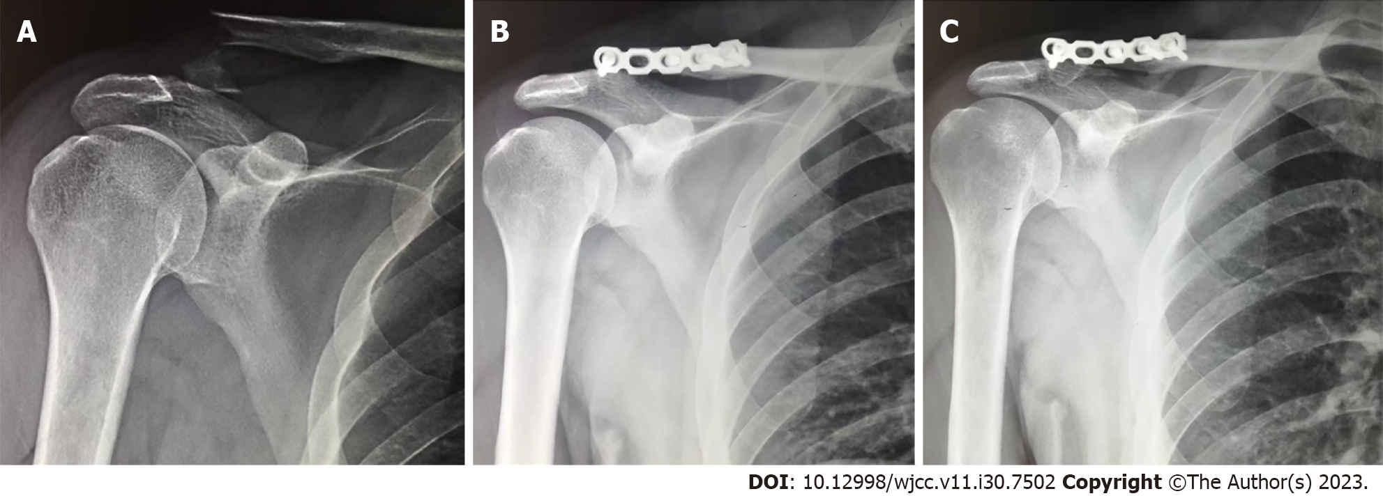
Case 1: The radiograph (Figure 1) showed a fracture of the distal right clavicle.
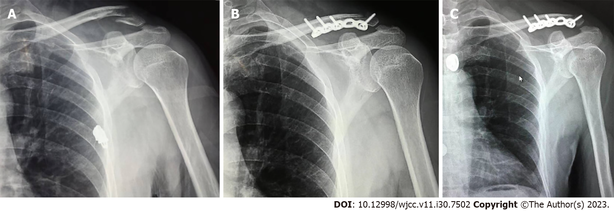
Case 2: The radiograph (Figure 2) showed a fracture of the distal left clavicle.
A fracture of the distal right clavicle.
A fracture of the distal left clavicle.
The operation was performed 2 d after injury. The patient was placed in the beach-chair position, subjected to brachial plexus block anesthesia. A parallel incision was made along the lateral lower edge of the clavicle. After the fracture was exposed, a molded anterior-inferior reconstruction plate (Baide Medical, Jiangsu, China) was placed while the reduction was maintained by an assistant pressing the proximal end of clavicle fracture. The distal hole of the plate was placed in a proper position so that a screw could be accurately placed at the distal fragment. Under fluoroscopy, a single screw with length of 3.5 cm and diameter of 3.5 mm was inserted to form a double cortical fixation at the distal fragment of the fracture. Firm control force was felt while the screw was tightened. 3 screws were inserted at the proximal end of the fracture subsequently. The right clavicle exhibited no displacement within itself while moving right shoulder fully, which indicated firm and reliable fixation effect had been achieved. The wound was irrigated and sutured while the ligaments were not treated during the operation. The patient was encouraged to start shoulder movement after the pain subsided. He was not allowed to load the operated shoulder for 6 wk.
The operation was performed 8 h after injury with the same operation procedure as that in case 1.
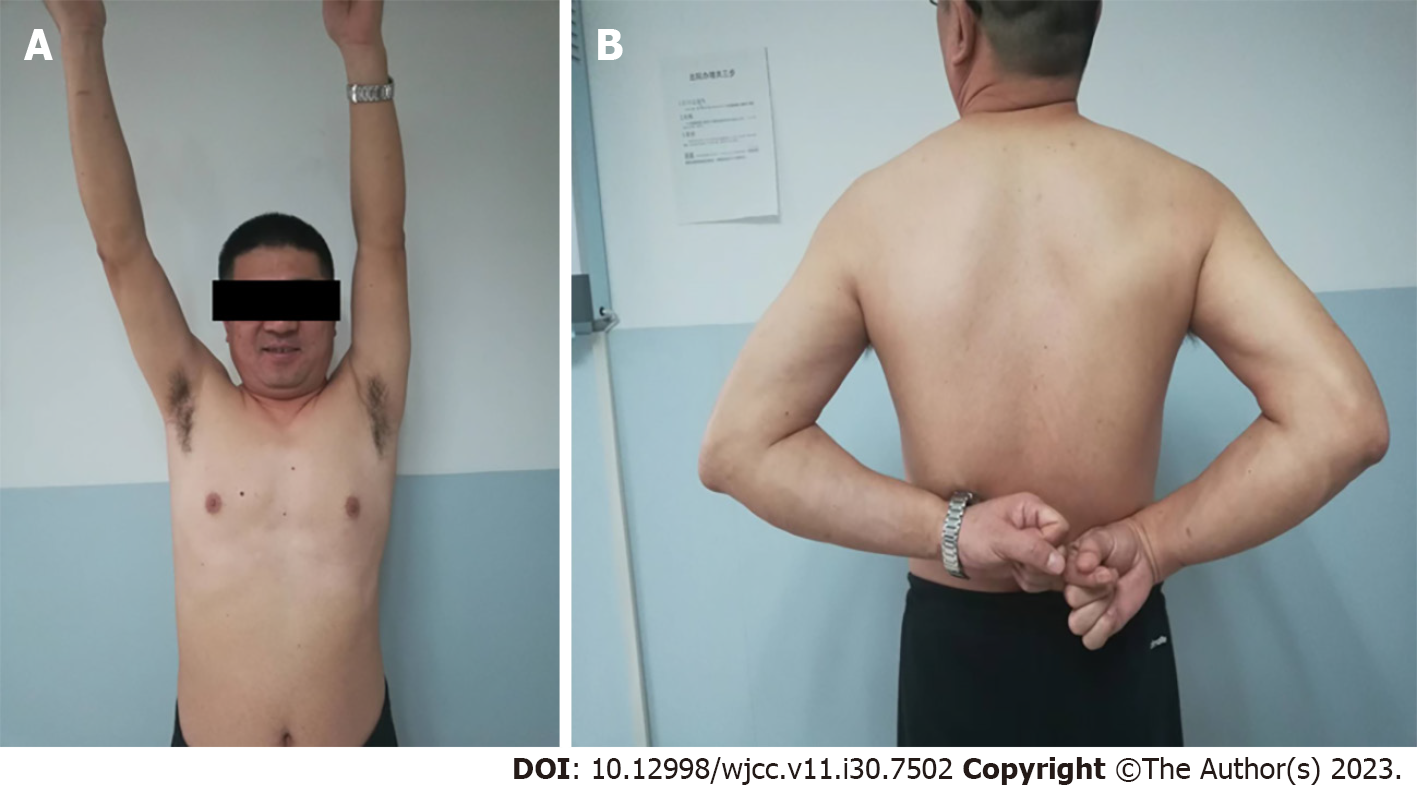
At follow-up examination after 1 year post operation, X-ray (Figure 1) showed that fracture reduction was not lost and union was achieved. At six months post operation, the patient was already pain free and back to his previous work with his right shoulder moving fully free. He felt comfortable after operation and was very satisfied with the local appearance and function in the right shoulder (Figure 3).
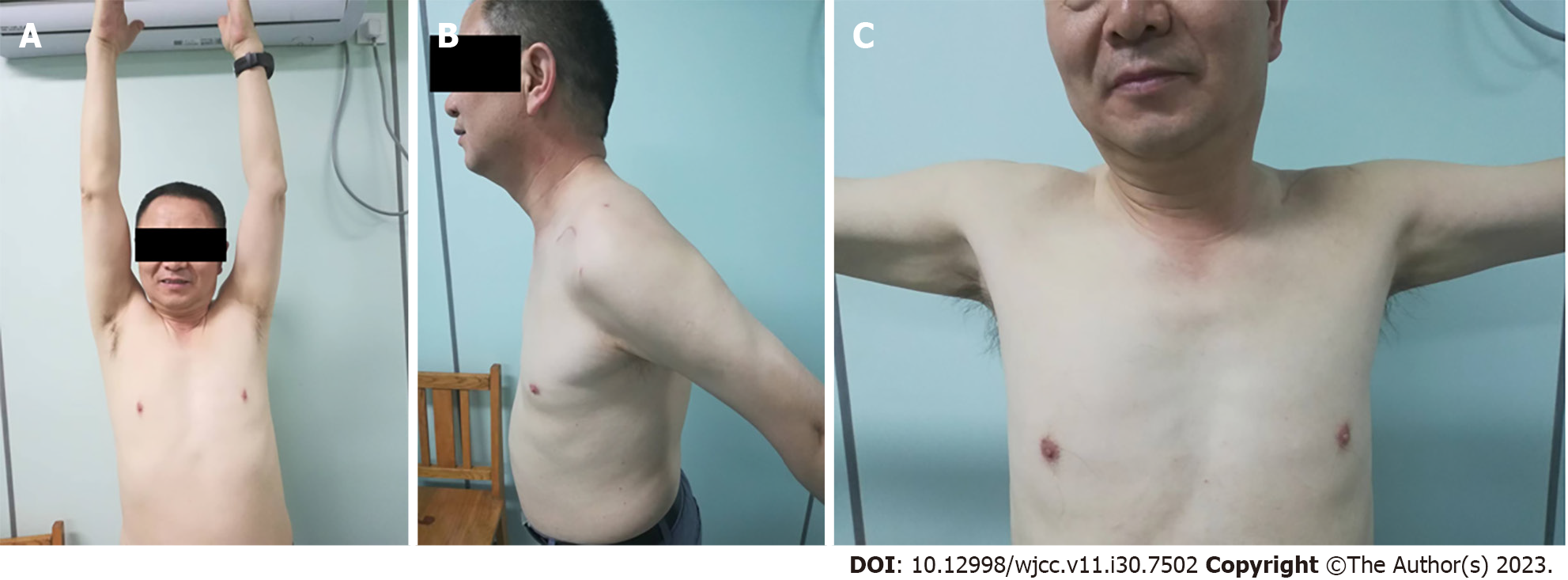
At follow-up examination after 6 mo post operation, X-ray (Figure 2) showed that the reduction at the fracture site remained in good condition and the left clavicle was well healed. He was satisfied with both the local appearance and function in the left shoulder (Figure 4). He resumed most of his activities before the injury at 6 mo after the operation.
Surgical treatment of unstable distal clavicle fractures can greatly promote fracture healing and reduce related complications. Because the distal fragment of the fracture is small and flat, it is difficult to fix the fracture directly. When the plate is positioned on the superior surface of the clavicle, the screws tend to be quite short. If the number of screws at the distal fragment of the fracture is small, the fixing effect might be disturbing. Different approaches have been invented so as to strengthen the fixation effect of superior plating. For example, Kaipe et al[13] placed a second plate on the anterior surface of the clavicle while Yoo et al[14] added several cerclage wires. We made full use of the anatomical advantage of the greater anterior-posterior diameter of the distal clavicle and placed the plate on the anteroinferior surface of the clavicle, where the length of the screw at the distal fragment could be significantly much longer. With just one single screw placed at the distal fragment, the grip force increases significantly, achieving satisfactory fixing effect while there is no need to repair the ligaments. It is also possible to fix smaller fracture fragment with our method. Anteroinferior plating does not need to interfere with acromioclavicular joint and postoperative patients feel more comfortable. The upwarping of the proximal end of the distal clavicle fracture is the main harmful stress potentially causing fixation failure. Our method of anteroinferior plating may prevent screw evulsion since the single anterior-posterior screw is perpendicular to the unfavorable upwarping stress. Furthermore, the screw drilling direction is upward and backward, which can potentially reduce the damage to subclavian nerves and vessels. The anteroinferior plate is relatively well concealed and covered by soft tissues, which maximally reduces plate protrusion as well as patient’s discomfort leading to less demand for plate removal. The site of surgical incision could move relatively more downward, which is also advantageous in cosmetic sense. Some of these advantages have been noticed by scholars in the treatment of midshaft clavicular fractures using anteroinferior plating method[15,16].
Based on our experience, it is feasible to treat unstable distal clavicle fractures by anteroinferior plating with a single screw placed at the distal fragment, which is simple and reliable. A long anterior-posterior screw alone could effectively control the smaller distal fragment, which, in our view, is the first. Due to the small number of cases, the effectiveness of this method awaits more observation and verification.
We thank the patients and their families.
Provenance and peer review: Unsolicited article; Externally peer reviewed.
Peer-review model: Single blind
Specialty type: Surgery
Country/Territory of origin: China
Peer-review report’s scientific quality classification
Grade A (Excellent): 0
Grade B (Very good): B
Grade C (Good): 0
Grade D (Fair): 0
Grade E (Poor): 0
P-Reviewer: Rezus E, Romania S-Editor: Liu JH L-Editor: A P-Editor: Yu HG
| 1. | Neer CS 2nd. Fractures of the distal third of the clavicle. Clin Orthop Relat Res. 1968;58:43-50. [PubMed] |
| 2. | Robinson CM, Cairns DA. Primary nonoperative treatment of displaced lateral fractures of the clavicle. J Bone Joint Surg Am. 2004;86:778-782. [RCA] [PubMed] [DOI] [Full Text] [Cited by in Crossref: 169] [Cited by in RCA: 149] [Article Influence: 7.1] [Reference Citation Analysis (0)] |
| 3. | Nordqvist A, Petersson C, Redlund-Johnell I. The natural course of lateral clavicle fracture. 15 (11-21) year follow-up of 110 cases. Acta Orthop Scand. 1993;64:87-91. [RCA] [PubMed] [DOI] [Full Text] [Cited by in Crossref: 173] [Cited by in RCA: 146] [Article Influence: 4.6] [Reference Citation Analysis (0)] |
| 4. | Banerjee R, Waterman B, Padalecki J, Robertson W. Management of distal clavicle fractures. J Am Acad Orthop Surg. 2011;19:392-401. [RCA] [PubMed] [DOI] [Full Text] [Cited by in Crossref: 105] [Cited by in RCA: 96] [Article Influence: 6.9] [Reference Citation Analysis (0)] |
| 5. | Kashii M, Inui H, Yamamoto K. Surgical treatment of distal clavicle fractures using the clavicular hook plate. Clin Orthop Relat Res. 2006;447:158-164. [RCA] [PubMed] [DOI] [Full Text] [Cited by in Crossref: 149] [Cited by in RCA: 140] [Article Influence: 7.4] [Reference Citation Analysis (0)] |
| 6. | Tambe AD, Motkur P, Qamar A, Drew S, Turner SM. Fractures of the distal third of the clavicle treated by hook plating. Int Orthop. 2006;30:7-10. [RCA] [PubMed] [DOI] [Full Text] [Cited by in Crossref: 69] [Cited by in RCA: 61] [Article Influence: 3.1] [Reference Citation Analysis (0)] |
| 7. | Blake MH, Lu MT, Shulman BS, Glaser DL, Huffman GR. Arthroscopic Cortical Button Stabilization of Isolated Acute Neer Type II Fractures of the Distal Clavicle. Orthopedics. 2017;40:e1050-e1054. [RCA] [PubMed] [DOI] [Full Text] [Cited by in Crossref: 17] [Cited by in RCA: 17] [Article Influence: 2.1] [Reference Citation Analysis (0)] |
| 8. | Hsu KH, Tzeng YH, Chang MC, Chiang CC. Comparing the coracoclavicular loop technique with a hook plate for the treatment of distal clavicle fractures. J Shoulder Elbow Surg. 2018;27:224-230. [RCA] [PubMed] [DOI] [Full Text] [Cited by in Crossref: 28] [Cited by in RCA: 33] [Article Influence: 4.7] [Reference Citation Analysis (0)] |
| 9. | Hessmann M, Gotzen L, Kirchner R, Gehling H. [Therapy and outcome of lateral clavicular fractures]. Unfallchirurg. 1997;100:17-23. [RCA] [PubMed] [DOI] [Full Text] [Cited by in Crossref: 16] [Cited by in RCA: 14] [Article Influence: 0.5] [Reference Citation Analysis (0)] |
| 10. | Tiefenboeck TM, Boesmueller S, Binder H, Bukaty A, Tiefenboeck MM, Joestl J, Hofbauer M, Ostermann RC. Displaced Neer Type IIB distal-third clavicle fractures-Long-term clinical outcome after plate fixation and additional screw augmentation for coracoclavicular instability. BMC Musculoskelet Disord. 2017;18:30. [RCA] [PubMed] [DOI] [Full Text] [Full Text (PDF)] [Cited by in Crossref: 7] [Cited by in RCA: 7] [Article Influence: 0.9] [Reference Citation Analysis (0)] |
| 11. | Nandra R, Kowalski T, Kalogrianitis S. Innovative use of single-incision internal fixation of distal clavicle fractures augmented with coracoclavicular stabilisation. Eur J Orthop Surg Traumatol. 2017;27:1057-1062. [RCA] [PubMed] [DOI] [Full Text] [Cited by in Crossref: 3] [Cited by in RCA: 5] [Article Influence: 0.6] [Reference Citation Analysis (0)] |
| 12. | Kwak SH, Lee YH, Kim DW, Kim MB, Choi HS, Baek GH. Treatment of Unstable Distal Clavicle Fractures With Multiple Steinmann Pins-A Modification of Neer's Method: A Series of 56 Consecutive Cases. J Orthop Trauma. 2017;31:472-478. [RCA] [PubMed] [DOI] [Full Text] [Cited by in Crossref: 4] [Cited by in RCA: 6] [Article Influence: 0.8] [Reference Citation Analysis (0)] |
| 13. | Kaipel M, Majewski M, Regazzoni P. Double-plate fixation in lateral clavicle fractures-a new strategy. J Trauma. 2010;69:896-900. [RCA] [PubMed] [DOI] [Full Text] [Cited by in Crossref: 18] [Cited by in RCA: 21] [Article Influence: 1.4] [Reference Citation Analysis (1)] |
| 14. | Yoo JH, Chang JD, Seo YJ, Shin JH. Stable fixation of distal clavicle fracture with comminuted superior cortex using oblique T-plate and cerclage wiring. Injury. 2009;40:455-457. [RCA] [PubMed] [DOI] [Full Text] [Cited by in Crossref: 17] [Cited by in RCA: 18] [Article Influence: 1.1] [Reference Citation Analysis (0)] |
| 15. | Hulsmans MH, van Heijl M, Houwert RM, Timmers TK, van Olden G, Verleisdonk EJ. Anteroinferior vs superior plating of clavicular fractures. J Shoulder Elbow Surg. 2016;25:448-454. [RCA] [PubMed] [DOI] [Full Text] [Cited by in Crossref: 26] [Cited by in RCA: 32] [Article Influence: 3.6] [Reference Citation Analysis (0)] |
| 16. | Baltes TPA, Donders JCE, Kloen P. What is the hardware removal rate after anteroinferior plating of the clavicle? A retrospective cohort study. J Shoulder Elbow Surg. 2017;26:1838-1843. [RCA] [PubMed] [DOI] [Full Text] [Cited by in Crossref: 14] [Cited by in RCA: 13] [Article Influence: 1.6] [Reference Citation Analysis (0)] |


