Published online Oct 26, 2023. doi: 10.12998/wjcc.v11.i30.7492
Peer-review started: August 4, 2023
First decision: September 19, 2023
Revised: October 6, 2023
Accepted: October 16, 2023
Article in press: October 16, 2023
Published online: October 26, 2023
Processing time: 81 Days and 21.4 Hours
Gout is a common type of inflammatory arthritis caused by the deposition of monosodium urate crystals in the joints and surrounding tissues. It typically appears with abrupt and intense pain, redness, and swelling in the affected joint. It frequently targets the lower extremities, such as the big toe. However, rarely, gout can manifest in atypical locations, including the hands, leading to an uncommon presentation known as gouty tenosynovitis. However, it can result in significant morbidity owing to the potential for severe complications, such as myonecrosis and compartment syndrome.
An 82-year-old male patient with a history of hypertension, cerebral infarction, Parkinson's disease, and recurrent gout attacks sought medical attention because of progressive pain and swelling in the right hand. Imaging findings revealed forearm swelling, raising concerns of possible tenosynovitis, bursitis, septic arthritis, and compartment syndrome. A fasciotomy was performed to decom
Septic-like complications can occur in the absence of infection in severe gout attacks with pus-like discharges due to compartment syndrome and myonecrosis. Cultures can be used to differentiate between gouty attacks, septic arthritis, and infectious tenosynovitis. Involvement of the flexor and extensor muscles, as in this case, is rare. This study contributes to the literature by reporting a rare case of successful fasciotomy and serial debridement in an elderly patient with multiple comorbidities.
Core Tip: This study reports three novel findings, which may contribute to the existing literature. First, there was an uncommon lesion in an area that was different from the usual site of a gout attack. Second, the gout attack was severe enough to cause compartment syndrome. Third, it was a very rare case involving the flexor and extensor tendons. The successful management of elderly patients highlights the importance of prompt recognition, interdisciplinary collaboration, and tailored treatment strategies for optimal patient outcomes.
- Citation: Lee DY, Eo S, Lim S, Yoon JS. Gouty tenosynovitis with compartment syndrome in the hand: A case report. World J Clin Cases 2023; 11(30): 7492-7496
- URL: https://www.wjgnet.com/2307-8960/full/v11/i30/7492.htm
- DOI: https://dx.doi.org/10.12998/wjcc.v11.i30.7492
Gout is a systemic inflammatory disorder characterized by the deposition of monosodium urate crystals in the joints and soft tissues, leading to recurrent acute arthritis attacks. The accumulation of urate crystals in tissues triggers a significant inflammatory reaction that can potentially lead to severe complications, such as acute septic arthritis or compartment syndrome. In well-established tophi, a granulomatous-like pattern characterized by histiocytic and foreign-body giant cell responses surrounds the deposited crystals, whereas acute gout attacks manifest as neutrophil exudates. Tophi may also form in the ligaments, muscles, and tendons, posing the risk of inducing injuries, such as myonecrosis or tendon rupture over time[1]. Although it commonly affects the lower extremities, atypical presentations, including gouty tenosynovitis of the hands, may occur. Gout manifestations in the upper extremities are less common and include subcutaneous tophi, arthritis, tenosynovitis, and nerve entrapment[1-5].
This report describes the challenging case of an elderly male patient with a history of multiple comorbidities who presented with severe swelling, erythema, and excruciating pain in the right hand, and was ultimately diagnosed with gouty tenosynovitis complicated by myonecrosis and compartment syndrome.
An 82-year-old male patient with a medical history of hypertension, cerebral infarction, Parkinson's disease, and recurrent gout attacks presented to the hospital with a chief complaint of a swollen and painful right hand.
Gout diagnosis in the patient was confirmed at the Rheumatology Department 4 years ago, and was characterized by the presence of positive monosodium urate and elevated serum uric levels.
Furthermore, the patient had a documented history of three separate gout attacks, specifically involving the right great toe. His hand symptoms progressively worsened over the past 4 d and were extremely tender, even to the slightest touch.
The patient denied a recent history of hand trauma.
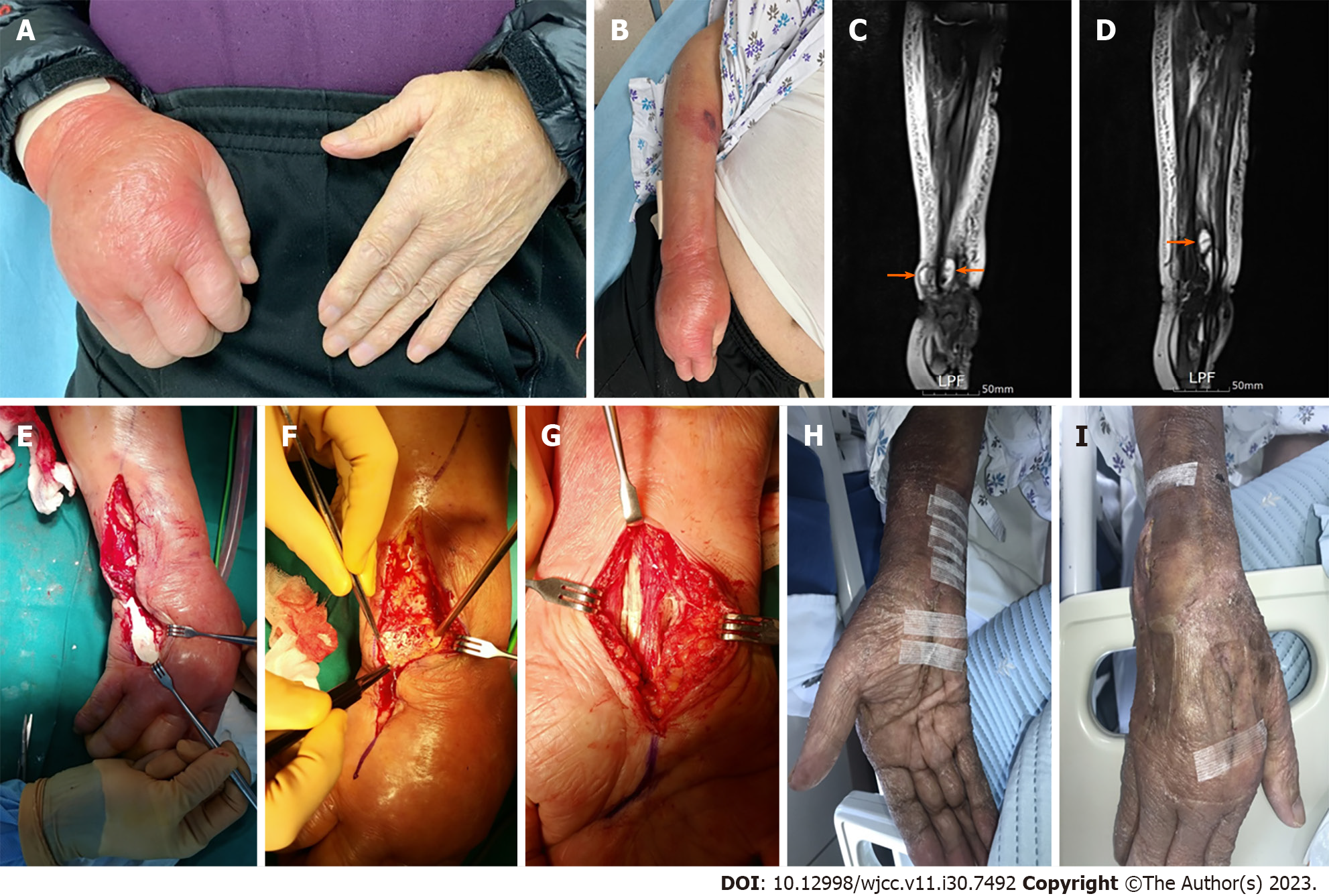
Physical examination revealed marked erythema and swelling of the right hand (Figure 1A and B). His systemic blood pressure/diastolic blood pressure decreased to 90/60 mmHg and the heart rate increased to 138 beats/min. He presented with a fever of 38.1℃.
Laboratory investigations revealed elevated C-reactive protein (CRP) (26.97 mg/L) levels and white blood cell (WBC) count (14020 cells/μL). Considering the patient's septic condition, the presence of systemic inflammatory response syndrome was deemed plausible.
Moreover, magnetic resonance imaging (MRI) of the hand and forearm revealed findings indicative of extensive subcutaneous swelling involving the hand and forearm, suggesting cellulitis and tenosynovitis affecting the flexor and extensor tendons of the wrist, with synovial fluid of the flexor digitorum tendons extending to the ulnar bursa at the distal forearm (Figure 1C). Enhanced synovial proliferation was also observed, implying ulnocarpal joint arthritis with concurrent bursitis (Figure 1D). There were concerns regarding septic arthritis involving the ulnocarpal joints.
The patient underwent urgent compartment fasciotomy because of clinically diagnosed compartment syndrome, indicated by tense volar compartments, reduced sensation in all fingers, reduced capillary refill, and severe pain with passive finger stretching.
Incisions were made along the forearm, carpal tunnel, and palmar crease. Intraoperatively, diffuse tenosynovitis surrounding the flexor tendons and tophi with a milk-like pus-like appearance around the carpal tunnel were observed (Figure 1E and F). Additionally, the extensor tendon compartment of the dorsum of the hand and ulnocarpal joint space were found to be involved. A hand dorsal fasciotomy incision extending to the ulnar joint and the distal forearm was made for drainage (similar to a flexor incision) (Figure 1G). Intravenous administration of a first-generation cephalosporin (cefazedone, 2 g) was initiated immediately following primary fasciotomy. Microbiological investigations, including cultures for acid-fast bacilli, tuberculosis, and non-tuberculous mycobacteria, yielded negative results. Culture specimens were collected before the commencement of empirical antibiotic treatment. Empirical antibiotics were administered for 10 d at 12-h intervals and were discontinued promptly upon confirmation of negative culture results, with suspicion of gout tenosynovitis. Additionally, another surgical debridement was performed within a 1-wk interval, and further cultures of the same types were obtained. Again, the results were negative. Pathological examination of the bone and soft tissues confirmed the presence of inflammatory fibrinoids and necrotic exudates.
The patient was ultimately diagnosed with severe gouty attack with compartment syndrome and myonecrosis, but septic arthritis or infectious flexor tenosynovitis was ruled out. Following the initial surgery, serial debridement was performed, and medications, including colchicine and nonsteroidal anti-inflammatory drugs (NSAIDs), were administered to control inflammation and gout. Staged closure with a skin graft was performed to promote wound healing and optimize functional outcomes.
The wound was fully closed during the 3rd postoperative week following primary fasciotomy. The patient was successfully discharged after recovery in the 6th postoperative week (Figure 1H and I).
Gout is a well-known inflammatory arthritis that primarily affects joints, commonly the big toes. However, it can also manifest with atypical presentations, such as gouty tenosynovitis of the hand. The deposition of monosodium urate crystals within the flexor tendons and synovial sheaths triggers an acute inflammatory response, resulting in swelling, redness, and severe pain in the affected hand[2-4]. Previous studies have emphasized that recognizing gouty tenosynovitis of the hand is vital for early intervention, as a delayed diagnosis can lead to severe complications, as observed in our case[3].
Gouty tenosynovitis of the hand can present a diagnostic challenge because its clinical features may overlap with those of infective flexor tenosynovitis or septic arthritis[4]. Although both conditions share signs of inflammation and pain, certain distinctive characteristics can aid in diagnosis. Laboratory investigations, including the measurement of the serum uric acid levels, CRP levels, and WBC counts, play a pivotal role in providing valuable insights. Elevated serum uric acid levels are indicative of gout, whereas the CRP levels and WBC counts can help gauge the severity of inflammation. Additionally, advanced imaging modalities, particularly MRI, offer valuable information by identifying the hallmark features of gouty tenosynovitis, such as the presence of tophi[1,2].
Moreover, it is important to acknowledge that gout attacks are occasionally accompanied by septic arthritis[5]. In multivariate analysis, patients with gout were found to be 2.6 times more likely to be diagnosed with septic arthritis than controls. The knee joint is the most commonly affected site in adults, followed by the hips, ankles, elbows, wrists, and shoulders. Synovial fluid analysis, particularly a WBC count > 50000 cells/mL, indicates septic arthritis. To rule out concomitant septic arthritis, patients with gout should undergo fluid aspiration for Gram staining and bacterial culture. Notably, Gram staining has varying sensitivities, ranging from 29% to 50%. Hence, high clinical suspicion and diligent follow-up are imperative in cases where gout and infective tenosynovitis coexist[4,5]. Notably, as a limitation of this study, the absence of monosodium urate crystals in the biopsy of the bone and soft tissue from the patient's hand indicates the possibility of concurrent gout-related inflammation and bacterial infections. Consequently, the potential for coinfection with atypical bacteria cannot be definitively ruled out. Our case underscores the importance of conducting a comprehensive assessment to exclude infectious etiologies and establish a conclusive diagnosis.
Medication is typically used as the primary approach for the initial treatment of gouty tenosynovitis. However, in specific situations, such as uncontrolled inflammation, severe cases, or cases accompanied by secondary bacterial infection, surgical management, such as aggressive debridement or fasciotomy, decompresses the affected hand and forearm. In our case, the patient developed compartment syndrome, a life-threatening condition characterized by increased pressure within anatomical compartments, leading to compromised blood flow and nerve function. Timely recognition of compartment syndrome and emergency fasciotomy play critical roles in preventing irreversible tissue damage and preserving hand function[5-7].
In addition to surgical management, medical treatment is essential for the treatment of gouty tenosynovitis. NSAIDs and colchicine are commonly used to control pain and inflammation during acute attacks[8-10]. Additionally, pharmacological agents, such as corticosteroids, may be considered to manage severe inflammation. In elderly patients with multiple comorbidities, careful consideration of drug interactions and potential adverse effects is necessary during treatment planning[10].
Another unusual aspect of this case was that the gout attack involved both the flexor and extensor muscles, which is uncommon in gout attacks involving only the flexor tendon. It is commonly associated with articular, synovial, nerve, and renal depositions. Flexor tendon tenosynovitis is a rare manifestation of gout and has recently been described in a case series of three relatively older men with a 7–30-year history of gout. To our knowledge, this is the first reported case of severe gout involving both flexor and extensor tenosynovitis.
This study reports three novel findings, which may contribute to the existing literature. First, there was an uncommon lesion in an area that was different from the usual site of a gout attack. Second, the gout attack was severe enough to cause compartment syndrome. Third, it was a very rare case involving the flexor and extensor tendons. The successful management of elderly patients highlights the importance of prompt recognition, interdisciplinary collaboration, and tailored treatment strategies for optimal patient outcomes.
Provenance and peer review: Unsolicited article; Externally peer reviewed.
Peer-review model: Single blind
Specialty type: Surgery
Country/Territory of origin: South Korea
Peer-review report’s scientific quality classification
Grade A (Excellent): 0
Grade B (Very good): 0
Grade C (Good): C
Grade D (Fair): D
Grade E (Poor): 0
P-Reviewer: Jatuworapruk K, Thailand S-Editor: Liu JH L-Editor: A P-Editor: Yu HG
| 1. | Moore JR, Weiland AJ. Gouty tenosynovitis in the hand. J Hand Surg Am. 1985;10:291-295. [RCA] [PubMed] [DOI] [Full Text] [Cited by in Crossref: 61] [Cited by in RCA: 54] [Article Influence: 1.4] [Reference Citation Analysis (0)] |
| 2. | Fairhurst RJ, Schwartz AM, Rozmaryn LM. Gouty Tenosynovitis of the Distal Biceps Tendon Insertion Complicated by Partial Rupture: First Case and Review of the Literature. Hand (N Y). 2017;12:NP1-NP5. [RCA] [PubMed] [DOI] [Full Text] [Cited by in Crossref: 8] [Cited by in RCA: 8] [Article Influence: 1.0] [Reference Citation Analysis (0)] |
| 3. | Tzanis P, Klavdianou K, Lazarini A, Theotikos E, Balanika A, Fanouriakis A, Elezoglou A. Septic Arthritis Complicating a Gout Flare: Report of Two Cases and Review of the Literature. Mediterr J Rheumatol. 2022;33:75-80. [RCA] [PubMed] [DOI] [Full Text] [Full Text (PDF)] [Cited by in RCA: 2] [Reference Citation Analysis (0)] |
| 4. | Yu KH, Luo SF, Liou LB, Wu YJ, Tsai WP, Chen JY, Ho HH. Concomitant septic and gouty arthritis--an analysis of 30 cases. Rheumatology (Oxford). 2003;42:1062-1066. [RCA] [PubMed] [DOI] [Full Text] [Cited by in Crossref: 136] [Cited by in RCA: 110] [Article Influence: 5.0] [Reference Citation Analysis (0)] |
| 5. | Skedros JG, Smith JS, Henrie MK, Finlinson ED, Trachtenberg JD. Upper Extremity Compartment Syndrome in a Patient with Acute Gout Attack but without Trauma or Other Typical Causes. Case Rep Orthop. 2018;2018:3204714. [RCA] [PubMed] [DOI] [Full Text] [Full Text (PDF)] [Cited by in Crossref: 1] [Cited by in RCA: 1] [Article Influence: 0.1] [Reference Citation Analysis (0)] |
| 6. | Akram Q, Hughes M, Muir L. Coexistent digital gouty and infective flexor tenosynovitis. BMJ Case Rep. 2016;2016. [RCA] [PubMed] [DOI] [Full Text] [Cited by in Crossref: 1] [Cited by in RCA: 1] [Article Influence: 0.1] [Reference Citation Analysis (0)] |
| 7. | Cochrane E, Sandler RD, Dargan D, Hughes M, Caddick J. Gout Presenting as Acute Flexor Tenosynovitis Mimicking Infection. J Clin Rheumatol. 2021;27:e236-e237. [RCA] [PubMed] [DOI] [Full Text] [Reference Citation Analysis (0)] |
| 8. | Meyer Zu Reckendorf G, Dahmam A. Hand involvement in gout. Hand Surg Rehabil. 2018;. [RCA] [PubMed] [DOI] [Full Text] [Cited by in Crossref: 5] [Cited by in RCA: 3] [Article Influence: 0.4] [Reference Citation Analysis (0)] |
| 9. | Pirker IFJ, Rein P, von Kempis J. Important differential diagnosis in acute tenosynovitis. BMJ Case Rep. 2019;12. [RCA] [PubMed] [DOI] [Full Text] [Cited by in Crossref: 1] [Cited by in RCA: 2] [Article Influence: 0.3] [Reference Citation Analysis (0)] |
| 10. | Holbrook HS, Calandruccio JH. Management of Gout in the Hand and Wrist. Orthop Clin North Am. 2023;54:299-308. [RCA] [PubMed] [DOI] [Full Text] [Reference Citation Analysis (0)] |


