Published online Oct 26, 2023. doi: 10.12998/wjcc.v11.i30.7386
Peer-review started: August 15, 2023
First decision: September 13, 2023
Revised: September 22, 2023
Accepted: October 8, 2023
Article in press: October 8, 2023
Published online: October 26, 2023
Processing time: 70 Days and 18.9 Hours
Comminuted manubrium sterni fractures are rare, and internal fixation methods are limited. This report explored a practical and feasible method of internal fixation for comminuted manubrium sterni fractures.
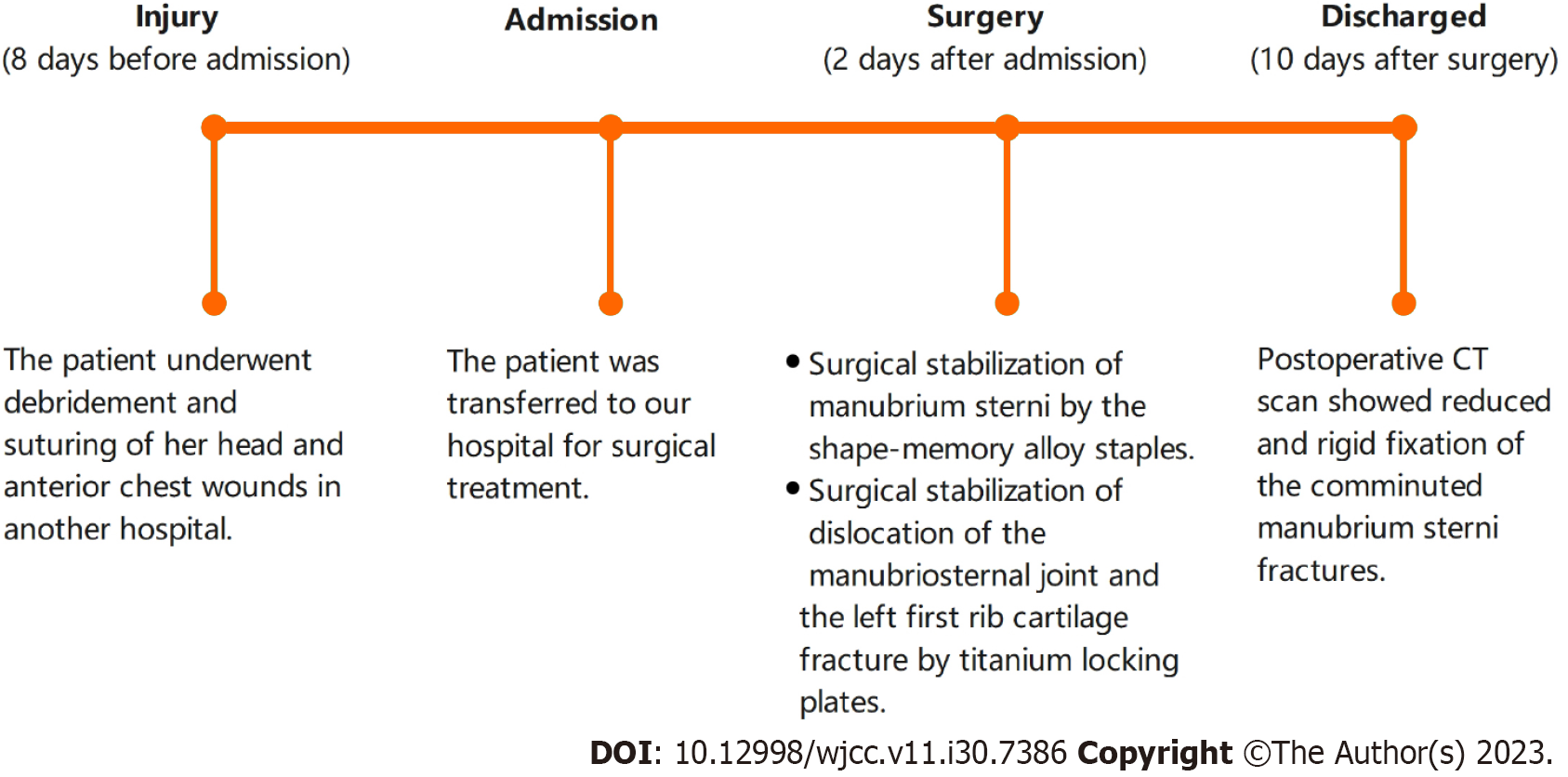
A 17-year-old female was injured in a car accident for which she underwent debridement and suturing of her head and anterior chest wounds in another hospital. Eight days later, the patient was transferred to our hospital for surgical treatment. The manubrium sterni was found intraoperatively to be split into three irregular fragments with obvious overlap and separation displacement. Mean
Shape-memory alloy staples had the advantage of being safe and effective during the repositioning and internal fixation of comminuted manubrium sterni fractures. Therefore, they provided a new surgical option for comminuted manubrium sterni fractures.
Core Tip: Manubrium sterni fractures are rare. Plate osteosynthesis is usually used for unstable transverse and oblique fractures. However, the surgical approach for comminuted manubrium sterni fractures has yet to be reported in the literature. In this case, comminuted manubrium sterni fractures, manubriosternal joint dislocation, and a left first rib cartilage fracture co-occurred in the patient. A creative attempt was made to use shape-memory alloy staples for repositioning and primary fixation of the manubrium sterni fragments. A postoperative computed tomography scan showed reduced and rigid fixation of the comminuted manubrium sterni fractures. This successful attempt provides a new solution for comminuted manubrium sterni fractures.
- Citation: Zhang M, Jiang W, Wang ZX, Zhou ZM. Using shape-memory alloy staples to treat comminuted manubrium sterni fractures: A case report. World J Clin Cases 2023; 11(30): 7386-7392
- URL: https://www.wjgnet.com/2307-8960/full/v11/i30/7386.htm
- DOI: https://dx.doi.org/10.12998/wjcc.v11.i30.7386
The incidence of manubrium sterni fractures is low, and most stable manubrium sterni fractures are treated conservatively. Unstable manubrium sterni fractures, which usually have significant clinical symptoms, such as persistent severe chest pain and chronic nonunion, often require plate osteosynthesis[1]. Comminuted manubrium sterni fractures are usually caused by a combination of severe direct and indirect violence in the anterior region of the thorax. They are usually accompanied with peripheral tissue injuries that are infrequently observed clinically. Repositioning and internal fixation for complex comminuted manubrium sterni fractures are very difficult with conventional methods. In this report, a patient presented with complex comminuted manubrium sterni fractures combined with manubriosternal joint dislocation and a left first rib cartilage fracture. Shape-memory alloy staples (Lanzhou Seemine Shape Memory Alloy Co., Ltd, China) were used for repositioning and primary fixation of the manubrium sterni fragments, followed by two titanium locking plates (DePuy Synthes, United States) for longitudinal fixation across the manubriosternal joint to enhance stability. Then, the left first rib cartilage fracture was fixed. The publication of this case report and the accompanying images was obtained with the patient’s written consent.
A 17-year-old female injured her head and anterior chest in a car accident.
The patient was admitted to another hospital with severe pain and bleeding in her front chest. She underwent debridement and suturing of her head and anterior chest wounds. Then, the patient was transferred to our hospital for surgical treatment.
The patient had no other underlying diseases.
The patient had no other relevant personal or family history.
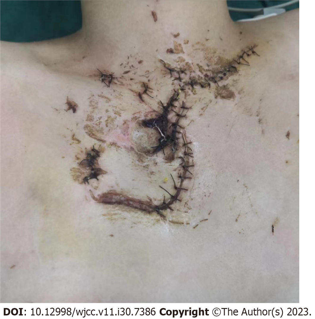
Physical examination revealed chest deformity and paradoxical breathing movements of the anterior chest wall. An irregular anterior chest wound with signs of necrosis and infection was observed (Figure 1).
Most of the laboratory examinations were in the normal range, except the hemogram, which indicated that her white blood cells were 10.0 × 10⁹/L.
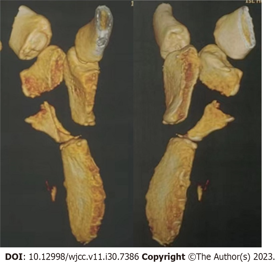
Her computed tomography three-dimensional reconstruction revealed a comminuted fracture of the manubrium sterni with dislocation of the manubriosternal joint (Figure 2).
The final diagnosis was comminuted fracture of the manubrium sterni, dislocation of the manubriosternal joint, left first rib cartilage fracture, left hemothorax, and lung contusion.
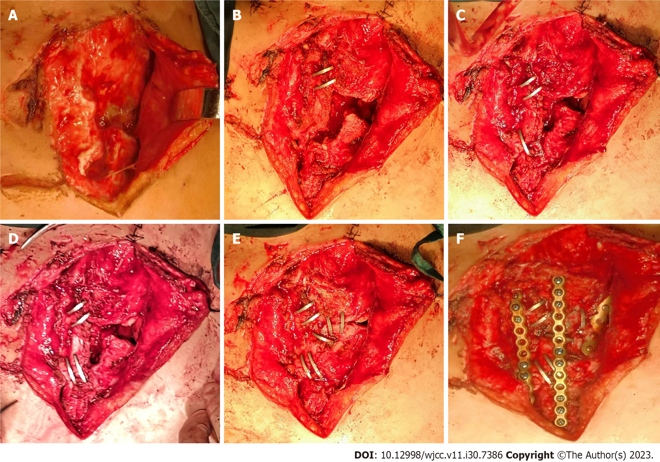
After examination and preoperative evaluation, the patient was administered general anesthesia on the second day of admission. The crushed manubrium sterni was revealed after removal of the sutures (Figure 3A). The manubrium sterni was found to be split into three irregular fragments with obvious overlap and separation displacement, and the fracture sections were wrapped in developing fibrous tissue. Meanwhile, dislocation of the manubriosternal joint and the left first rib cartilage fracture were observed. The developing fibrous tissue wrapped around the fracture sections was removed, and then the fracture fragments were pulled together using the retraction force of the shape-memory alloy staples (Figure 3B-E). Two more titanium locking plates were used to fix the manubrium sterni and corpus sterni longitudinally. The left first rib cartilage fracture was repositioned and fixed with a titanium locking plate with one end fixed to the first rib and the other to the more stable lower left fragment of the manubrium sterni (Figure 3F). Finally, after a thorough debridement, one subcutaneous drainage tube and one left chest tube were placed, and the incision was re-sutured layer by layer.
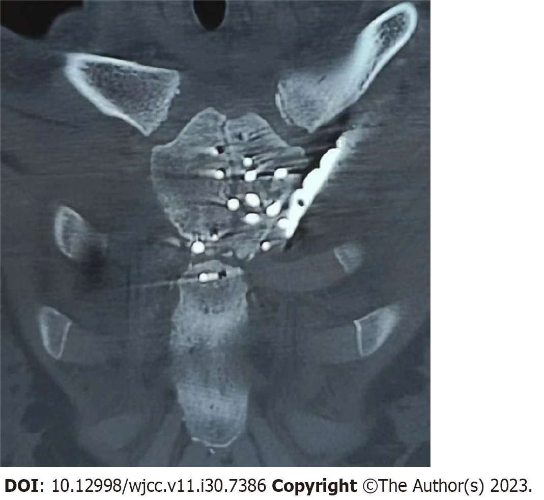
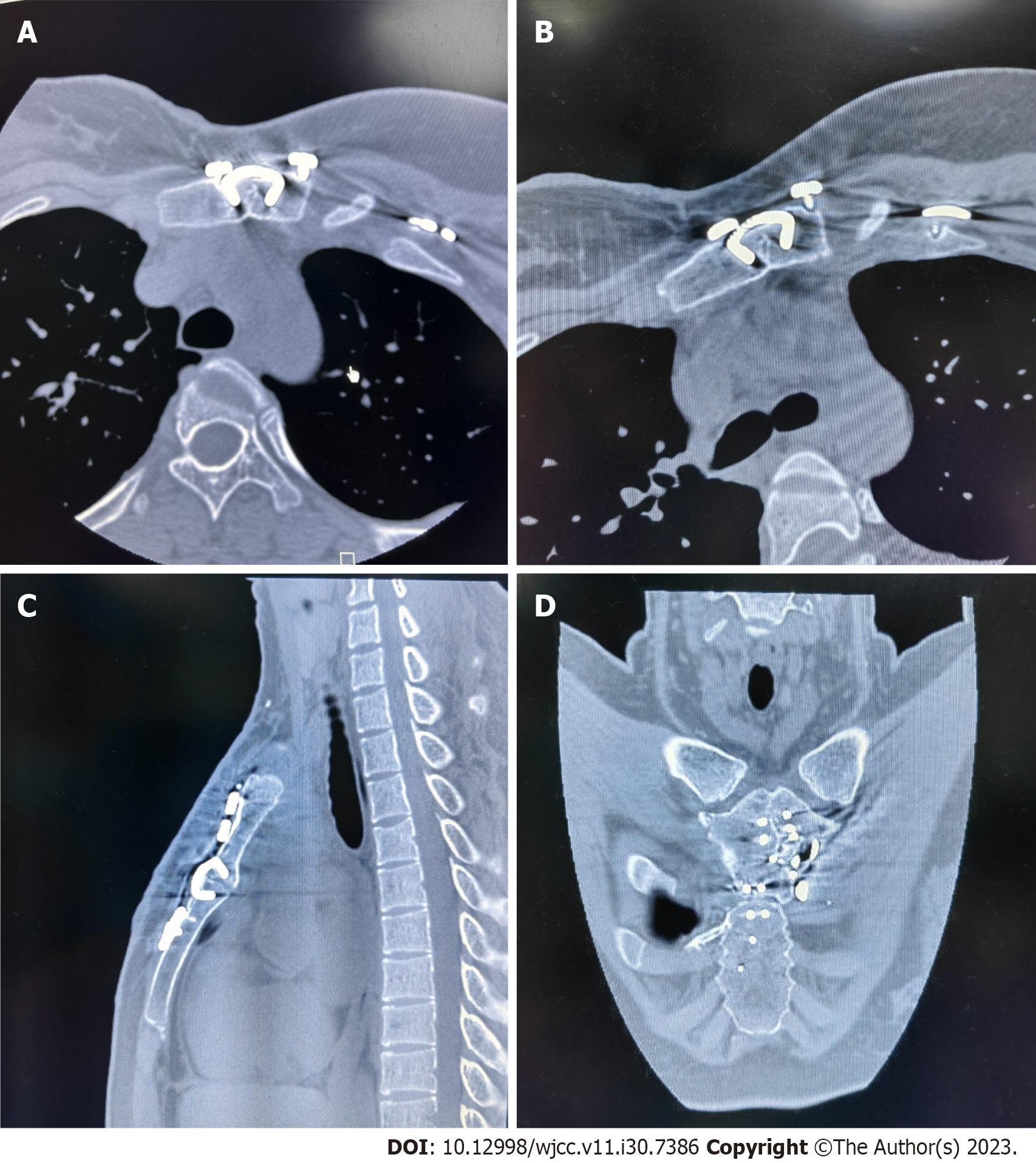
A postoperative computed tomography scan showed reduced and rigid fixation of the comminuted manubrium sterni fractures (Figure 4). The patient was discharged 10 d postoperatively after the stitches had been removed. The patient had a 24-mo follow-up to evaluate recovery. At this time, a computed tomography scan was performed (Figure 5). No radiographic evidence of staple loosening was observed. The patient recovered well with no significant complaint of discomfort (Figure 6).
Manubrium sterni fractures are rare. Schulz-Drost et al[2] classified manubrium sterni fractures into three types: transverse, oblique, and comminuted. Good results are usually obtained for unstable transverse and oblique fractures using titanium locking plate fixation after repositioning. Takeda et al[3] achieved satisfactory results using a mesh plate for oblique fractures of the manubrium sterni. For comminuted manubrium sterni fractures, Schulz-Drost et al[2] used T- or H-shaped plates for internal surgical fixation. Compared to their cases, the case presented here is more complex. The patient’s manubrium sterni fragments were more irregular and had significant separation and overlapping displacement. Worse, the patient had a combination of manubriosternal joint dislocation and left first rib cartilage fracture, making it challenging to complete fixation using the internal fixation described in the literature.
The shape-memory alloy staples are not new to orthopedic fracture surgery. The earliest clinical review of fracture fixation using shape-memory alloy staples was reported by Yang and colleagues[4] in 1987. They documented 51 cases of fractures and arthrodeses by shape-memory alloy staples. Most of the cases were small bones in the foot and ankle. The popularity of shape-memory alloy staples has increased, and they have been widely used in osteotomies, arthrodeses, and fracture fixation. Recent reports about shape-memory alloy staple fixation have included scapula fractures[5], intercarpal fusions[6], patellar fractures[7], first metatarsophalangeal joint arthrodeses[8] and spinal surgeries[9].
There are many advantages of shape-memory alloy staples. First, they can complete the reduction and internal fixation by their retraction when traditional techniques are difficult or impossible. In this case, the shape-memory alloy staples have demonstrated their advantages in challenging fracture patterns. Secondly, shape-memory alloy staples may provide strong preliminary fixation and excellent stability through their strong retraction force, especially when multiple shape-memory alloy staples are used[10]. In this patient, shape-memory alloy staples were creatively used to restore the separated and overlapping fragmented manubrium sterni to their original shape. This was possible due to the strong retraction force of the staples. Furthermore, the staples offered the possibility of later fixation of the dislocated manubriosternal joint and left first rib cartilage fracture with a titanium locking plate because this implant is small and takes up little space. Despite the obvious advantages, there are still some limitations of using shape-memory alloy staples. For example, shape-memory alloy staples cannot be used in osteoporotic bone and highly comminuted fractures. In addition, galvanic corrosion is an issue when different metals are used in the same area.
This positive experience of surgical treatment using shape-memory alloy staples may help establish new surgical approaches in cases of comminuted manubrium sterni fractures.
We thank the patient and staff who participated in this work.
Provenance and peer review: Unsolicited article; Externally peer reviewed.
Peer-review model: Single blind
Specialty type: Surgery
Country/Territory of origin: China
Peer-review report’s scientific quality classification
Grade A (Excellent): 0
Grade B (Very good): B
Grade C (Good): 0
Grade D (Fair): 0
Grade E (Poor): 0
P-Reviewer: Sa-Ngasoongsong P, Thailand S-Editor: Liu JH L-Editor: A P-Editor: Zhao S
| 1. | Harston A, Roberts C. Fixation of sternal fractures: a systematic review. J Trauma. 2011;71:1875-1879. [RCA] [PubMed] [DOI] [Full Text] [Cited by in Crossref: 33] [Cited by in RCA: 37] [Article Influence: 2.6] [Reference Citation Analysis (0)] |
| 2. | Schulz-Drost S, Krinner S, Oppel P, Grupp S, Schulz-Drost M, Hennig FF, Langenbach A. Fractures of the manubrium sterni: treatment options and a possible classification of different types of fractures. J Thorac Dis. 2018;10:1394-1405. [RCA] [PubMed] [DOI] [Full Text] [Cited by in Crossref: 7] [Cited by in RCA: 9] [Article Influence: 1.3] [Reference Citation Analysis (0)] |
| 3. | Takeda S, Yamamoto M, Mitsuya S, Hashimoto K, Hirata H, Yamauchi KI. Surgical fixation by mesh plate and intraoperative safe techniques for the manubrium sterni. Trauma Case Rep. 2021;33:100462. [RCA] [PubMed] [DOI] [Full Text] [Full Text (PDF)] [Reference Citation Analysis (0)] |
| 4. | Yang PJ, Zhang YF, Ge MZ, Cai TD, Tao JC, Yang HP. Internal fixation with Ni-Ti shape memory alloy compressive staples in orthopedic surgery. A review of 51 cases. Chin Med J (Engl). 1987;100:712-714. [PubMed] [DOI] [Full Text] |
| 5. | Dunn J, Kusnezov N, Fares A, Mitchell J, Pirela-Cruz M. The Scaphoid Staple: A Systematic Review. Hand (N Y). 2017;12:236-241. [RCA] [PubMed] [DOI] [Full Text] [Cited by in Crossref: 14] [Cited by in RCA: 20] [Article Influence: 2.5] [Reference Citation Analysis (0)] |
| 6. | Toby EB, McGoldrick E, Chalmers B, McIff T. Rotational stability for intercarpal fixation is enhanced by a 4-tine staple. J Hand Surg Am. 2014;39:880-887. [RCA] [PubMed] [DOI] [Full Text] [Cited by in Crossref: 2] [Cited by in RCA: 1] [Article Influence: 0.1] [Reference Citation Analysis (0)] |
| 7. | Schnabel B, Scharf M, Schwieger K, Windolf M, Pol Bv, Braunstein V, Appelt A. Biomechanical comparison of a new staple technique with tension band wiring for transverse patella fractures. Clin Biomech (Bristol, Avon). 2009;24:855-859. [RCA] [PubMed] [DOI] [Full Text] [Cited by in Crossref: 33] [Cited by in RCA: 47] [Article Influence: 2.9] [Reference Citation Analysis (0)] |
| 8. | Willmott H, Al-Wattar Z, Halewood C, Dunning M, Amis A. Evaluation of different shape-memory staple configurations against crossed screws for first metatarsophalangeal joint arthrodesis: A biomechanical study. Foot Ankle Surg. 2018;24:259-263. [RCA] [PubMed] [DOI] [Full Text] [Cited by in Crossref: 5] [Cited by in RCA: 6] [Article Influence: 0.9] [Reference Citation Analysis (0)] |
| 9. | Yaszay B, Doan JD, Parvaresh KC, Farnsworth CL. Risk of Implant Loosening After Cyclic Loading of Fusionless Growth Modulation Techniques: Nitinol Staples Versus Flexible Tether. Spine (Phila Pa 1976). 2017;42:443-449. [RCA] [PubMed] [DOI] [Full Text] [Cited by in Crossref: 3] [Cited by in RCA: 2] [Article Influence: 0.3] [Reference Citation Analysis (0)] |
| 10. | McKnight RR, Lee SK, Gaston RG. Biomechanical Properties of Nitinol Staples: Effects of Troughing, Effective Leg Length, and 2-Staple Constructs. J Hand Surg Am. 2019;44:520.e1-520.e9. [RCA] [PubMed] [DOI] [Full Text] [Cited by in Crossref: 8] [Cited by in RCA: 14] [Article Influence: 2.3] [Reference Citation Analysis (0)] |


