Published online Oct 16, 2023. doi: 10.12998/wjcc.v11.i29.7242
Peer-review started: August 19, 2023
First decision: September 4, 2023
Revised: September 7, 2023
Accepted: September 18, 2023
Article in press: September 18, 2023
Published online: October 16, 2023
Processing time: 55 Days and 0.5 Hours
Spontaneous cerebrospinal fluid (CSF) leaks associated with cervical spondylosis are rare. To our knowledge, only a few cases have been reported in which treat
A 70-year-old female patient who was treated for a cerebral infarction, presented with complains of weakness in the right lower extremity and a feeling of stepping on cotton. The patient underwent regular neck massage and presented with neck and right shoulder pain radiating to the right upper extremity one-month ago. Magnetic resonance imaging showed a strip of leaking cerebrospinal fluid posterior to the C1-4 vertebrae, and computed tomography showed a “sickle-shaped” disc prolapse with calcification in C4/5. We chose to perform an anterior cervical discectomy. When the prolapsed C4/5 disc was scraped, clear fluid leakage was observed, and exploration revealed a 1 mm diameter rupture in the anterior aspect of the dura mater, which was compressed continuously with cotton patties, with no significant cerebrospinal fluid leakage after 1 h.
Three months after surgery, the patient was asymptomatic and follow-up imaging demonstrated complete resolution.
Core Tip: We report a 70-year-old female patient who had been treated for cerebral infarction. She complained of weakness in the right lower extremity and a feeling of stepping on cotton. Magnetic resonance imaging showed a strip of leaking cerebrospinal fluid posterior to the C1-4 vertebrae, and computed tomography showed a "sickle-shaped" disc prolapse with calcification in C4/5. We chose to perform an anterior cervical discectomy. When the prolapsed C4/5 disc was scraped, clear fluid leakage was seen and exploration revealed a 1 mm diameter rupture in the anterior aspect of the dura mater, with no significant cerebrospinal fluid leakage after 1 h.
- Citation: Yu Z, Zhang HFZ, Wang YJ. Surgical treatment of mixed cervical spondylosis with spontaneous cerebrospinal fluid leakage: A case report. World J Clin Cases 2023; 11(29): 7242-7247
- URL: https://www.wjgnet.com/2307-8960/full/v11/i29/7242.htm
- DOI: https://dx.doi.org/10.12998/wjcc.v11.i29.7242
Spontaneous cerebrospinal fluid (CSF) leakage refers to CSF leakage in the absence of major trauma, forceful external impact, or a history of surgery (such as anterior cervical, posterior cervical, or lumbar puncture)[1]. Clinically, the incidence of spontaneous CSF leakage is relatively low, and cervical spondylosis combined with spontaneous CSF leakage has rarely been reported. Here, we report a case of mixed-type cervical spondylosis with spontaneous CSF leakage and present a literature review.
The patient, a 70-year-old female, developed numbness and weakness in her right lower limb for no apparent reason 18 mo ago, with the sensation of “walking on cotton” in her right foot.
She did not experience postural headache, nausea, vomiting, neck and shoulder pain, or radiating pain. The patient was treated with physiotherapy and massage of her lower back and lower limbs at a local hospital for the sequelae of cerebral infarction. Treatment was ineffective, and her symptoms continued to worsen. One year prior, she had fallen twice due to unstable walking and developed pain in her neck and right shoulder, radiating to her right upper limb. The patient did not visit the hospital on time due to the coronavirus disease 2019 pandemic and was treated with traditional Chinese medicine. She underwent 10 neck massages, 8 acupuncture sessions, and 10 modulated medium-frequency electrotherapy sessions at a local clinic, but her symptoms were not relieved. Recently, these symptoms had worsened, affecting her everyday life and work.
Straightening of normal cervical lordosis and muscle spasms on the right side of the neck and shoulder. Hypoesthesia far to the right of the sternal angle; diminished abdominal wall reflexes; bilateral biceps, knee, and Achilles tendon hyperreflexia; bilateral positive ankle clonus, positive patellar clonus, positive Hoffmann sign, and positive Babinski sign. There was increased muscle tone in the right lower limb, with grade 3 muscle strength in the right anterior tibial muscle and grade 4 strength in the rest. The Japanese Orthopaedic Association score was 10, the score was 6, and the Cervical Dysfunction Index Scale score was 37.5%.
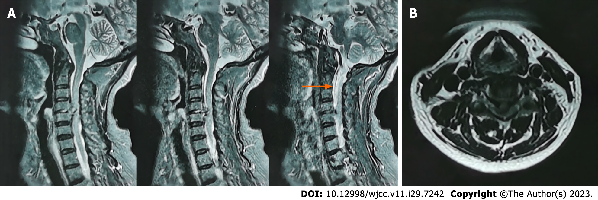
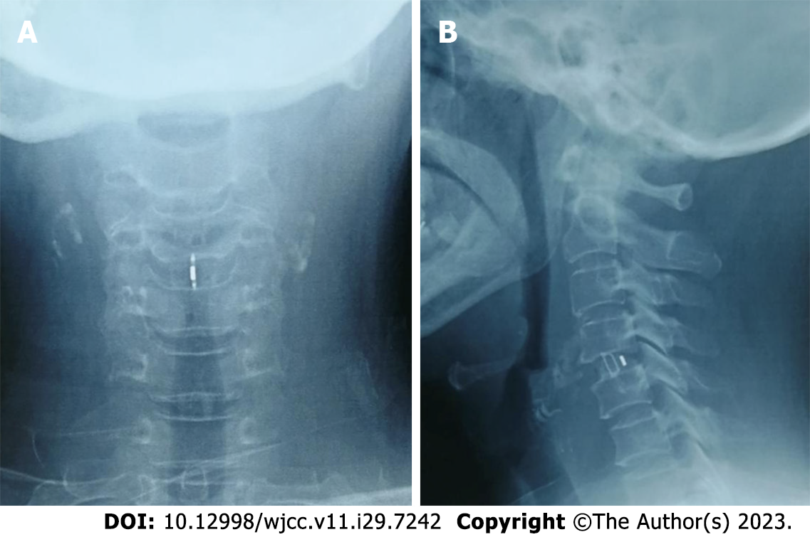
Magnetic resonance imaging (MRI) of the cervical spine showed protrusion of the cervical spine 4/5 (C4/5) disc into the spinal canal, stenosis of the spinal canal at the level of the corresponding disc, a strip of leaking cerebrospinal fluid at the level of the posterior C1-4 vertebrae, loss of epidural space, compression of the spinal cord, and a high-signal area in the medulla (Figure 1). Computed tomography (CT) of the cervical spine revealed “sickle-shaped” disc prolapse and calcification at the level of C4/5, small sagittal diameter of the cervical spinal canal, and loss of epidural fat (Figure 2).
Mixed-type cervical spondylosis with spontaneous CSF leakage. The patient experienced severe neurogenic pain and conservative treatment was ineffective. The spinal cord and nerve roots are significantly compressed, with neurological dysfunction affecting everyday life and work, which is indicative of surgery. After a thorough investigation, the author decided to perform anterior cervical discectomy decompression and fusion with bone grafting and if necessary, dural repair.
The patient was placed in the supine position and general anesthesia was administered after endotracheal intubation. A soft leather cushion bolster was placed under the shoulder and a cylindrical soft bolster was placed under the neck. The target segment was identified by X-ray fluoroscopy. The surgical site was disinfected using povidone-iodine. A transverse incision approximately 2 cm in length was made from the medial border of the right sternocleidomastoid muscle to the anterior midline. The skin, subcutaneous tissue, and broad cervical muscle were incised layer-by-layer, and the superficial cervical fascia between the carotid sheath and tracheoesophagus was bluntly separated to reach the anterior fascia. The C4/5 segment was identified using fluoroscopy, and a Caspar cervical retractor was placed parallel to the vertebral endplate to fully expose the intervertebral space. When the prolapsed C4/5 disc was scraped, clear fluid leakage was observed, and exploration revealed a 1 mm diameter rupture in the anterior aspect of the dura mater, which was compressed continuously with cotton patties, with no significant cerebrospinal fluid leakage after 1 h. After adequate decompression, the adjacent upper and lower vertebral end plates were decorticated for effective bone grafting. The height and width of the decompressed intervertebral space were measured using a trial mold. A cervical fusion cage with a height of 6.9 mm (Fidji Cervical Cage, Zimmer, Bordeaux, France) loaded with a homogeneous bone graft (Bio-Gene, Beijing, China) was inserted into the C4/5 intervertebral space. A drainage tube was placed, and the incision was closed layer-by-layer.
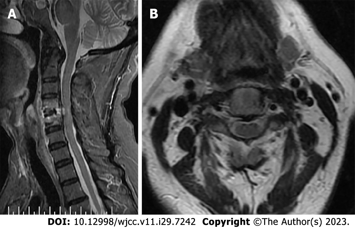
On the first post-operative day, approximately 45 mL of sanguineous drainage was observed. Pain and numbness in the right upper limb were significantly reduced, muscle tone in the right lower limb was also reduced, and there was no headache. On the second post-operative day, about 10 mL of sanguineous fluid was drained, pain in the right upper limb was relieved, numbness was further reduced, heaviness in the right lower limb was relieved, and the right patellar clonus and ankle clonus disappeared. There was almost no drainage on the third postoperative day, and the muscle strength almost returned to normal. After six days of bed rest, the patient gradually sat up and went down to the floor and walked smoothly with no headache or dizziness. The patient still had slight numbness in the right upper limb, weakly positive Hoffman’s sign, slightly high muscle tension in the right lower limb, and weakly positive Bartholomew’s sign. The sutures were removed one week after surgery as the wound healed completely. Half a month after the operation, the muscle tone of the right lower limb has returned to normal, the feeling of “walking on cotton” disappeared, the muscle strength was normal, and there were no more pathological signs. Mild heaviness was observed in the right lower limb. Three months after the operation, recovery was nearly complete (Figures 3 and 4). The clinical results are shown in Table 1.
| JOA | VAS | NDI (%) | |
| Preoperative | 10 | 6 | 37.50 |
| The first d after surgery | 12 | 4 | 20.60 |
| Fourteen d after surgery | 14 | 3 | 16.30 |
| Three mon after surgery | 16 | 2 | 3.06 |
To our knowledge, six cases of spontaneous CSF leakage associated with cervical spondylosis have been reported in the literature (Table 2)[1-4]. It often occurs at the level of the C4/5 and C5/6 intervertebral spaces, mostly in combination with an osteophyte at the posterior edge of the vertebral body or a herniated disc. The symptoms include postural headaches. It is generally believed that the cause of spontaneous CSF leakage is related to weak spinal structures, and dural weakness can induce a dural defect, allowing cerebrospinal fluid to leak into the epidural space; a variety of dural defects can be observed intraoperatively[5]. Another less common cause is dural rupture associated with degenerative spinal pathologies, such as disc herniation, bone hypergrowth, or bone spur formation[6-8]. We believe that the cause of spontaneous CSF leakage in this case was related to the old age of the patient, weak dural structure, repeated cervical massages, acupuncture, and modulated medium-frequency electrotherapy at a local clinic, which caused the herniated and calcified intervertebral disc to repeatedly rub against the dura mater, resulting in dural rupture and spontaneous CSF leakage.
| Ref. | Jounal | Age and gender | Intervention | Clinical outcome | Follow-up |
| Witiw et al[2] | Eur Spine, 2012 | 46/F | C4-5 anterior discectomy and resection of the posterior osteophyte | Asymptomatic | 2 mo |
| Arai et al[4] | No Shinkei Geka, 2016 | 69/F | Conservative treatment | Subjective improvement in orthostatic headaches | 36 mo |
| Vishteh et al[3] | Journal of Neurosurgery, 1998 | 32/M | C5-6 anterior discectomy and osteophytectomy with suture closure of the dura | Asymptomatic | Unknown |
| Eross et al[1] | Cephalalgia, 2002 | 44/F | C5-6 anterior discectomy and osteophtectomy, C6 and C7 corpectomy and lumboperitoneal shunt | Persistent debilitating orthostatic headache | 32 mo |
| Eross et al[1] | Cephalalgia, 2002 | 39/F | C7-T1 anterior discectomy and osteophytectomy with suture closure of the dura | 50% subjective improvement in orthostatic headaches | 1 mo |
| Eross et al[1] | Cephalalgia, 2002 | 46/M | Lumbar autologous epidural blood patch and propoxyphrene | Subjective improvement in orthostatic headaches | 2 mo |
Spontaneous CSF leakage may present with typical postural headache, nausea, and vomiting, etc[9]. Concerning imaging study, CT myelography remains the most reliable and accurate imaging method for detecting the exact location of spinal fluid leakage. Myelography clearly shows the extent of cerebrospinal fluid leakage characterized by the posterior aspect of the vertebral canal, where the contrast agent is in contact with the cerebrospinal fluid. In addition, MRI of the spine following intrathecal gadolinium injection can help identify the site of cerebrospinal fluid leakage[10,11]. Our patient did not have the typical symptoms of postural headache, nausea, or vomiting; therefore, it was considered that the cerebrospinal fluid leakage occurred insidiously and in a small amount. The current treatment of choice for cerebrospinal fluid leaks is one-stage suture repair of the injured dural site[12]. When the dural defect is too large for direct suturing or cannot be observed, it can be repaired by muscle/fascia graft, gelatin sponge, artificial dura, or fibrin glue, etc[13,14]. In our case, owing to the small size of the rupture, we opted for compression with cotton patties; however, after 1 h of compression, the cerebrospinal fluid leakage stopped, and the postoperative cerebrospinal fluid leakage was less. If intraoperative cerebral cotton compression stopped the leakage of cerebrospinal fluid, or if there was more postoperative CSF leakage, we would have chosen to leave a lumbar pool drain in place.
If spinal cervical spondylosis occurs, even in the absence of obvious trauma, spontaneous CSF leakage may occur because of active neck exercise or passive massage movement. A combination of disc herniation and bone loss was also observed. Alternatively, postural headache as the main feature of cerebrospinal fluid leakage may not be present[4]. Treatment can be conservative or surgical, depending on the specific circumstances of the CSF leak.
Provenance and peer review: Unsolicited article; Externally peer reviewed.
Peer-review model: Single blind
Specialty type: Medicine, research and experimental
Country/Territory of origin: China
Peer-review report’s scientific quality classification
Grade A (Excellent): 0
Grade B (Very good): B
Grade C (Good): 0
Grade D (Fair): 0
Grade E (Poor): 0
P-Reviewer: Chrastina J, Czech Republic S-Editor: Qu XL L-Editor: A P-Editor: Yu HG
| 1. | Eross EJ, Dodick DW, Nelson KD, Bosch P, Lyons M. Orthostatic headache syndrome with CSF leak secondary to bony pathology of the cervical spine. Cephalalgia. 2002;22:439-443. [RCA] [PubMed] [DOI] [Full Text] [Cited by in Crossref: 54] [Cited by in RCA: 53] [Article Influence: 2.3] [Reference Citation Analysis (0)] |
| 2. | Witiw CD, Fallah A, Muller PJ, Ginsberg HJ. Surgical treatment of spontaneous intracranial hypotension secondary to degenerative cervical spine pathology: a case report and literature review. Eur Spine J. 2012;21 Suppl 4:S422-S427. [RCA] [PubMed] [DOI] [Full Text] [Cited by in Crossref: 19] [Cited by in RCA: 14] [Article Influence: 1.0] [Reference Citation Analysis (0)] |
| 3. | Vishteh AG, Schievink WI, Baskin JJ, Sonntag VK. Cervical bone spur presenting with spontaneous intracranial hypotension. Case report. J Neurosurg. 1998;89:483-484. [RCA] [PubMed] [DOI] [Full Text] [Cited by in Crossref: 67] [Cited by in RCA: 67] [Article Influence: 2.5] [Reference Citation Analysis (0)] |
| 4. | Arai A, Miyamoto H, Shiomi R, Tatsumi S, Kohmura E. [A Case of Spontaneous Cerebrospinal Fluid Leak Associated with Cervical Spondylosis]. No Shinkei Geka. 2016;44:767-772. [RCA] [PubMed] [DOI] [Full Text] [Reference Citation Analysis (0)] |
| 5. | Cohen-Gadol AA, Mokri B, Piepgras DG, Meyer FB, Atkinson JL. Surgical anatomy of dural defects in spontaneous spinal cerebrospinal fluid leaks. Neurosurgery. 2006;58:ONS-238. [RCA] [PubMed] [DOI] [Full Text] [Cited by in Crossref: 13] [Cited by in RCA: 23] [Article Influence: 1.2] [Reference Citation Analysis (0)] |
| 6. | Binder DK, Sarkissian V, Dillon WP, Weinstein PR. Spontaneous intracranial hypotension associated with transdural thoracic osteophyte reversed by primary. dural repair. Case report. J Neurosurg Spine. 2005;2:614-618. [RCA] [PubMed] [DOI] [Full Text] [Cited by in Crossref: 38] [Cited by in RCA: 39] [Article Influence: 2.0] [Reference Citation Analysis (0)] |
| 7. | Winter SC, Maartens NF, Anslow P, Teddy PJ. Spontaneous intracranial hypotension due to thoracic disc herniation. Case report. J Neurosurg. 2002;96:343-345. [RCA] [PubMed] [DOI] [Full Text] [Cited by in Crossref: 6] [Cited by in RCA: 11] [Article Influence: 0.5] [Reference Citation Analysis (0)] |
| 8. | Rapport RL, Hillier D, Scearce T, Ferguson C. Spontaneous intracranial hypotension from intradural thoracic disc herniation. Case report. J Neurosurg. 2003;98:282-284. [RCA] [PubMed] [DOI] [Full Text] [Cited by in Crossref: 4] [Cited by in RCA: 10] [Article Influence: 0.5] [Reference Citation Analysis (0)] |
| 9. | Guerin P, El Fegoun AB, Obeid I, Gille O, Lelong L, Luc S, Bourghli A, Cursolle JC, Pointillart V, Vital JM. Incidental durotomy during spine surgery: incidence, management and complications. A retrospective review. Injury. 2012;43:397-401. [RCA] [PubMed] [DOI] [Full Text] [Cited by in Crossref: 155] [Cited by in RCA: 191] [Article Influence: 14.7] [Reference Citation Analysis (0)] |
| 10. | Jinkins JR, Rudwan M, Krumina G, Tali ET. Intrathecal gadolinium-enhanced MR cisternography in the evaluation of clinically suspected cerebrospinal fluid rhinorrhea in humans: early experience. Radiology. 2002;222:555-559. [RCA] [PubMed] [DOI] [Full Text] [Cited by in Crossref: 58] [Cited by in RCA: 59] [Article Influence: 2.6] [Reference Citation Analysis (0)] |
| 11. | Tali ET, Ercan N, Krumina G, Rudwan M, Mironov A, Zeng QY, Jinkins JR. Intrathecal gadolinium (gadopentetate dimeglumine) enhanced magnetic resonance myelography and cisternography: results of a multicenter study. Invest Radiol. 2002;37:152-159. [RCA] [PubMed] [DOI] [Full Text] [Cited by in Crossref: 71] [Cited by in RCA: 73] [Article Influence: 3.2] [Reference Citation Analysis (0)] |
| 12. | Faltings L, Kulason KO, Du V, Schneider JR, Chakraborty S, Kwan K, Pramanik B, Boockvar J. Early Epidural Blood Patch to Treat Intracranial Hypotension after Iatrogenic Cerebrospinal Fluid Leakage from Lumbar Tubular Microdiscectomy. Cureus. 2018;10:e3633. [RCA] [PubMed] [DOI] [Full Text] [Full Text (PDF)] [Cited by in Crossref: 1] [Cited by in RCA: 5] [Article Influence: 0.7] [Reference Citation Analysis (0)] |
| 13. | Yokota H, Yokoyama K, Nishioka T, Iwasaki S. Active cerebrospinal fluid leakage after resolution of postdural puncture headache. J Anesth. 2012;26:318-319. [RCA] [PubMed] [DOI] [Full Text] [Cited by in Crossref: 1] [Cited by in RCA: 1] [Article Influence: 0.1] [Reference Citation Analysis (0)] |
| 14. | Sun X, Sun C, Liu X, Liu Z, Qi Q, Guo Z, Leng H, Chen Z. The frequency and treatment of dural tears and cerebrospinal fluid leakage in 266 patients with thoracic myelopathy caused by ossification of the ligamentum flavum. Spine (Phila Pa 1976). 2012;37:E702-E707. [RCA] [PubMed] [DOI] [Full Text] [Cited by in Crossref: 58] [Cited by in RCA: 65] [Article Influence: 5.0] [Reference Citation Analysis (0)] |


