Published online Oct 16, 2023. doi: 10.12998/wjcc.v11.i29.7150
Peer-review started: June 19, 2023
First decision: August 9, 2023
Revised: August 15, 2023
Accepted: September 25, 2023
Article in press: September 25, 2023
Published online: October 16, 2023
Processing time: 111 Days and 13.9 Hours
Complicated cataract surgery is challenging, especially in cases of hard nuclear cataract with severe anterior capsule organization. It is important to avoid the risk of surgery and improve the surgical skills of surgeons.
A 60-year-old man presented with severe cataract and visual impairment. The anterior capsule of the lens was irregularly organized and pulled to the surrounding capsule, and white porcelain organized cord and brown-black lens nucleus were clearly visible. In phacoemulsification, maintaining the anterior capsule round and intact plays a key role in a successful surgery. In this case, if the conventional capsule treatment method was used, the anterior capsule would be torn. Therefore, we adopted a segmented anterior capsule treatment method, and a blasting method to release energy when dealing with the lens nucleus, and achieved good surgical results.
Complicated cataract surgery is challenging and requires precise skills. Operation plans should be made reasonably to predict the risk of surgery, and improve the visual quality of the patients.
Core Tip: This report aims to guide clinicians on the issues they should pay attention to when dealing with complex cataracts, and on the surgical techniques. We report a 60-year-old male patient with severe cataract and visual impairment. The severe anterior capsular organization and the hard lens nucleus made the operation difficult. We adopted an ingenious surgery which mainly focused on two aspects. First, when dealing with the anterior capsule of the lens, the ingenious operation is applied to avoid the anterior capsule tear and serious complications. Second, when dealing with extremely hard nuclear cataract, the surgical parameters are adjusted, which greatly protects the intraocular tissue while removing the cataract.
- Citation: Wang LW, Fang SF. Surgical treatment of severe anterior capsular organized hard core cataract: A case report. World J Clin Cases 2023; 11(29): 7150-7155
- URL: https://www.wjgnet.com/2307-8960/full/v11/i29/7150.htm
- DOI: https://dx.doi.org/10.12998/wjcc.v11.i29.7150
Cataract is the world's first blinding eye disease, and surgical removal is the only treatment. To safely and effectively remove the turbid lens is the main purpose of the operation. With the development of cataract surgery technology, patients’ requirements for visual quality after cataract surgery are increasing, and cataract surgery is becoming easier in some complex cataract cases. We here report a case of grade IV cataract surgery for severe organization of the anterior capsule in a 60-year-old male patient, in an attempt to provide a reference for clinical treatment of this disease.
A 60-year-old man presented with progressive and painless blurred vision of the right eye which appeared 3 years ago.
The patient had painless loss of vision in the right eye for 3 years.
He had no previous history of chronic and ocular disease, and no history of ocular trauma.
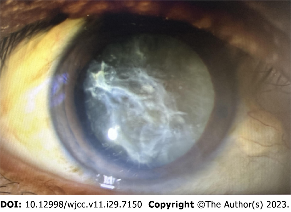
Physical examination showed a visual acuity of the right eye (VOD) manual/10 cm, visual acuity of the left eye 0.5, revised visual acuity was 0.6 ( + 2.00 DS/- 2.50 DC 100°); retinal vision of the right eye was lightperception, retinal vision of the left eye was 0.63, non-hyperemic conjunctiva in both eyes, with transparent cornea, normal anterior chamber depth round pupil, with a diameter of 3 mm, photosensitive, and the anterior capsule of the lens in the right eye was obvious, 10 to 4 points formed a dense organic film, 6 points of the organic membrane bulge, the lower capsule was stretched in the direction of the cornea and was “hilly”.
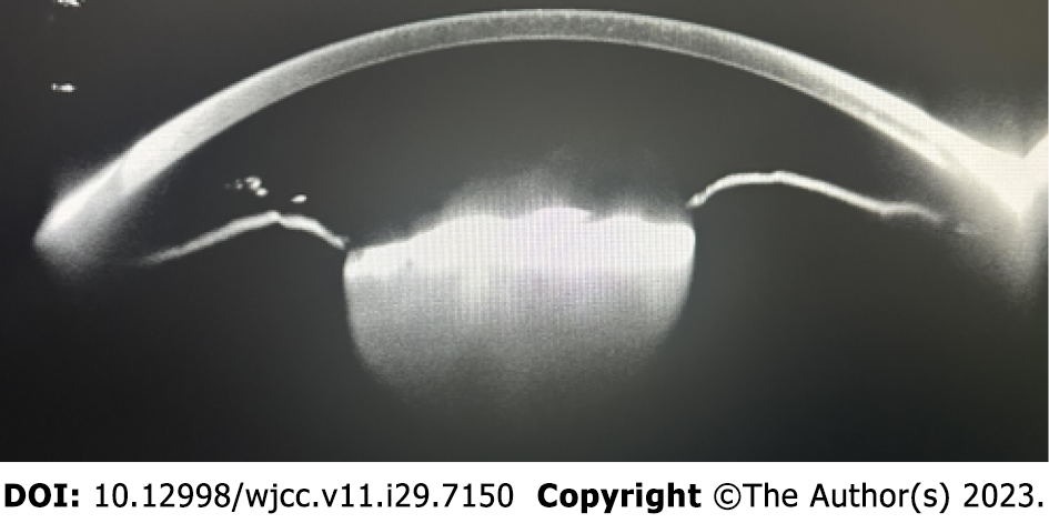
The Pentacam anterior segment analysis system revealed that the lens was a dense mass with strong reflection, the front surface of the lens was uneven, locally “hilly”, and the posterior surface echo was unclear (Figure 1).
The lens was densely white and cloudy, nuclear grade IV (Figure 2), and vitreous and fundus structures were invisible. Mild opacity of the lens was seen in the left eye, with C2N1P1, mild opacity of the vitreous, positive boundary of optic disc color, c/d = 0.3, and the flat retina was orange.
The patient was diagnosed with bilateral senile cataract.
The patient underwent surgery. We twisted the ultrasound mode with the Centurion phacoemulsifier, adjusted the parameters before surgery, increased the energy to 80%, negative pressure to 500 mmHg, flow rate to 35 mL/min, and prepared DisCoVisc to protect the corneal endothelium. After staining the anterior capsule, it was found that the anterior capsule was closely adhered to its lower tissue, forming a dense organic barrier, the tearing capsule operation was difficult, the tearing capsule started at 3 points, the intraocular shear cut the organic tissue in the direction of the extended tear, the position of the organic tissue was deep and was difficult to cut, a small amount of viscoelastic agent was injected under the organic cord to gently provoke it, and it was cut by homeopathic shearing, and it was found that the anterior capsule was torn to the periphery. The anterior chamber was injected with viscoelastic agent again, and the tearing capsule forceps continued to tear the capsule in the vertical direction of the tear. Surgery was repeated to maintain the integrity of the capsule, maintain the anterior capsule tear capsule mouth of approximately 5.2 mm, and the tearing capsule mouth was continuous, with no further tearing. The phacoemulsification needle was placed obliquely downward, the interception-splitting method was used to split the lens nucleus into several pieces, the blasting mode and the main control fluid flow mode were turned on, the diffuse viscoelastic DisCoVisc was injected into the anterior chamber many times, and the fragmented nucleus was sucked out by emulsification intermittently (Figure 3). The lens cortex was completely removed by I/A, the viscoelastic agent was injected, and the intraocular lens was implanted into the capsule bag (Figure 4). No severe tearing of the anterior capsule occurred during surgery, and posterior capsule rupture and intraocular tissue damage were not observed. One day after surgery, the VOD FC/10 cm, obvious corneal edema was seen in the right eye, no intraocular lens shift was noted, and mild inflammation was seen in the anterior chamber.
One week after surgery, VOD was 0.8, right corneal edema was absorbed, intraocular lens position was good, and a clean surface was noted. No abnormalities were observed in the fundus and the patient was satisfied with the outcome.
Phacoemulsification cataract removal is a mature and advanced cataract surgery and the main treatment method for cataracts[1]. The surgical method involves inserting a phacoemulsification needle using a small incision in the cornea or sclera to crush the turbid lens into fragments, and then suck out the fragments with the help of a suction perfusion system, while maintaining anterior chamber filling, and finally implant an intraocular lens, which can be performed under epianesthesia. This method has the advantages of a small incision, small surgical astigmatism, fast recovery, and few complications[2]. The surgical method of using ultrasound energy to emulsify the turbid lens nucleus and cortex, and then aspirate it while preserving the posterior capsule of the lens is an important means of treating hard nuclear cataracts[3]. However, overripe hard core cataract removal is extremely difficult and risky, causing intraoperative complications such as tearing the capsule, posterior capsule rupture and lens nucleus falling into the vitreous cavity. In patients accompanied by severe organization of the anterior capsule and unclear echo of the posterior capsular membrane, the surgical difficulty increases, and there are few available clinical studies on this technique. The continuous circumferential tearing of the anterior capsule centered on the anterior capsule is the key to the success of phacoemulsification of cataracts, and the pressure exerted by the continuous circumferential tearing capsule during water separation, phacoemulsification, irrigation and aspiration, and intraocular lens implantation is distributed along the smooth capsule margin, and radial tearing does not usually occur[4]. After anterior capsule staining, the shape and distribution of the organic cord can be identified easily, and capsule shears can be used with manual tearing[5]. It is not easy to use capsular scissors, the capsule needs to be within a certain size range. The position of the main incision is fixed, placing the intraocular scissors in the different points of the required direction is difficult. After the start of the tearing sac, inject a small amount of viscoelastic material under the cord to upturn or fill it to the required range of the intraocular shear, and cut it homeopathically to ensure the continuity of the anterior capsule. In addition, phacoemulsification of the hard core cataract is critical, and an improper technique can lead to serious damage of the corneal endothelium or even decom
In summary, severe anterior capsular organized hard core cataract is a rare disorder, surgical treatment is complicated, and the incidence of capsular tear is high. Attention should be paid to surgical skills, protecting the capsule and corneal endothelium, improving the quality of surgery, and reducing patient pain.
Provenance and peer review: Unsolicited article; Externally peer reviewed.
Peer-review model: Single blind
Specialty type: Medicine, research and experimental
Country/Territory of origin: China
Peer-review report’s scientific quality classification
Grade A (Excellent): 0
Grade B (Very good): 0
Grade C (Good): C
Grade D (Fair): 0
Grade E (Poor): 0
P-Reviewer: Sotelo J, Mexico S-Editor: Lin C L-Editor: Ma JY P-Editor: Lin C
| 1. | Bamdad S, Bolkheir A, Sedaghat MR, Motamed M. Changes in corneal thickness and corneal endothelial cell density after phacoemulsification cataract surgery: a double-blind randomized trial. Electron Physician. 2018;10:6616-6623. [RCA] [PubMed] [DOI] [Full Text] [Full Text (PDF)] [Cited by in Crossref: 13] [Cited by in RCA: 20] [Article Influence: 2.9] [Reference Citation Analysis (0)] |
| 2. | Lu JX, Zhang Y. Effect of phacoemulsification with different incision on corneal endothelial cells. Int J Ophthalmol. 2020;20:1578-1582. [DOI] [Full Text] |
| 3. | Shen XC, Zhong XN. Effects of phacoemulsification cataract extraction in the treatment of hard nuclear cataract and its influence on postoperative dry eye. Zhongguo Yikan. 2022;57:782-785. |
| 4. | Xu Y. Clinical study of anterior capsule fixation with intraocular lens implantation in the treatment of cataract. Zhongguo Yiyao Zhinan. 2019;17:131-132. |
| 5. | Dong S G, Guo FF, Meng XX, Yan SL. Retrospective analysis of the causes of anterior capsular tear in early femtosecond laser-assisted cataract surgery. Shandong Daxue Erbihouyan Xuebao. 2023;37:110-114. |
| 6. | Zhang HY, Sun L, Song XD, Song HX. Effects of two kinds of viscoelastics and phacoemulsification time on corneal endothelial cells and corneal thickness in phacoemulsification combined with intraocular lens implantation. Zhonghua Yanke Yixue Zazhi (Dianziban). 2020;10:357-362. |
| 7. | Wang SM. The application experience of phacoemulsification for hypermature hard nuclear cataract. Zhongguo Yejin Gongye Yixue Zazhi. 2017;34:448-449. |


