Copyright
©The Author(s) 2023.
World J Clin Cases. Sep 6, 2023; 11(25): 5926-5933
Published online Sep 6, 2023. doi: 10.12998/wjcc.v11.i25.5926
Published online Sep 6, 2023. doi: 10.12998/wjcc.v11.i25.5926
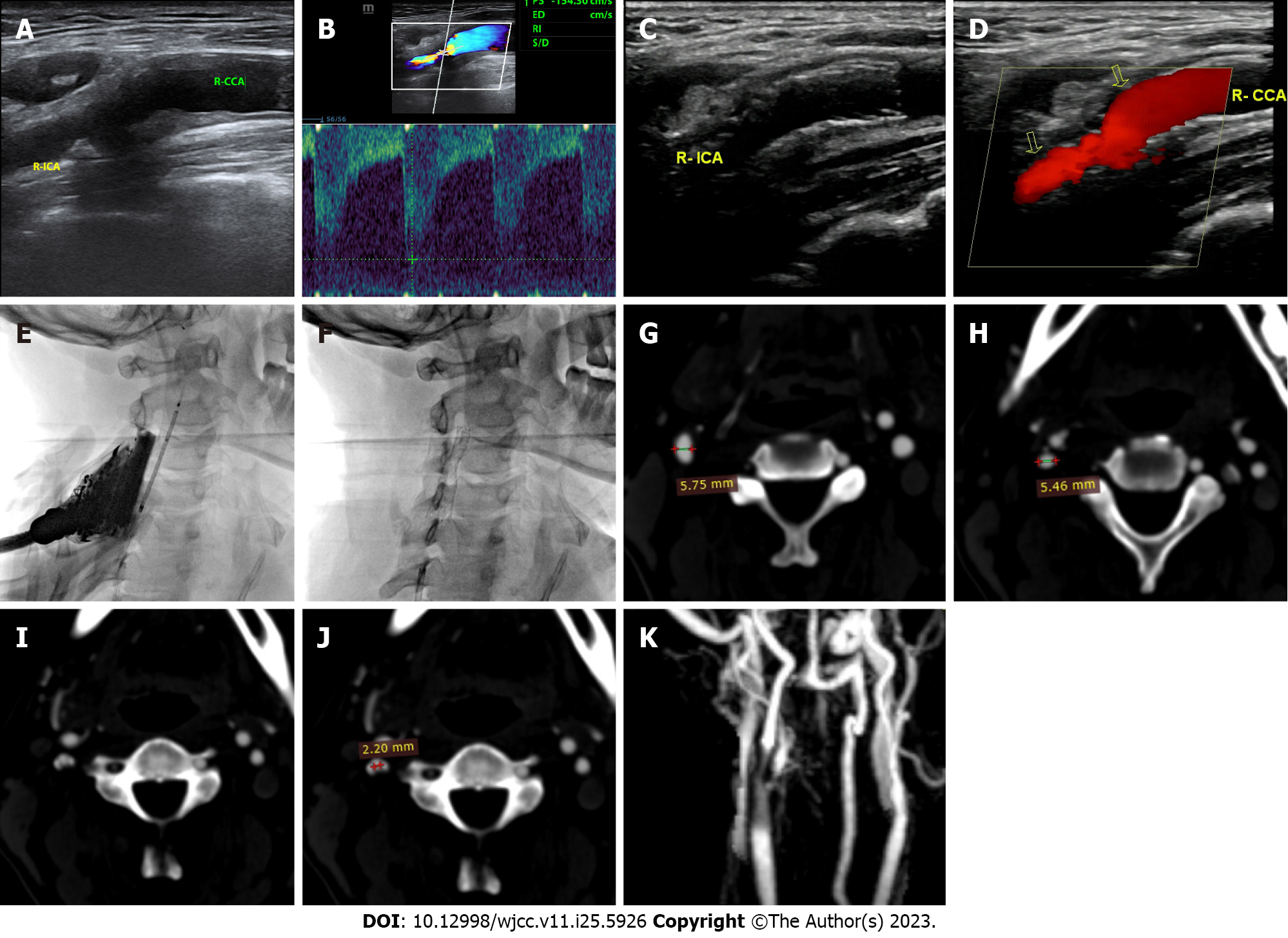
Figure 2 Images before and after carotid artery stent implantation.
A: Ultrasound showing stenosis of the right internal carotid artery (R-ICA) before surgery; B: Ultrasound showing blood flow in the region of R-ICA stenosis before surgery; C: Ultrasound showing good stent shape of the R-ICA after surgery; D: Ultrasound showing good blood flow in the region of R-ICA stent after surgery; E: X-ray showing the shape of the stent before stenting; F: X-ray showing the shape of the stent after stenting; G: Preoperative computed tomography angiography (CTA) showing a proximal diameter at the site of R-ICA stenosis of 5.75 mm; H: Preoperative CTA showing a distal diameter at the site of R-ICA stenosis of 5.46 mm; I: CTA showing a cross-section of the site of R-ICA stenosis. The vascular lumen is in the middle, and calcified plaques are present at both ends; J: CTA indicating a lumen diameter at the site of R-ICA stenosis of 2.20 mm; K: Magnetic resonance angiography showing good stent shape after surgery. R-SCA: Right subclavian artery; R-ICA: Right internal carotid artery.
- Citation: Li L, Wang ZY, Liu B. Ultrasound-guided carotid angioplasty and stenting in a patient with iodinated contrast allergy: A case report. World J Clin Cases 2023; 11(25): 5926-5933
- URL: https://www.wjgnet.com/2307-8960/full/v11/i25/5926.htm
- DOI: https://dx.doi.org/10.12998/wjcc.v11.i25.5926


