Published online Aug 16, 2023. doi: 10.12998/wjcc.v11.i23.5622
Peer-review started: June 15, 2023
First decision: July 4, 2023
Revised: July 7, 2023
Accepted: July 24, 2023
Article in press: July 24, 2023
Published online: August 16, 2023
Processing time: 61 Days and 12.5 Hours
Liver abscess due to foreign body-induced gastrointestinal tract perforation is a rare event that could be misdiagnosed due to low suspicion. Less than 100 cases have been reported to date.
We report a case of a 53-year old female patient with pyogenic liver abscess secondary to ingestion of a toothpick with penetration through the lesser curvature of the stomach. The patient presented with persistent epigastric pain. Abdominal computed tomography demonstrated the presence of a linear radiopaque object associated with abscess formation in the left liver lobe. Inflammatory changes in the lesser curvature of the stomach indicated gastric wall penetration by the object. As the abscess was refractory to antibiotic treatment, laparoscopic liver resection was performed to remove the foreign body and adjacent liver parenchyma. Following surgery, symptoms fully resolved without any sequelae.
This rare case demonstrates the importance of considering foreign body penetration as a cause of pyogenic liver abscess, particularly in abscesses of unknown origin that are resistant to antibiotic therapy. Clinical suspicion, early diagnosis, and prompt removal of the foreign body could lead to improved outcomes in these patients.
Core Tip: Most ingested foreign bodies can be managed without intervention. In rare occasions, sharp objects might directly penetrate from the gastrointestinal tract into the liver. In such cases, early diagnosis and proper surgical management are necessary. We present a rare case of pyogenic liver abscess secondary to penetration of the stomach by an ingested toothpick. After administration of systemic antibiotics, laparoscopic removal of the foreign body was pursued. As the foreign body was not visible from the liver surface, left lateral sectionectomy was performed. Postoperative recovery was uneventful. In refractory liver abscesses, clinical suspicion for foreign body ingestion should be maintained.
- Citation: Park Y, Han HS, Yoon YS, Cho JY, Lee B, Kang M, Kim J, Lee HW. Pyogenic liver abscess secondary to gastric perforation of an ingested toothpick: A case report. World J Clin Cases 2023; 11(23): 5622-5627
- URL: https://www.wjgnet.com/2307-8960/full/v11/i23/5622.htm
- DOI: https://dx.doi.org/10.12998/wjcc.v11.i23.5622
Foreign body ingestion is often encountered in clinical practice, particularly in the emergency department. Most foreign bodies either pass through the gastrointestinal (GI) tract uneventfully or can be successfully removed by endoscopy. Less than 1% of patients require surgical management due to severe complications including GI perforation or obstruction[1].
Depending on composition, foreign bodies may be difficult to detect on plain radiography or computed tomography (CT) scans due to their radiolucent nature. This may contribute to delays in diagnosis or definitive treatment. Rarely, recurrent events of intra-abdominal abscess, peritonitis, or even sepsis may later be attributed to foreign body ingestion and consequent GI complications[2]. Herein, we present a case of hepatic abscess secondary to toothpick-induced gastric perforation with direct penetration into adjacent liver tissue.
A 53-year-old female patient was admitted to our emergency department for evaluation of an intrahepatic foreign body.
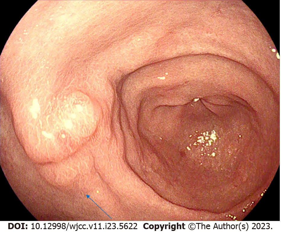
The patient had attended an outside hospital on the same day with a two-week history of persistent epigastric pain. Abdominal CT had revealed a needle-like structure in the left liver lobe. Esophagogastroduodenoscopy had demonstrated a fungating mass-like lesion with central depression on the anterior wall of the lesser curvature of the stomach without ulceration or perforation (Figure 1). At the time of evaluation at our center, she complained of persistent epigastric pain accompanied by vomiting. She denied any history of fever or chills.
The patient denied any past illnesses.
The patient denied any personal or family history of related diseases.
On arrival at the emergency department, her vital signs were stable with a blood pressure of 126/81 mmHg, pulse rate of 80 beats per minutes, respiratory rate of 18 breaths per minutes, and body temperature of 36.9 ℃. Physical examination revealed no signs of peritonitis, with a soft, flat abdomen without any focal tenderness.
Laboratory studies demonstrated a white blood cell count of 13120/µL with 83.4% neutrophils, a hemoglobin level of 13.4 g/dL, and a serum C-reactive protein (CRP) concentration of 17.70 mg/dL. Mild increases in serum liver enzyme levels were observed with a serum aspartate transaminase level of 45 IU/L and serum alanine transaminase level of 46 IU/L. Blood cultures yielded no bacterial growth.
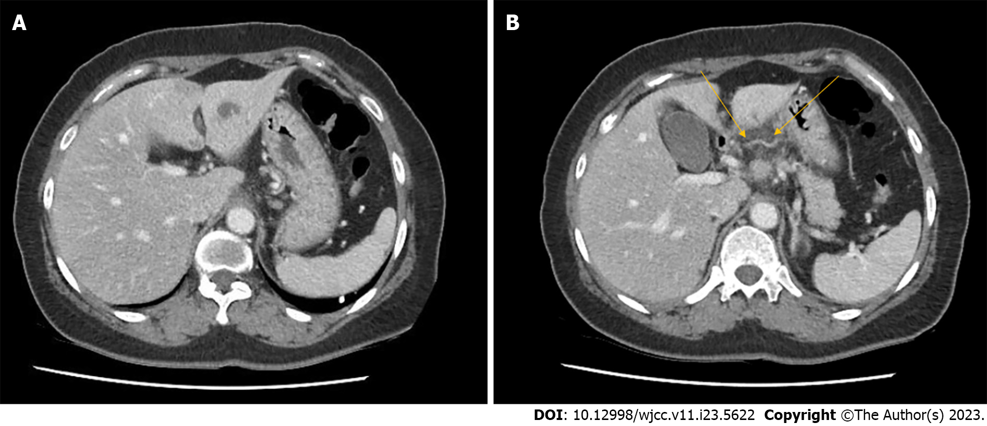
Chest and abdominal plain radiography were unremarkable. CT images from the outside hospital were re-evaluated by our radiologists and demonstrated a thin linear radiopaque lesion with an adjacent low attenuating lesion in S3 of the liver (Figure 2). Mild infiltration was observed along the lesser curvature of the stomach adjacent to the S3 Lesion. Accordingly, foreign body penetration from the stomach to the liver was suspected. The patient was unable to recall the event of foreign body ingestion. She was admitted for conservative management including intravenous antibiotic therapy (ceftriaxone 2 g/d and metronidazole 500 mg every 8 h) and analgesia. Follow-up CT was performed on the second day of admission. An interval increase in size of the adjacent complicated fluid collection was noted, indicating formation of a liver abscess.
The patient was diagnosed with pyogenic liver abscess secondary to foreign body penetration from stomach to liver.
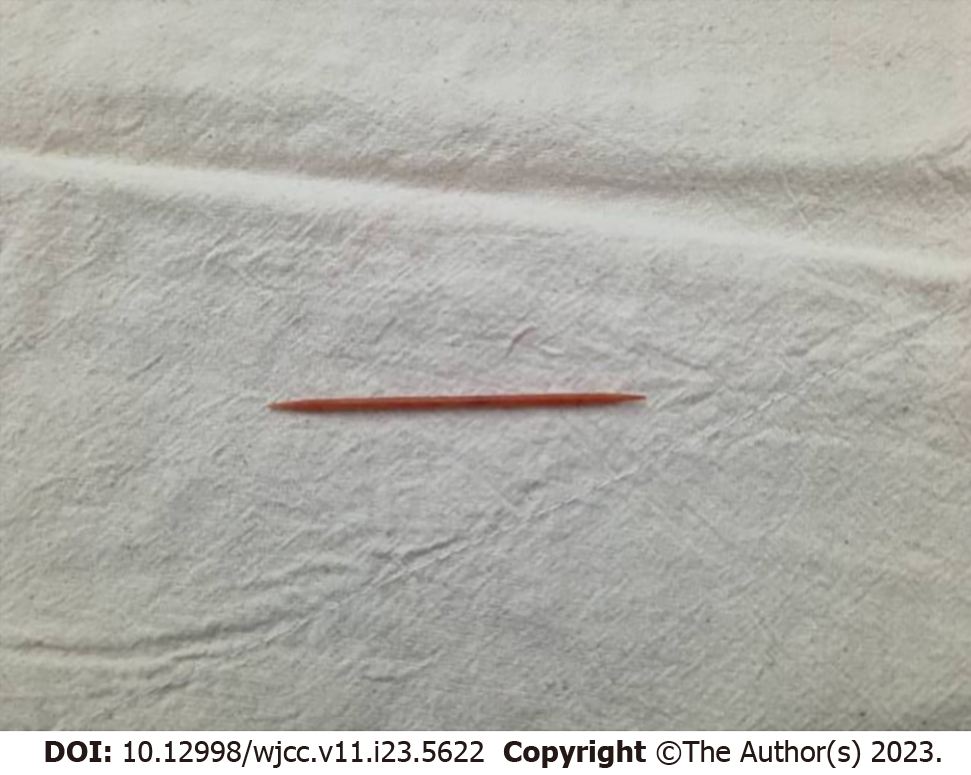
As the patient developed fever, antibiotic therapy was changed to ampicillin/sulbactam 3000 mg every 6 h. Due to the development of sepsis, a delayed operation was planned for removal of the foreign body. Fever persisted for four days, and the operation was performed one week after admission as the patients’ body temperature was maintained in normal range. On laparoscopic inspection, the foreign body could not be identified from the liver surface on the suspected segment. Laparoscopic left lateral sectionectomy of the liver was therefore performed. Upon removal of the specimen, the foreign body was identified as a wooden toothpick (Figure 3). The patient was discharged on the fifth postoperative day without complications. Treatment with an oral antibiotic (cefpodoxime 200 mg bid) was continued for a further week.
Outpatient clinic follow-up confirmed the resolution of all symptoms and the return of serum CRP levels to within normal limits.
Most ingested foreign bodies are excreted without injury to the GI tract, and foreign body-induced GI tract perforation is a rare event occurring in less than 1% of all cases[3]. Liver abscess formation due to foreign body-induced GI perforation is even rarer, with less than 100 cases reported to date[4,5]. As most patients fail to recall the event of foreign body ingestion, foreign bodies migrating to the liver often remain unnoticed until signs of systemic infection develop. Delayed diagnosis may increase the risk of morbidity and mortality, with foreign objects occasionally found only upon autopsy after patients have died from sepsis with an uncertain focus[6].
Although the type of foreign body ingested varies according to personal dietary habits, sociocultural features, and established psychiatric illnesses, fish bones and toothpicks are the most commonly discovered objects[7,8]. However, the identification of ingested radiolucent foreign bodies remains a radiologic challenge compared to metallic objects as they are not detectable by plain abdominal radiography[7,9]. Ultrasonography and CT are the preferred modalities in such cases and should always be considered when there is clinical suspicion of foreign body ingestion[10,11]. In the present case, the patient had ingested an entire wooden toothpick without any marks of mastication. Although the exact mechanism by which the toothpick entered the patients’ GI tract was unclear, this involuntary ingestion may have been related to the Korean food culture of drinking soup. The toothpick may have been mixed in with soup ingredients and then accidentally swallowed.
The key aspects of treatment for hepatic abscesses caused by foreign body-induced GI perforation is early diagnosis and prompt removal of the foreign body[2,10]. Simple percutaneous drainage of the abscess may transiently alleviate symptoms; however, recurrent episodes of sepsis might follow and surgical removal of the foreign body is likely to be required in the end[12]. The removal of foreign bodies from the liver can be performed by endoscopic procedures, laparoscopic surgery, or open surgery[13]. In cases where the foreign body is in transit between the GI tract and the liver, the foreign body may be visible and therefore removed by endoscopy[14]. When penetration into liver tissue is suspected, surgical removal accompanied by abscess drainage may be necessary[2]. Intra-operative ultrasonography may have utility in identifying the location of intra-parenchymal foreign bodies[14]. For objects located deep in the liver pa
We report a case of successful treatment of a hepatic abscess caused by foreign body-induced GI perforation with laparoscopic liver resection. Hepatic abscesses caused by migration of an ingested toothpick are extremely rare, with less than 20 cases officially reported. Diagnosis is often delayed due to patients being unable to recall foreign body ingestion and low awareness of this rare condition among clinicians. The present case emphasizes the importance of considering ingested foreign bodies in the differential diagnosis of cases of pyogenic liver abscess, particularly in previously healthy individuals presenting with left-sided abscesses refractory to aspiration and antibiotic therapy.
Provenance and peer review: Unsolicited article; Externally peer reviewed.
Peer-review model: Single blind
Specialty type: Medicine, research and experimental
Country/Territory of origin: South Korea
Peer-review report’s scientific quality classification
Grade A (Excellent): 0
Grade B (Very good): B
Grade C (Good): 0
Grade D (Fair): D
Grade E (Poor): 0
P-Reviewer: Cerwenka H, Austria; Sudhamshu K, Nepal S-Editor: Qu XL L-Editor: A P-Editor: Yu HG
| 1. | Eisen GM, Baron TH, Dominitz JA, Faigel DO, Goldstein JL, Johanson JF, Mallery JS, Raddawi HM, Vargo JJ 2nd, Waring JP, Fanelli RD, Wheeler-Harbough J; American Society for Gastrointestinal Endoscopy. Guideline for the management of ingested foreign bodies. Gastrointest Endosc. 2002;55:802-806. [RCA] [PubMed] [DOI] [Full Text] [Cited by in Crossref: 399] [Cited by in RCA: 361] [Article Influence: 15.7] [Reference Citation Analysis (0)] |
| 2. | Bandeira-de-Mello RG, Bondar G, Schneider E, Wiener-Stensmann IC, Gressler JB, Kruel CRP. Pyogenic Liver Abscess Secondary to Foreign Body (Fish Bone) Treated by Laparoscopy: A Case Report. Ann Hepatol. 2018;17:169-173. [RCA] [PubMed] [DOI] [Full Text] [Cited by in Crossref: 10] [Cited by in RCA: 16] [Article Influence: 2.3] [Reference Citation Analysis (0)] |
| 3. | Natsuki S, Iseki Y, Nagahara H, Fukuoka T, Shibutani M, Ohira M. Liver abscess caused by fish bone perforation of Meckel's diverticulum: a case report. BMC Surg. 2020;20:121. [RCA] [PubMed] [DOI] [Full Text] [Full Text (PDF)] [Reference Citation Analysis (0)] |
| 4. | Santos SA, Alberto SC, Cruz E, Pires E, Figueira T, Coimbra E, Estevez J, Oliveira M, Novais L, Deus JR. Hepatic abscess induced by foreign body: case report and literature review. World J Gastroenterol. 2007;13:1466-1470. [RCA] [PubMed] [DOI] [Full Text] [Full Text (PDF)] [Cited by in CrossRef: 78] [Cited by in RCA: 97] [Article Influence: 5.4] [Reference Citation Analysis (0)] |
| 5. | Pérez Saborido B, Bailón Cuadrado M, Velasco López R. A liver abscess secondary to a toothpick: a rare complication of accidental foreign body ingestion. Rev Esp Enferm Dig. 2019;111:167-168. [RCA] [PubMed] [DOI] [Full Text] [Cited by in Crossref: 3] [Cited by in RCA: 5] [Article Influence: 1.0] [Reference Citation Analysis (0)] |
| 6. | Masoodi I, Alsayari K, Al Mohaimeed K, Ahmad S, Almtawa A, Alomair A, Alqutub A, Khan S. Fish bone migration: an unusual cause of liver abscess. BMJ Case Rep. 2012;2012. [RCA] [PubMed] [DOI] [Full Text] [Cited by in Crossref: 6] [Cited by in RCA: 13] [Article Influence: 1.0] [Reference Citation Analysis (0)] |
| 7. | Martin S, Petraszko AM, Tandon YK. A case of liver abscesses and porto-enteric fistula caused by an ingested toothpick: A review of the distinctive clinical and imaging features. Radiol Case Rep. 2020;15:273-276. [RCA] [PubMed] [DOI] [Full Text] [Full Text (PDF)] [Cited by in Crossref: 4] [Cited by in RCA: 10] [Article Influence: 2.0] [Reference Citation Analysis (0)] |
| 8. | Luo CF, Xu J, Lu YQ. Hepatic abscess resulted from a toothpick piercing the gastric wall into the liver. Hepatobiliary Pancreat Dis Int. 2020;19:502-504. [RCA] [PubMed] [DOI] [Full Text] [Cited by in Crossref: 1] [Cited by in RCA: 4] [Article Influence: 0.8] [Reference Citation Analysis (0)] |
| 9. | Bekki T, Fujikuni N, Tanabe K, Amano H, Noriyuki T, Nakahara M. Liver abscess caused by fish bone perforation of stomach wall treated by laparoscopic surgery: a case report. Surg Case Rep. 2019;5:79. [RCA] [PubMed] [DOI] [Full Text] [Full Text (PDF)] [Cited by in Crossref: 11] [Cited by in RCA: 18] [Article Influence: 3.0] [Reference Citation Analysis (0)] |
| 10. | Abu-Wasel B, Eltawil KM, Keough V, Molinari M. Liver abscess caused by toothpick and treated by laparoscopic left hepatic resection: case report and literature review. BMJ Case Rep. 2012;2012. [RCA] [PubMed] [DOI] [Full Text] [Cited by in Crossref: 8] [Cited by in RCA: 15] [Article Influence: 1.2] [Reference Citation Analysis (0)] |
| 11. | Kishawi S, Anderson MJ, Chavin K. Toothpick in the porta: Recurrent liver abscesses secondary to transgastric migration of a toothpick with successful surgical exploration retrieval. Ann Hepatobiliary Pancreat Surg. 2020;24:362-365. [RCA] [PubMed] [DOI] [Full Text] [Full Text (PDF)] [Cited by in Crossref: 1] [Cited by in RCA: 1] [Article Influence: 0.2] [Reference Citation Analysis (0)] |
| 12. | Grayson N, Shanti H, Patel AG. Liver abscess secondary to fishbone ingestion: case report and review of the literature. J Surg Case Rep. 2022;2022:rjac026. [RCA] [PubMed] [DOI] [Full Text] [Full Text (PDF)] [Cited by in RCA: 4] [Reference Citation Analysis (0)] |
| 13. | Glick WA, Simo KA, Swan RZ, Sindram D, Iannitti DA, Martinie JB. Pyogenic hepatic abscess secondary to endolumenal perforation of an ingested foreign body. J Gastrointest Surg. 2012;16:885-887. [RCA] [PubMed] [DOI] [Full Text] [Cited by in Crossref: 20] [Cited by in RCA: 20] [Article Influence: 1.5] [Reference Citation Analysis (0)] |
| 14. | Carver D, Bruckschwaiger V, Martel G, Bertens KA, Abou-Khalil J, Balaa F. Laparoscopic retrieval of a sewing needle from the liver: A case report. Int J Surg Case Rep. 2018;51:376-378. [RCA] [PubMed] [DOI] [Full Text] [Full Text (PDF)] [Cited by in Crossref: 8] [Cited by in RCA: 9] [Article Influence: 1.3] [Reference Citation Analysis (0)] |


