Published online Aug 16, 2023. doi: 10.12998/wjcc.v11.i23.5595
Peer-review started: May 22, 2023
First decision: June 19, 2023
Revised: June 27, 2023
Accepted: July 25, 2023
Article in press: July 25, 2023
Published online: August 16, 2023
Processing time: 85 Days and 18.8 Hours
Osteochondroma is one of the most common benign bone tumors, and it may cause bone and joint deformities and limited range of motion of an adjacent joint. The pes anserinus region is one of the most frequent sites of osteochondroma, but knee locking caused by osteochondromas in the pes anserinus region is extremely rare.
We describe a 13-year-old Japanese girl’s extra-articular knee locking that occurred when the semitendinosus tendon got caught in osteochondroma that had developed in the pes anserinus region. The osteochondroma was surgically resected. The postoperative outcome has been excellent, with no recurrence of knee locking or tumor one-year post-surgery.
When a young person develops knee locking, the possibility of extra-articular as well as intra-articular locking should be considered. Osteochondroma, one of the causes of extra-articular locking, can be treated with surgery with good posto
Core Tip: Osteochondroma is one of the most common benign bone tumors, but extra-articular knee locking caused by osteochondroma in the pes anserinus region is extremely rare. We report a case of extra-articular knee locking that occurred when the semitendinosus tendon got caught in osteochondroma that had developed in the pes anserinus region. The osteochondroma was surgically resected. The postoperative outcome has been excellent with no recurrence of knee locking or tumor 3 mo post-surgery.
- Citation: Sonobe T, Hakozaki M, Matsuo Y, Takahashi Y, Yoshida K, Konno S. Knee locking caused by osteochondroma of the proximal tibia adjacent to the pes anserinus: A case report. World J Clin Cases 2023; 11(23): 5595-5601
- URL: https://www.wjgnet.com/2307-8960/full/v11/i23/5595.htm
- DOI: https://dx.doi.org/10.12998/wjcc.v11.i23.5595
Osteochondroma is one of the most common benign bone tumors, and it accounts for 35% of benign bone lesions and 8% of all bone tumors[1,2]. The average age at the diagnosis of solitary osteochondroma is 18 years (median 15 years; range 2-77 years), with a male predominance (65% vs 35%)[2]. Osteochondromas have been identified at various part of the body. Since osteochondroma frequently occurs in the metaphysis of the long bone of an extremity, it may cause bone and joint deformities and a limited range of motion of an adjacent joint.
The pes anserinus region is one of the most frequent sites of osteochondroma in young individuals[3], but knee locking caused by osteochondromas in the pes anserinus region is extremely rare. We describe a case of extra-articular knee locking that occurred when the semitendinosus tendon got caught in an osteochondroma that had developed in the pes anserinus region. This case was treated surgically, and the patient’s postsurgical functional outcome has been excellent.
The patient was a 13-year-old Japanese girl. While she was playing badminton, her left knee was locked after the flexion, and the locking later resolved spontaneously.
Three months after that incident, left knee was locked again when she got up from a couch at home. At a primary hospital, osteochondroma was suspected based on radiological findings, and she was referred to our hospital for surgical treatment.
On admission, she was in good health with no previous history of other diseases or injuries.
She had no remarkable personal or family medical history.
The knee locking had released. Ballottement of the patella of the left knee was negative, and a bony protuberance was palpated in the same region as the pes anserinus. The active range of motion (ROM) of the left knee was from 0° to 145°, and left knee pain and anxiety were caused during flexion and extension of the knee, but locking was not reproduced.
Laboratory test results were all negative.
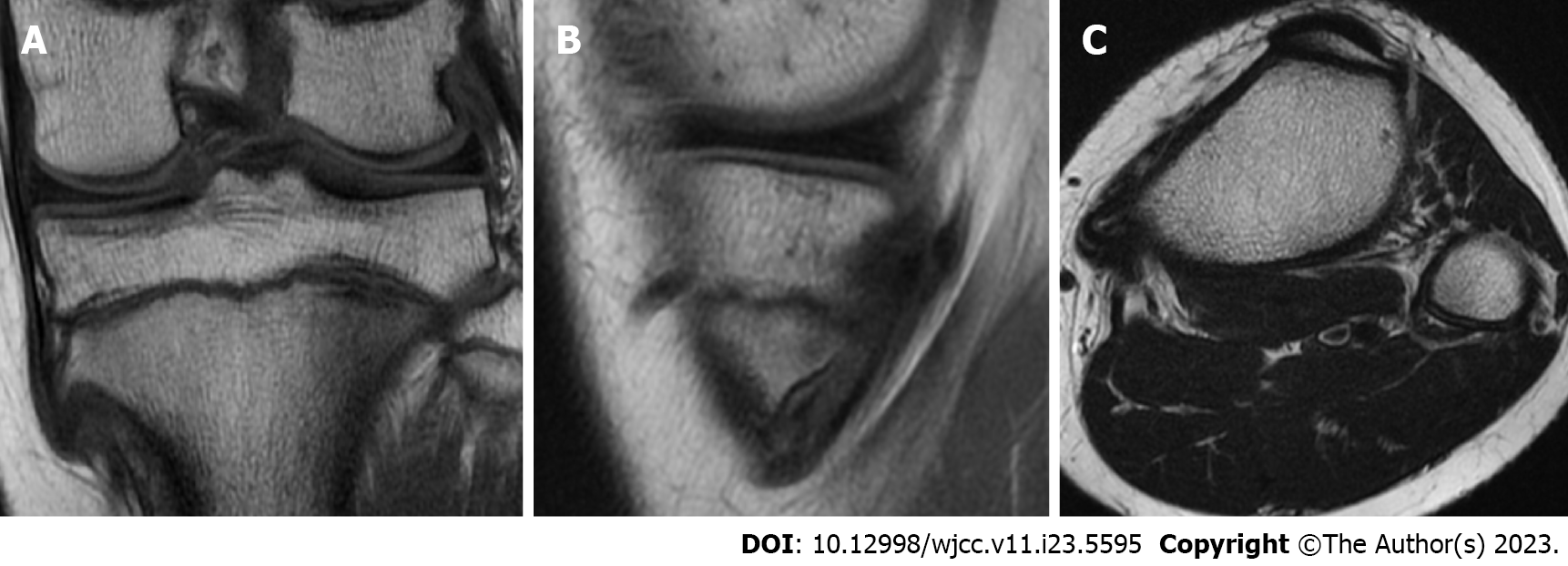
Plain radiographs showed a bony protuberance on the medial side of the proximal tibia (Figure 1A and B). Computed tomography showed a hook-shaped bony protuberance extending distally. Axial sections showed bone marrow continuity within the lesion (Figure 2). T2-weighted magnetic resonance imaging (MRI) showed that the pes anserinus directly covered the bony protuberance, and a high signal change was observed in the semitendinosus tendon (ST). MRI also showed bone marrow continuity within the lesion (Figure 3).
The final diagnosis was extra-articular knee locking due to an osteochondroma of the pes anserinus region.
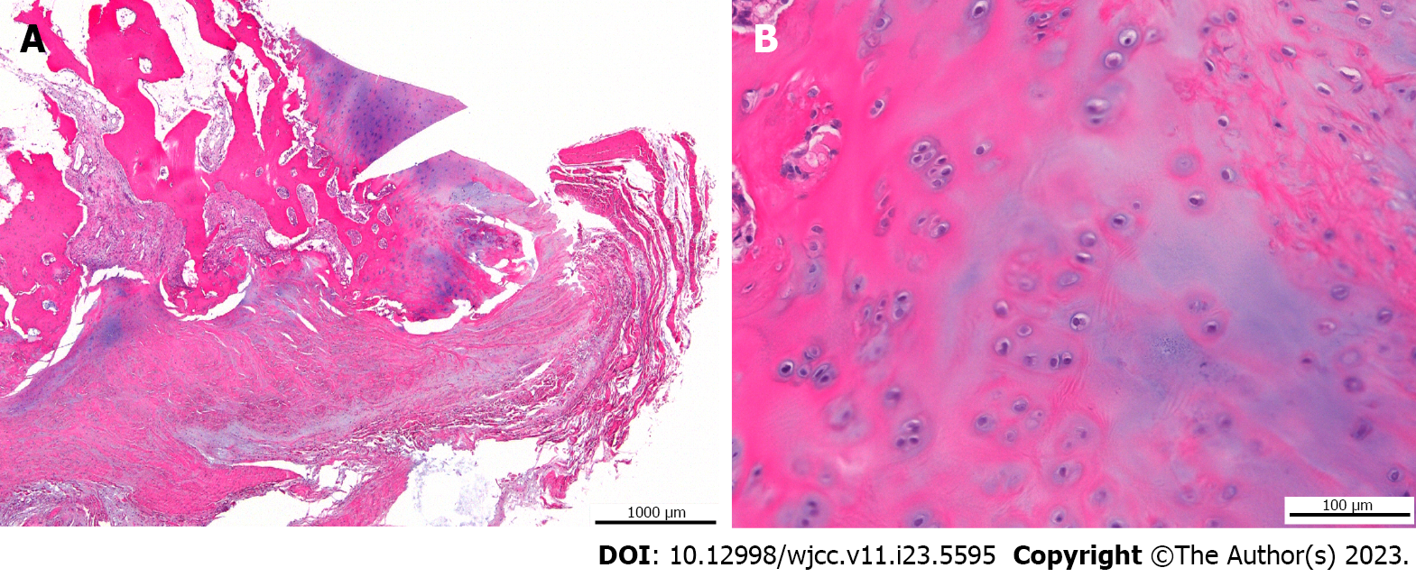
An incision was made directly above the pes anserinus region, and a bony protuberance covered by periosteum was identified under the pes anserinus. Flexion and extension of the left knee joint reproduced impingement and snapping of the ST and bony prominence, but knee locking did not occur. A longitudinal incision was made in the sartorial fascia directly above the ST. The ST was retracted proximally, and the superficial medial collateral ligament (MCL) was confirmed. By making a longitudinal incision through the MCL, the whole aspect of the tumor and the cartilage cap at the apex could be observed. The tumor was resected with a luer and chisel, taking care to avoid under-resection. After the tumor resection, the left knee joint was flexed, and it was observed that the impingement with the ST had disappeared. Postoperative plain radiographs demonstrated that the osteochondroma was sufficiently resected (Figure 1C and D). In the pathology examination, a coating of vitreous cartilage which thought to be a cartilage cap was observed on the surface of the lesion (Figure 4), and the tumor was thus pathologically diagnosed as osteochondroma.
Full weight-bearing and ROM exercise with no restriction was initiated, and at one-year post-surgery, the patient had no recurrence of osteochondroma or knee locking (Figure 1E and F). In addition, the active ROM of the left knee was from 0° to 150°, with no limitation of joint ROM.
Knee locking develops suddenly and causes significant hinderance of daily life due to pain and limitations of activities. The most common cause of knee locking is intra-articular lesions; meniscus injuries are the most frequent, followed by ligament injuries and intra-articular free bodies[4]. Synovial hemangiomas, tenosynovial giant cell tumors, gouty arthritis, lipomas, and intra-articular ganglions are less common causative diseases[5-7]. Conversely, knee locking due to extra-articular disease is uncommon; post-fracture deformity and osteochondroma have been reported as causative conditions[8-10]. Abnormal bone shapes may cause knee locking due to the incarceration or constraint of musculotendons around the knee joint.
In terms of knee pain, the differential diagnosis includes apophysitis, such as Osgood-Schlatter disease[11] and Sinding-Larsen-Johansson disease[12], jumper’s knee[13], and bipartite patella[14]; these disorders are more common in children and adolescents. In the present case, the final diagnosis was established based on the injury mechanism, the localization of tenderness, and radiological findings.
Osteochondroma that develops in the pes anserinus region causes impingement with pes anserinus. Muscle tendons and soft tissues are subjected to repeated mechanical irritation, which can cause tendonitis, tendon rupture, and bursitis[15]. Clinical symptoms such as pain, swelling, snapping, and catching are generally referred to as pes anserinus syndrome[16]. Pes anserinus syndrome is more common in females after middle age and is often associated with anatomical deformity[17,18].
Only a few cases of knee locking due to osteochondroma have been reported. The possibility of knee locking is usually considered based on the patient’s clinical history, physical examination, and imaging findings, and the cause of locking is then identified and treated based on the cause. Our search of the relevant literature revealed only 10 prior cases of osteochondroma-causing extra-articular knee locking (Table 1)[8,10,19-21]. Among the cases in which the mechanism of injury was known, the knee locking occurred due to knee deep flexion in all cases.
| Ref. | Age in yr | Sex | Tumor location | Injury mechanism | Post- operative follow-up period | Clinical outcome |
| Kralik et al[20], 1951 | NA | NA | Distal medial femur | NA | NA | NA |
| NA | NA | Distal medial femur | NA | NA | NA | |
| Kernohan et al[10], 1984 | 19 | F | Proximal medial tibia | Knee deep flexion | NA | Symptom resolved |
| 19 | M | Proximal medial tibia | Crouching | NA | Symptom resolved | |
| 19 | F | Proximal medial tibia | After squat | NA | Symptom resolved | |
| 13 | F | Proximal medial tibia | During squat | NA | Symptom resolved | |
| Fraser et al[19], 1996 | NA | NA | Proximal medial tibia | NA | NA | Symptom resolved |
| NA | NA | Proximal medial tibia | NA | NA | Symptom resolved | |
| Sansone et al[8], 1999 | 23 | F | Proximal medial tibia | Knee deep flexion | 24 mo | No recurrence of osteochondroma and locking |
| Andrews et al[21], 2019 | 18 | F | Proximal medial tibia | During squat | 3 mo | No recurrence of osteochondroma and locking |
| Present case | 13 | F | Proximal medial tibia | Knee deep flexion | 3 mo | No recurrence of osteochondroma and locking |
The mechanism of extra-articular knee locking of the pes anserinus and osteochondroma was described as occurring when the pes anserinus, which is located anterior to the osteochondroma during knee extension, moves posterior to the osteochondroma during flexion, and is caught by the hook-shaped osteochondroma during re-extension[19]. Surgery was performed in all 10 of the previously reported cases, and no recurrence of knee locking has been reported (Table 1). Although there are two reports that repeated flexion-extension motion can release the knee locking, excessive flexion-extension motion may cause tendon and tendon-membrane injury[10,19]. Resection is thus recommended for clinically symptomatic osteochondromas.
In the present case, the knee locking could not be reproduced intraoperatively, but impingement and snapping were observed. Since no other factors inside or outside the knee joint were observed to have caused the locking, we concluded that the cause of the knee locking was osteochondroma. As in previous reports, the surgical resection of the patient’s osteochondroma showed good postoperative results without locking knee or tumor recurrence.
When a young patient develops knee locking, intra-articular locking may be suspected as the cause, but if there are no intra-articular findings that could cause the knee locking and extra-articular knee locking is not present, the patient may have recurrent pes anserinus syndrome and a musculotendinous injury may occur around the knee joint in the future. When clinicians encounter a young patient with extra-articular knee locking or pes anserinus syndrome, the possibility of osteochondroma should be considered.
Provenance and peer review: Unsolicited article; Externally peer reviewed.
Peer-review model: Single blind
Specialty type: Orthopedics
Country/Territory of origin: Japan
Peer-review report’s scientific quality classification
Grade A (Excellent): 0
Grade B (Very good): B
Grade C (Good): C
Grade D (Fair): 0
Grade E (Poor): 0
P-Reviewer: Mostafavinia A, Iran; Vyshka G, Albania S-Editor: Chen YL L-Editor: A P-Editor: Chen YL
| 1. | Unni KK, Inwards CY. Dahlin’s bone tumors: General aspects and data on 11087 cases. Sixth edition. 2009. [cited 2 July 2023]. Available from: https://www.wolterskluwer.com/ja-jp/solutions/ovid/dahlins-bone-tumors-3482. |
| 2. | Bovée JVMG, Bloem JL, Heymann D, Wuyts W. WHO Classification of Tumors Soft Tissue and Bone Tumors. Fifth edition. 2020. [cited 2 July 2023]. Available from: https://publications.iarc.fr/Book-And-Report-Series/Who-Classification-Of-Tumours/Soft-Tissue-And-Bone-Tumours-2020. |
| 3. | Chrisman OD, Goldenberg RR. Untreated solitary osteochondroma. Report of two cases. J Bone Joint Surg Am. 1968;50:508-512. [RCA] [PubMed] [DOI] [Full Text] [Cited by in Crossref: 20] [Cited by in RCA: 14] [Article Influence: 0.2] [Reference Citation Analysis (0)] |
| 4. | Allum RL, Jones JR. The locked knee. Injury. 1986;17:256-258. [RCA] [PubMed] [DOI] [Full Text] [Cited by in Crossref: 23] [Cited by in RCA: 27] [Article Influence: 0.7] [Reference Citation Analysis (0)] |
| 5. | Tzurbakis M, Mouzopoulos G, Morakis E, Nikolaras G, Georgilas I. Intra-articular knee haemangioma originating from the anterior cruciate ligament: a case report. J Med Case Rep. 2008;2:254. [RCA] [PubMed] [DOI] [Full Text] [Full Text (PDF)] [Cited by in Crossref: 11] [Cited by in RCA: 11] [Article Influence: 0.6] [Reference Citation Analysis (0)] |
| 6. | Sakti M, Usman MA, Lee J, Benjamin M, Maulidiah Q. Atypical musculoskeletal manifestations of gout in hyperuricemia patients. Open Access Rheumatol. 2019;11:47-52. [RCA] [PubMed] [DOI] [Full Text] [Full Text (PDF)] [Cited by in Crossref: 2] [Cited by in RCA: 6] [Article Influence: 1.0] [Reference Citation Analysis (0)] |
| 7. | Kim H, Shin DC, Lee KS, Jang IT, Lee K. Localized pigmented villonodular synovitis with hemorrhage arising from lateral meniscocapsular junction: A case report. Eklem Hastalik Cerrahisi. 2019;30:177-181. [RCA] [PubMed] [DOI] [Full Text] [Cited by in Crossref: 1] [Cited by in RCA: 3] [Article Influence: 0.5] [Reference Citation Analysis (0)] |
| 8. | Sansone V, De Ponti A, Ravasi F. An extra-articular cause of locking knee. Int Orthop. 1999;23:118-119. [RCA] [PubMed] [DOI] [Full Text] [Cited by in Crossref: 3] [Cited by in RCA: 4] [Article Influence: 0.2] [Reference Citation Analysis (0)] |
| 9. | Wood KB, Bradley JP, Ward WT. Pes anserinus interposition in a proximal tibial physeal fracture. A case report. Clin Orthop Relat Res. 1991;239-242. [PubMed] |
| 10. | Kernohan J, Blackburne JS. 'Locking knee' due to osteochondroma. Injury. 1984;15:239-241. [RCA] [PubMed] [DOI] [Full Text] [Cited by in Crossref: 3] [Cited by in RCA: 1] [Article Influence: 0.0] [Reference Citation Analysis (0)] |
| 11. | Osgood RB. Lesions of the tibial tubercle occurring during adolescence. 1903. Clin Orthop Relat Res. 1993;4-9. [PubMed] |
| 12. | Sinding-Lorsen CM. A Hitherto Unknown Affection of the Patella In Children. Acta Radiol. 2016;57:e42-e46. [RCA] [PubMed] [DOI] [Full Text] [Cited by in Crossref: 2] [Cited by in RCA: 6] [Article Influence: 0.7] [Reference Citation Analysis (0)] |
| 13. | Ferretti A, Puddu G, Mariani PP, Neri M. The natural history of jumper's knee. Patellar or quadriceps tendonitis. Int Orthop. 1985;8:239-242. [RCA] [PubMed] [DOI] [Full Text] [Cited by in Crossref: 82] [Cited by in RCA: 68] [Article Influence: 1.7] [Reference Citation Analysis (0)] |
| 14. | Bourne MH, Bianco AJ Jr. Bipartite patella in the adolescent: results of surgical excision. J Pediatr Orthop. 1990;10:69-73. [PubMed] [DOI] [Full Text] |
| 15. | de Souza AM, Bispo Júnior RZ. Osteochondroma: ignore or investigate? Rev Bras Ortop. 2014;49:555-564. [RCA] [PubMed] [DOI] [Full Text] [Full Text (PDF)] [Cited by in Crossref: 7] [Cited by in RCA: 31] [Article Influence: 2.8] [Reference Citation Analysis (0)] |
| 16. | Lee JH, Kim KJ, Jeong YG, Lee NS, Han SY, Lee CG, Kim KY, Han SH. Pes anserinus and anserine bursa: anatomical study. Anat Cell Biol. 2014;47:127-131. [RCA] [PubMed] [DOI] [Full Text] [Full Text (PDF)] [Cited by in Crossref: 20] [Cited by in RCA: 23] [Article Influence: 2.1] [Reference Citation Analysis (0)] |
| 17. | Helfenstein M Jr, Kuromoto J. Anserine syndrome. Rev Bras Reumatol. 2010;50:313-327. [RCA] [PubMed] [DOI] [Full Text] [Cited by in Crossref: 33] [Cited by in RCA: 25] [Article Influence: 1.7] [Reference Citation Analysis (0)] |
| 18. | Clapp A, Trecek J, Joyce M, Sundaram M. Pes anserine bursitis. Orthopedics. 2008;31:306, 407-408. [RCA] [PubMed] [DOI] [Full Text] [Cited by in Crossref: 5] [Cited by in RCA: 5] [Article Influence: 0.3] [Reference Citation Analysis (0)] |
| 19. | Fraser RK, Nattrass GR, Chow CW, Cole WG. Pes anserinus syndrome due to solitary tibial spurs and osteochondromas. J Pediatr Orthop. 1996;16:247-248. [PubMed] [DOI] [Full Text] |
| 20. | Kralik V, Polakova Z. Two cases of locking of the knee caused by an exostosis of the femur. Acta Chir Orthop Traumatol Cech. 1951;18:1-7. [PubMed] |
| 21. | Andrews K, Rowland A, Tank J. Knee locked in flexion: incarcerated semitendinosus tendon around a proximal tibial osteochondroma. J Surg Case Rep. 2019;2019:rjy346. [RCA] [PubMed] [DOI] [Full Text] [Full Text (PDF)] [Cited by in Crossref: 2] [Cited by in RCA: 2] [Article Influence: 0.3] [Reference Citation Analysis (0)] |


