Published online Jan 16, 2023. doi: 10.12998/wjcc.v11.i2.472
Peer-review started: November 15, 2022
First decision: November 25, 2022
Revised: December 5, 2022
Accepted: December 23, 2022
Article in press: December 23, 2022
Published online: January 16, 2023
Processing time: 58 Days and 6.2 Hours
Coronavirus disease 2019 (COVID-19) has spread rapidly, resulting in a pandemic in January 2020. Few studies have focused on the natural history and conse
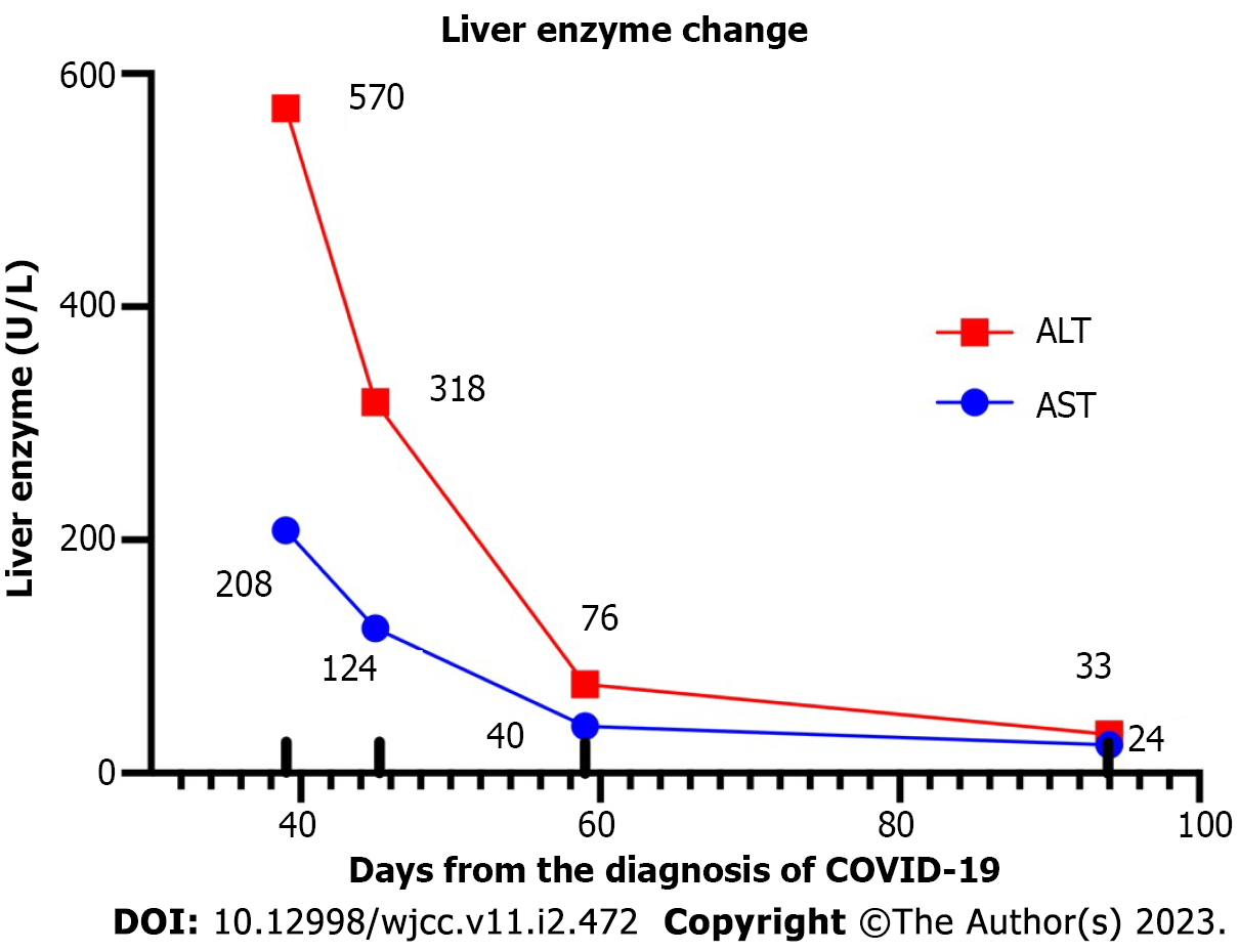
A 60-year-old woman without medical history or chronic illness received three COVID-19 vaccinations since the start of the pandemic. The patient was infected with severe acute respiratory syndrome coronavirus 2 (SARS-CoV-2) and presented with mild symptoms on July 12th, 2022. Post-recovery, she underwent an examination at our hospital on August 30th, 2022. AST and ALT levels in the liver function test were 207 U/L (normal value < 39, 5.3-fold increase) and 570 U/L (normal value < 52, 10.9-fold increase), respectively. The patient was diagnosed with ALI, and no treatment was prescribed. The following week, blood tests showed a reduction in both levels (ALT 124 U/L, AST 318 U/L). Two weeks later, AST and ALT levels had decreased to near the expected upper limits (ALT 40 U/L, AST 76 U/L).
Clinicians should pay attention to liver function testing during COVID-19 rec
Core Tip: Even though elevated aminotransferase levels and acute liver injury (ALI) are expected for severe coronavirus disease 2019 cases, here we report a rare case of ALI following a mild infection. We provide detailed information on ALI’s natural course in such patients.
- Citation: Lai PH, Ding DC. Acute liver injury in a COVID-19 infected woman with mild symptoms: A case report. World J Clin Cases 2023; 11(2): 472-478
- URL: https://www.wjgnet.com/2307-8960/full/v11/i2/472.htm
- DOI: https://dx.doi.org/10.12998/wjcc.v11.i2.472
Coronavirus disease 2019 (COVID-19), caused by severe acute respiratory syndrome coronavirus 2 (SARS-CoV-2), has spread rapidly, resulting in a pandemic in January 2020[1]. The major clinical symptoms of COVID-19 include respiratory disease and respiratory failure[2]. In addition to the characteristic respiratory symptoms, COVID-19 has extrapulmonary manifestations, including acute kidney injury, myocarditis, cardiac dysfunction, the risk of developing type 1 diabetes, thrombosis, and acute liver injury (ALI)[3-5].
A significant proportion of COVID-19 patients presenting with elevated liver enzymes have been reported worldwide[6]. The prevalence of ALI has ranged from 16% to 53% in COVID-19 patients[7]. Elevated aminotransferase levels are often mild [1-2 times the upper limit of normal (UNL)], while severe cases with much higher aminotransferase levels (UNL > 5) have also been observed[2,8]. Severe liver injury occurred in only 6.4% of the affected patients, and it is associated with poor clinical outcomes, including respiratory failure requiring intubation, renal replacement therapy, intensive care unit admission, and death[6,9]. Alterations in liver enzyme levels are usually transient. No deaths were directly related to hepatic decompensation in patients without pre-existing liver disease[10].
Most studies have addressed the prevalence of ALI and its association with clinical outcomes in patients hospitalized for COVID-19 pneumonia. However, few studies have focused on ALI’s natural history and consequences in patients with mild COVID-19 or asymptomatic carriers. Only one SARS-CoV-2 infection without respiratory symptoms presenting with acute hepatitis has been previously reported in the literature[11]. Here, we report a case of COVID-19 presenting mild respiratory symptoms and inadvertently discovered elevated alanine aminotransferase (ALT) and aspartate aminotransferase (AST) levels.
A 60-year-old woman was referred to the Department of Family Medicine of our hospital for further investigation of elevated liver enzymes during her health examination.
She was vaccinated three times (BNT162b2, Pfizer-BioNTech, New York, NY, United States) since the start of the COVID-19 pandemic and was infected with SARS-CoV-2 on July 12, 2022. After recovering from the infection, she underwent a health examination at our hospital on August 30, 2022. She recalled suffering only from a mild productive cough, without fever, chills, fatigue, or other discomforts. Hepatitis-associated symptoms were not observed, including nausea, vomiting, and changes in skin color, urine, or stool. There was no drug or alcohol abuse.
She had no remarkable medical history or chronic illness. Traditional Chinese medicine was self-administered for improved wellness during the past six months.
There was no significant personal or family history.
There was no significant finding of physical examination.
Laboratory blood liver function test revealed elevated AST level of 207 U/L (normal value < 39, UNL > 5.3) and ALT level of 570 U/L (normal value < 52, UNL > 10.9). Serum total bilirubin (TBI), alkaline phosphatase (ALP), and gamma-glutamyl transferase (GGT) levels were not elevated. There was no evidence of acute or chronic hepatitis A, B, and C (Table 1).
| Normal value | Day 39 | Day 45 | Day 59 | Day 94 | |
| ALP | 34-104 IU/L | 87.00 | 88 | 80 | 79 |
| AST (GOT) | 40-124 U/L | 207.00 | 124 | 40 | 24 |
| ALT (GPT) | 7-52 U/L | 570.00 | 318 | 76 | 33 |
| TBI | 0.3-1.0 mg/dL | 1.00 | 0.8 | 0.6 | 0.6 |
| DBI | < 0.2 mg/dL | 0.20 | 0.2 | 0.1 | 0.1 |
| GGT | 9-64 U/L | 84.00 | 68 | 57 | 47 |
| TP | 6.4-8.9 g/dL | 7.50 | |||
| ALB | 3.5-5.7 g/dL | 4.60 | |||
| GLO | 2.90 | ||||
| A/G ratio | 1.60 | ||||
| BUN | 7-25 mg/dL | 8.00 | |||
| UA | 2.3-6.6 mg/dL | 6.70 | |||
| CRE | 0.6-1.2 mg/dL | 0.67 | |||
| eGFR | > 90 | 95.40 | |||
| TCH | < 200 mg/dL | 200.00 | |||
| TG | < 150 mg/dL | 84.00 | |||
| GLU-AC | 70-100 mg/dL | 82.00 | |||
| Na | 136-145 mmol/L | 139.00 | |||
| K | 3.5-5.1 mmol/L | 3.70 | |||
| Ca | 2.2-2.6 mmol/L | 2.40 | |||
| HDL | > 50 mg/dL | 47.00 | |||
| HbA1c | 4%-6% | 6.00 | |||
| eA GLU | mg/dL | 125.00 | |||
| LDL | < 100 mg/dL | 143.00 | |||
| Anti-mitochondria Ab | < 1:20 | Negative | |||
| HAV IgM | Negative | ||||
| Anti-HAV | Reactive | ||||
| HBs Ag | Nonreactive | ||||
| Anti-HBs | Negative | ||||
| Anti-HCV | Nonreactive | ||||
| AFP | < 9 ng/mL | 1.30 | |||
| CEA | < 3 ng/mL | 2.90 | |||
| FT4 | 0.59-1.43 ng/mL | 1.29 | |||
| T3 | 72-172 ng/mL | 130.00 | |||
| TSH | 0.38-5.33 uIU/mL | 1.41 | |||
| CA125 | < 35 U/mL | 4.20 | |||
| CA19-9 | < 35 U/mL | 2.40 | |||
| ESR | < 20 mm/h | 28.00 | |||
| WBC | 3.5-11.0× 10³/μL | 4.90 | |||
| RBC | 4.0-5.2 × 10³/μL | 4.73 | |||
| PLT | 150-400 × 103/μL | 208.00 |
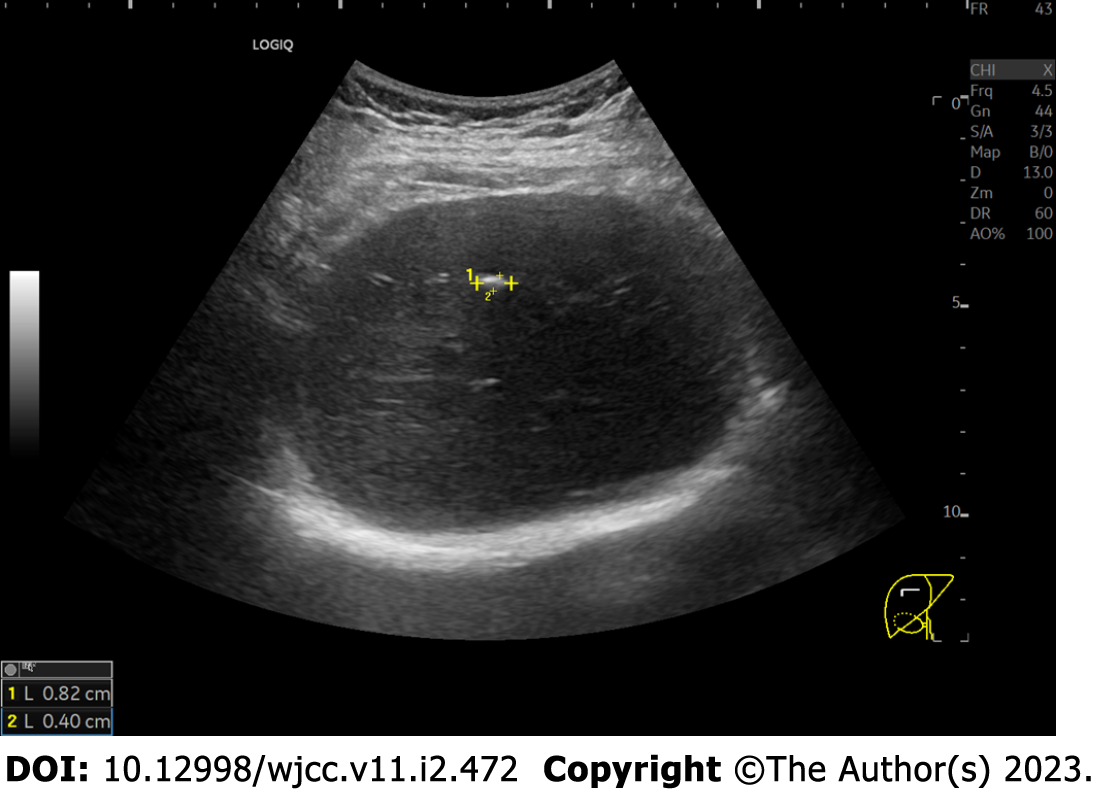
Abdominal ultrasound showed a calcified nodule in the liver (Figure 1), with no severe abnormalities in the liver, gallbladder, or other abdominal viscera.
The patient was diagnosed as ALI after mild COVID-19.
The patient was treated as expectant management. No medication was prescribed.
ALI was closely monitored for asymptomatic features and an unknown etiology. The following week, blood tests showed a reduction in both AST and ALT levels (ALT 124 U/L, AST 318 U/L). Two weeks later, they had decreased to near the expected UNL (ALT 40 U/L, AST 76 U/L) (Figure 2).
Our case highlights the importance of monitoring unusual liver injury after mild COVID-19. We demonstrated that recovery from liver injury could be achieved by natural course. No medication or intervention was applied in this case.
Gastrointestinal (GI) symptoms associated with COVID-19 include diarrhea, vomiting, nausea, and decreased appetite[12,13]. The coronavirus infection in the intestinal tissue causes these GI symptoms[12]. Additionally, gut-brain interaction might also induce GI tract discomfort[13]. The complete blood count (CBC) in most SARS-CoV-2-infected patients with liver injury shows erythrocytes, platelets, and leukocytes within normal limits[13]. Our patient showed the same CBC pattern.
Patients with COVID-19 usually present elevated liver enzymes in liver function tests, including ALT and AST. However, severe liver injury is uncommon, even in severe COVID-19 cases[13]. SARS-CoV-2 enters the cells via the angiotensin-converting enzyme 2 (ACE2) receptor[14]. Cholangiocytes express the ACE2 receptor and may be invaded by SARS-CoV-2, causing elevated GGT[15]. Our patient also presented elevated GGT initially, decreasing after day 59. A previous study also showed hypoalbuminemia and elevated AST levels in critical COVID-19 cases[13]. It was suggested that albumin and AST could be liver function biomarkers in patients with COVID-19. AST elevation was noted in the case presented here, but no hypoalbuminemia was detected.
The mechanism by which COVID-19 triggers acute hepatitis still remains unclear. Sun et al[10] suggested several possible explanations, such as the combination of the immune-mediated inflammatory response, direct cytotoxic injury due to viral replication, hypoxic hepatitis, drug-induced liver injury, or reactivation of pre-existing liver disease. ACE2 expression in the biliary and hepatic endothelial cells can explain the observed liver injury[16]. Hypoxia, drugs, or pre-existing liver disease were disregarded in our case because of the patient’s narrative history.
The molecular biology behind hepatic impairment is currently being explored since researchers are starting to recognize that ALI emerges as a clinically significant consequence of COVID-19. The hyperinflammation resulting from the cytokine storm and immune dysfunction provoked by COVID-19 contributes to ALI[17,18]. Pathological evidence of typical viral infection lesions with scarce CD4+ and CD8+ lymphocytes indicates that SARS-CoV-2 directly infects the liver[19]. Moreover, others believe that the virus binds to ACE2-positive cholangiocytes, not hepatocytes, and that cholangiocyte dysfunction induces liver injury[16].
COVID-19-induced liver injury may indicate that SARS-CoV-2 infection could cause multiple organ dysfunction. A previous pathological study of liver injury in a patient with COVID-19 showed mild lobular and portal activity and moderate steatosis[20]. Regarding imaging, computed tomography (CT) scans show typical liver injury characteristics in COVID-19 patients, including liver hypodensity and pericholecystic fat stranding[13]. Findings of liver hypodensity in imaging may also indicate liver steatosis[13]. Unfortunately, in our case, a liver CT scan was not performed due to the patient’s mild symptoms.
Severe COVID-19 correlates with severe hepatic, renal, cardiovascular, and coagulation complications[9]. Liver injury is an independent predictor of severe COVID-19 and even hospitalization and death in critically ill COVID-19 patients[21]. Our case illustrates that patients with mild COVID-19 could present variable degrees of organ impairment. These events may be subclinical and not anticipated solely by the severity of COVID-19. Thus, prompt surveillance and special care should be provided to treat these patients, and long-term outcomes should be monitored. Fortunately, COVID-19-related ALI has been reported to be self-limiting, and the outcomes have been satisfactory. The long-term effects on liver function remain unclear[10].
Previous studies documented that an active lifestyle[22] and healthy dietary patterns[23] may decrease the COVID-19 severity. There was no information about these confounding factors in the current case study.
This case report describes a patient with mild COVID-19 complicated by acute liver injury. This case is noteworthy because substantially elevated aminotransferase levels were discovered in a mild COVID-19 case, proving that not only patients with severe disease can develop ALI. Clinicians should pay attention to liver function testing during the COVID-19 treatment regardless of disease severity. Patients with similar characteristics should be identified to establish clinical significance and treatment principles and provide information to determine the, still unclear, long-term impact of COVID-19 on liver function.
Provenance and peer review: Unsolicited article; Externally peer reviewed.
Peer-review model: Single blind
Specialty type: Infectious diseases
Country/Territory of origin: Taiwan
Peer-review report’s scientific quality classification
Grade A (Excellent): A, A
Grade B (Very good): 0
Grade C (Good): 0
Grade D (Fair): D
Grade E (Poor): 0
P-Reviewer: Rahmati M, Iran; Saeed MAM, Egypt; Shariati MBH, Iran S-Editor: Chen YL L-Editor: A P-Editor: Chen YL
| 1. | Sharma A, Tiwari S, Deb MK, Marty JL. Severe acute respiratory syndrome coronavirus-2 (SARS-CoV-2): a global pandemic and treatment strategies. Int J Antimicrob Agents. 2020;56:106054. [RCA] [PubMed] [DOI] [Full Text] [Full Text (PDF)] [Cited by in Crossref: 446] [Cited by in RCA: 367] [Article Influence: 73.4] [Reference Citation Analysis (0)] |
| 2. | Wang D, Hu B, Hu C, Zhu F, Liu X, Zhang J, Wang B, Xiang H, Cheng Z, Xiong Y, Zhao Y, Li Y, Wang X, Peng Z. Clinical Characteristics of 138 Hospitalized Patients With 2019 Novel Coronavirus-Infected Pneumonia in Wuhan, China. JAMA. 2020;323:1061-1069. [RCA] [PubMed] [DOI] [Full Text] [Cited by in Crossref: 14113] [Cited by in RCA: 14747] [Article Influence: 2949.4] [Reference Citation Analysis (0)] |
| 3. | Gupta A, Madhavan MV, Sehgal K, Nair N, Mahajan S, Sehrawat TS, Bikdeli B, Ahluwalia N, Ausiello JC, Wan EY, Freedberg DE, Kirtane AJ, Parikh SA, Maurer MS, Nordvig AS, Accili D, Bathon JM, Mohan S, Bauer KA, Leon MB, Krumholz HM, Uriel N, Mehra MR, Elkind MSV, Stone GW, Schwartz A, Ho DD, Bilezikian JP, Landry DW. Extrapulmonary manifestations of COVID-19. Nat Med. 2020;26:1017-1032. [RCA] [PubMed] [DOI] [Full Text] [Cited by in Crossref: 2419] [Cited by in RCA: 2038] [Article Influence: 407.6] [Reference Citation Analysis (2)] |
| 4. | Rahmati M, Koyanagi A, Banitalebi E, Yon DK, Lee SW, Il Shin J, Smith L. The effect of SARS-CoV-2 infection on cardiac function in post-COVID-19 survivors: A systematic review and meta-analysis. J Med Virol. 2022;. [RCA] [PubMed] [DOI] [Full Text] [Cited by in Crossref: 1] [Cited by in RCA: 18] [Article Influence: 9.0] [Reference Citation Analysis (0)] |
| 5. | Rahmati M, Keshvari M, Mirnasuri S, Yon DK, Lee SW, Il Shin J, Smith L. The global impact of COVID-19 pandemic on the incidence of pediatric new-onset type 1 diabetes and ketoacidosis: A systematic review and meta-analysis. J Med Virol. 2022;94:5112-5127. [RCA] [PubMed] [DOI] [Full Text] [Full Text (PDF)] [Cited by in Crossref: 86] [Cited by in RCA: 98] [Article Influence: 32.7] [Reference Citation Analysis (2)] |
| 6. | Phipps MM, Barraza LH, LaSota ED, Sobieszczyk ME, Pereira MR, Zheng EX, Fox AN, Zucker J, Verna EC. Acute Liver Injury in COVID-19: Prevalence and Association with Clinical Outcomes in a Large U.S. Cohort. Hepatology. 2020;72:807-817. [RCA] [PubMed] [DOI] [Full Text] [Full Text (PDF)] [Cited by in Crossref: 201] [Cited by in RCA: 271] [Article Influence: 54.2] [Reference Citation Analysis (2)] |
| 7. | Lee IC, Huo TI, Huang YH. Gastrointestinal and liver manifestations in patients with COVID-19. J Chin Med Assoc. 2020;83:521-523. [RCA] [PubMed] [DOI] [Full Text] [Full Text (PDF)] [Cited by in Crossref: 144] [Cited by in RCA: 128] [Article Influence: 25.6] [Reference Citation Analysis (2)] |
| 8. | Guan WJ, Ni ZY, Hu Y, Liang WH, Ou CQ, He JX, Liu L, Shan H, Lei CL, Hui DSC, Du B, Li LJ, Zeng G, Yuen KY, Chen RC, Tang CL, Wang T, Chen PY, Xiang J, Li SY, Wang JL, Liang ZJ, Peng YX, Wei L, Liu Y, Hu YH, Peng P, Wang JM, Liu JY, Chen Z, Li G, Zheng ZJ, Qiu SQ, Luo J, Ye CJ, Zhu SY, Zhong NS; China Medical Treatment Expert Group for Covid-19. Clinical Characteristics of Coronavirus Disease 2019 in China. N Engl J Med. 2020;382:1708-1720. [RCA] [PubMed] [DOI] [Full Text] [Full Text (PDF)] [Cited by in Crossref: 19202] [Cited by in RCA: 18838] [Article Influence: 3767.6] [Reference Citation Analysis (7)] |
| 9. | Wu C, Chen X, Cai Y, Xia J, Zhou X, Xu S, Huang H, Zhang L, Du C, Zhang Y, Song J, Wang S, Chao Y, Yang Z, Xu J, Chen D, Xiong W, Xu L, Zhou F, Jiang J, Bai C, Zheng J, Song Y. Risk Factors Associated With Acute Respiratory Distress Syndrome and Death in Patients With Coronavirus Disease 2019 Pneumonia in Wuhan, China. JAMA Intern Med. 2020;180:934-943. [RCA] [PubMed] [DOI] [Full Text] [Cited by in Crossref: 4960] [Cited by in RCA: 5508] [Article Influence: 1101.6] [Reference Citation Analysis (1)] |
| 10. | Sun J, Aghemo A, Forner A, Valenti L. COVID-19 and liver disease. Liver Int. 2020;40:1278-1281. [RCA] [PubMed] [DOI] [Full Text] [Cited by in Crossref: 200] [Cited by in RCA: 219] [Article Influence: 43.8] [Reference Citation Analysis (0)] |
| 11. | Bongiovanni M, Zago T. Acute hepatitis caused by asymptomatic COVID-19 infection. J Infect. 2021;82:e25-e26. [RCA] [PubMed] [DOI] [Full Text] [Full Text (PDF)] [Cited by in Crossref: 10] [Cited by in RCA: 18] [Article Influence: 3.6] [Reference Citation Analysis (0)] |
| 12. | Zhou Z, Zhao N, Shu Y, Han S, Chen B, Shu X. Effect of Gastrointestinal Symptoms in Patients With COVID-19. Gastroenterology. 2020;158:2294-2297. [RCA] [PubMed] [DOI] [Full Text] [Full Text (PDF)] [Cited by in Crossref: 151] [Cited by in RCA: 166] [Article Influence: 33.2] [Reference Citation Analysis (0)] |
| 13. | Lei P, Zhang L, Han P, Zheng C, Tong Q, Shang H, Yang F, Hu Y, Li X, Song Y. Liver injury in patients with COVID-19: clinical profiles, CT findings, the correlation of the severity with liver injury. Hepatol Int. 2020;14:733-742. [RCA] [PubMed] [DOI] [Full Text] [Full Text (PDF)] [Cited by in Crossref: 60] [Cited by in RCA: 50] [Article Influence: 10.0] [Reference Citation Analysis (0)] |
| 14. | Jackson CB, Farzan M, Chen B, Choe H. Mechanisms of SARS-CoV-2 entry into cells. Nat Rev Mol Cell Biol. 2022;23:3-20. [RCA] [PubMed] [DOI] [Full Text] [Full Text (PDF)] [Cited by in Crossref: 1831] [Cited by in RCA: 1878] [Article Influence: 626.0] [Reference Citation Analysis (0)] |
| 15. | Hoffmann M, Kleine-Weber H, Schroeder S, Krüger N, Herrler T, Erichsen S, Schiergens TS, Herrler G, Wu NH, Nitsche A, Müller MA, Drosten C, Pöhlmann S. SARS-CoV-2 Cell Entry Depends on ACE2 and TMPRSS2 and Is Blocked by a Clinically Proven Protease Inhibitor. Cell. 2020;181:271-280.e8. [RCA] [PubMed] [DOI] [Full Text] [Full Text (PDF)] [Cited by in Crossref: 11946] [Cited by in RCA: 14209] [Article Influence: 2841.8] [Reference Citation Analysis (0)] |
| 16. | Chai X, Hu L, Zhang Y, Han W, Lu Z, Ke A, Zhou J, Shi G, Fang N, Fan J, Cai J, Lan F. Specific ACE2 Expression in Cholangiocytes May Cause Liver Damage After 2019-nCoV Infection. [cited 14 Oct 2022]. Available from: https://www.biorxiv.org/content/10.1101/2020.02.03.931766v1. |
| 17. | Taneva G, Dimitrov D, Velikova T. Liver dysfunction as a cytokine storm manifestation and prognostic factor for severe COVID-19. World J Hepatol. 2021;13:2005-2012. [RCA] [PubMed] [DOI] [Full Text] [Full Text (PDF)] [Cited by in Crossref: 22] [Cited by in RCA: 21] [Article Influence: 5.3] [Reference Citation Analysis (0)] |
| 18. | Anirvan P, Narain S, Hajizadeh N, Aloor FZ, Singh SP, Satapathy SK. Cytokine-induced liver injury in coronavirus disease-2019 (COVID-19): untangling the knots. Eur J Gastroenterol Hepatol. 2021;33:e42-e49. [RCA] [PubMed] [DOI] [Full Text] [Cited by in Crossref: 14] [Cited by in RCA: 6] [Article Influence: 1.5] [Reference Citation Analysis (1)] |
| 19. | Wang Y, Liu S, Liu H, Li W, Lin F, Jiang L, Li X, Xu P, Zhang L, Zhao L, Cao Y, Kang J, Yang J, Li L, Liu X, Li Y, Nie R, Mu J, Lu F, Zhao S, Lu J, Zhao J. SARS-CoV-2 infection of the liver directly contributes to hepatic impairment in patients with COVID-19. J Hepatol. 2020;73:807-816. [PubMed] |
| 20. | Xu Z, Shi L, Wang Y, Zhang J, Huang L, Zhang C, Liu S, Zhao P, Liu H, Zhu L, Tai Y, Bai C, Gao T, Song J, Xia P, Dong J, Zhao J, Wang FS. Pathological findings of COVID-19 associated with acute respiratory distress syndrome. Lancet Respir Med. 2020;8:420-422. [RCA] [PubMed] [DOI] [Full Text] [Full Text (PDF)] [Cited by in Crossref: 5228] [Cited by in RCA: 5770] [Article Influence: 1154.0] [Reference Citation Analysis (2)] |
| 21. | Salık F, Uzundere O, Bıçak M, Akelma H, Akgündüz M, Korhan Z, Kandemir D, Kaçar CK. Liver function as a predictor of mortality in COVID-19: A retrospective study. Ann Hepatol. 2021;26:100553. [RCA] [PubMed] [DOI] [Full Text] [Full Text (PDF)] [Cited by in Crossref: 9] [Cited by in RCA: 15] [Article Influence: 3.8] [Reference Citation Analysis (0)] |
| 22. | Rahmati M, Shamsi MM, Khoramipour K, Malakoutinia F, Woo W, Park S, Yon DK, Lee SW, Shin JI, Smith L. Baseline physical activity is associated with reduced mortality and disease outcomes in COVID-19: A systematic review and meta-analysis. Rev Med Virol. 2022;32:e2349. [RCA] [PubMed] [DOI] [Full Text] [Full Text (PDF)] [Cited by in Crossref: 43] [Cited by in RCA: 37] [Article Influence: 12.3] [Reference Citation Analysis (0)] |
| 23. | Rahmati M, Fatemi R, Yon DK, Lee SW, Koyanagi A, Il Shin J, Smith L. The effect of adherence to high-quality dietary pattern on COVID-19 outcomes: A systematic review and meta-analysis. J Med Virol. 2022;e28298. [RCA] [PubMed] [DOI] [Full Text] [Cited by in Crossref: 1] [Cited by in RCA: 25] [Article Influence: 12.5] [Reference Citation Analysis (0)] |


