Published online Jul 6, 2023. doi: 10.12998/wjcc.v11.i19.4635
Peer-review started: December 29, 2022
First decision: February 17, 2023
Revised: March 19, 2023
Accepted: April 20, 2023
Article in press: April 20, 2023
Published online: July 6, 2023
Processing time: 183 Days and 2.9 Hours
Congenital glaucoma associated with Roberts syndrome (RS) is an unusual and unique condition. No previous report describes this association. A multidisciplinary approach including molecular studies were conducted to reach the final diagnosis.
We present a rare case of a 1-wk-old male with RS associated with bilateral congenital glaucoma, left ectopic kidney, and left-hand rudimentary digits. A comprehensive approach was applied by which bilateral non-penetrating glaucoma surgery was performed with good control of intraocular pressure for more than 6 mo. Cytogenetic and molecular testing were conducted and revealed normal measurements.
This report described a case of a male baby with clinical features of RS but with a negative molecular analysis, presenting with left-hand rudimentary digits, bilateral congenital glaucoma, and left ectopic kidney. To the best of our know
Core Tip: Roberts syndrome (RS) is an extremely rare disease characterized by a combination of deformities in the lower and/or upper extremities in association with other organ abnormalities. We provide here the first reported case of RS associated with bilateral congenital glaucoma. Bilateral non-penetrating glaucoma surgery was performed to control intraocular pressure and the outcome was excellent.
- Citation: Almulhim A, Almoallem B, Alsirrhy E, Osman EA. Unique Roberts syndrome with bilateral congenital glaucoma: A case report. World J Clin Cases 2023; 11(19): 4635-4639
- URL: https://www.wjgnet.com/2307-8960/full/v11/i19/4635.htm
- DOI: https://dx.doi.org/10.12998/wjcc.v11.i19.4635
Phocomelia with ocular and internal organ abnormalities are reported as part of Roberts syndrome (RS). RS (OMIM #268300; also known as Roberts-SC phocomelia syndrome) was reported by John Roberts in 1919 and is an autosomal recessive condition characterized by prenatal and postnatal growth retardation, skeletal malformation including tetraphocomelia or mesomelia, mental retardation, and craniofacial dysmorphic features[1].
The clinical presentation is widely variable, from mild cases that can be difficult to confirm without a molecular test to the severe spectrum of diseases that present with a typical phenotype. Babies born with severe RS usually die in utero or shortly postpartum, while mildly affected people can survive to middle age[2].
Herein, we report an unusual case of a male baby with clinical features of RS but with a negative molecular analysis, presenting with left-hand rudimentary digits, bilateral congenital glaucoma, and left ectopic kidney.
A 1-wk-old male baby was referred to our hospital in June 2021 with a picture of bilateral congenital glaucoma.
According to the parents, the deformed left hand with bilateral cloudy cornea were noticed immediately after birth.
The baby was the second child of a healthy non-consanguineous couple and a product of an uneventful pregnancy with spontaneous vaginal delivery at 40 wk. Birth weight was 3 kg. There was neither a family history of similar conditions nor a prenatal history of exposure to any known teratogenic medication.
On physical examination, left-hand rudimentary digits were noticed (Figure 1). The child was active and sucking well. Examinations of the central nervous system, cardiovascular system, abdomen, and genitalia showed all to be normal.
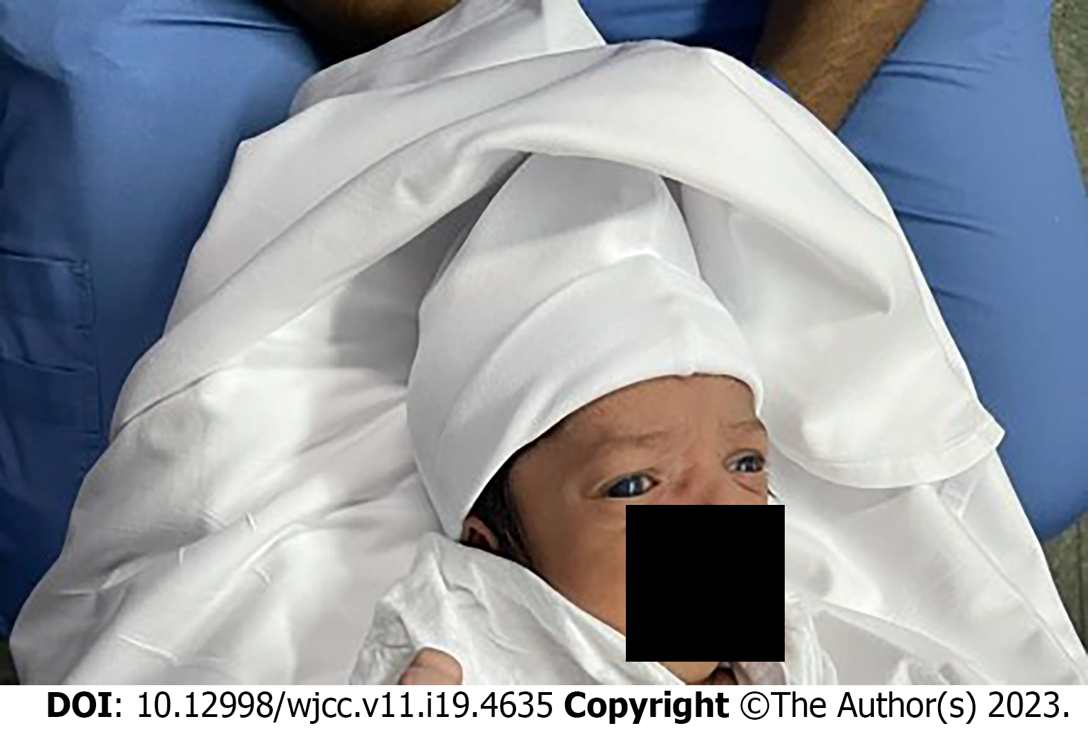
An ocular examination was performed under general anesthesia and showed bilateral buphthalmos (Figure 2), significant corneal edema, and corneal diameter of 11 mm right/11.5 mm left. The patient was treated with travaprost and combined dorzolamide and timolol, and the intraocular pressure (IOP) was 25 mmHg right/39 mmHg left. Fundus examination showed a cup disc ratio of about 0.2 in both eyes. Central corneal thickness ranged between 900 microns and 930 microns in both eyes.
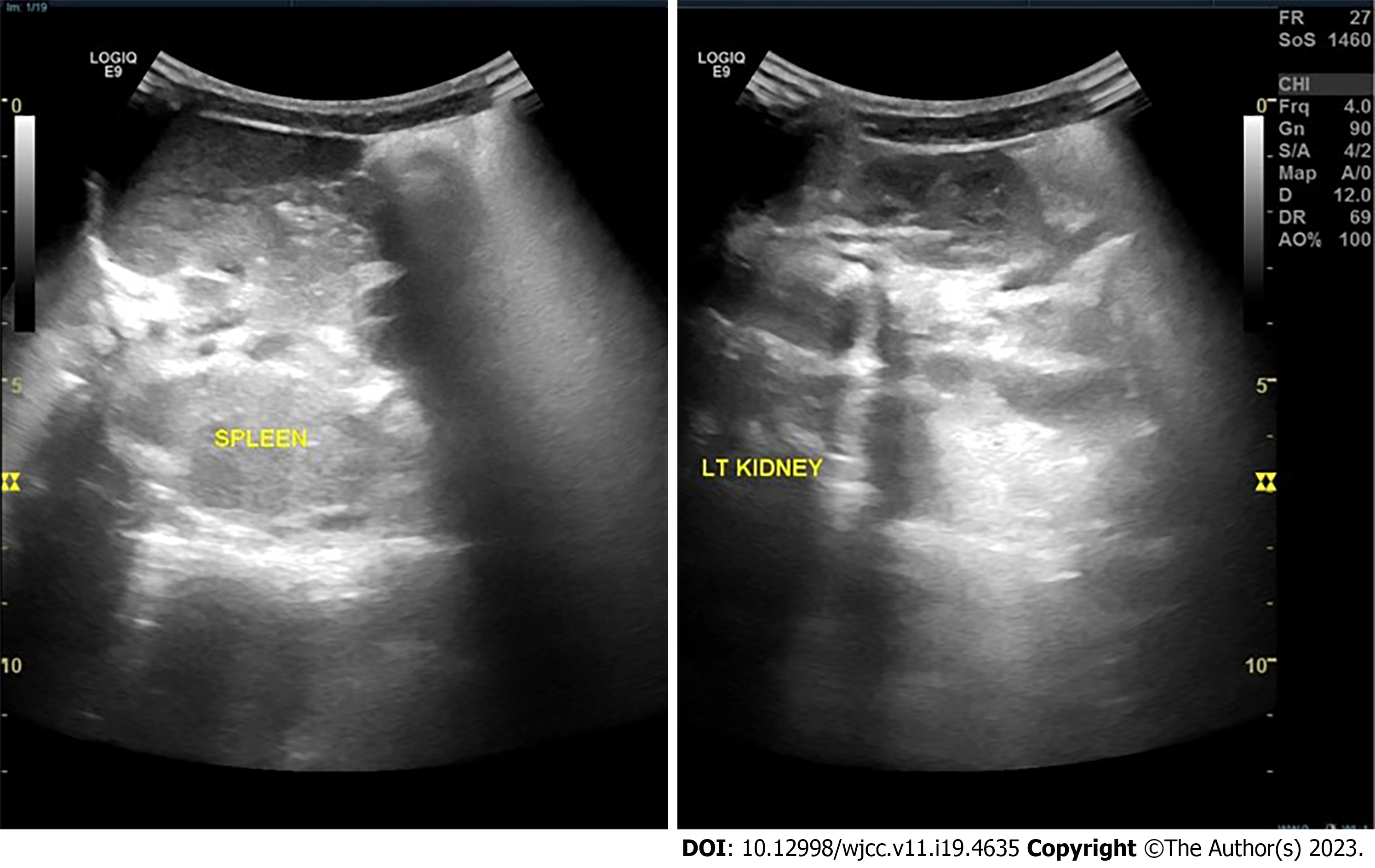
Echocardiogram findings were normal. Abdominal ultrasound showed a left ectopic kidney located in the ipsilateral pelvic region and not in the lumbar area (Figure 3). Both kidneys appeared normal in size and echogenicity.
Based on the given detailed ophthalmological and medical assessment, we applied comprehensive genetic testing by performing karyotyping and chromosomal microarray analysis. Karyotype showed normal male (46, XY) with no evidence of clinically significant numerical or structural chromosome abnormalities. Moreover, we performed whole-exome sequencing (WES) that allowed us to screen the whole exome and to focus in particular on the ESCO2 gene that is known to be linked with RS and other genes related to primary congenital glaucoma such as the CYP1B1 and LTBP2 genes. Despite our detailed review of the WES data, negative results were found, with no observed genetic variants or incidental findings.
Thalidomide-induced phocomelia; Holt-Oram syndrome; Thrombocytopenia with absent radius syndrome; sporadic phocomelia.
Based on the clinical and diagnostic findings, the final diagnosis was RS with bilateral congenital glaucoma and left ectopic kidney.
Bilateral non-penetrating deep sclerectomy plus mitomycin C was applied. The IOP was stable for more than 6 mo postoperatively.
Written informed consent was obtained from the parents of the baby. The Institutional Review Board reviewed and approved this study. Permission to publish photographs was also obtained.
At 6 mo, 9 mo, and 1 year postoperatively, the IOP was stable without anti-glaucoma medication.
RS is an extremely rare autosomal recessive condition that presents with widely variable prenatal and postnatal growth retardation, skeletal malformation, and mental retardation. It was first described by Roberts in 1919 in two children of consanguineous parents. The children presented with upper and lower limb anomalies, skull deformity, exophthalmos, cleft lip, and palate bilaterally[3]. In 1969, Afifi et al[1] reported a syndrome with milder features and labeled it as SC phocomelia syndrome.
A systematic review and severity scoring system was published by Van Den Berg and Francke[3] in 1993 that included 100 cases. Goh et al[2] and Zhou et al[4] conducted a review of the literature on RS in adult patients. None of the previously reported cases were associated with congenital glaucoma. Ocular manifestations reported in the literature to date have been hypertelorism in 86.7% of patients, exophthalmia in 69.4% of patients, cloudy cornea in 68.1% of patients, optic nerve cavernous hemangioma in 1 patient, and blue sclera with bilateral macular dysfunction in 1 patient. Renal abnormalities were reported in 50% of patients[1-4].
Worldwide about 157 cases of RS have been reported, and the most recent case was published in October 2020 regarding a 30-wk gestation stillborn male who had the typical phenotype of RS with a normal molecular analysis[5].
In our case, we applied comprehensive genetic testing by performing karyotyping and chromosomal microarray analysis. Chromosomal analysis revealed normal male (46, XY) without any detected abnormalities. WES revealed no abnormalities in the ESCO2 gene (linked to RS) or the CYP1B1 and LTBP2 genes (linked to primary congenital glaucoma). Although this patient had normal molecular study findings, the diagnosis of RS can be established based on the phenotype because RS cases without any molecular abnormality have been reported in the literature[5].
This report describes a case of a male baby with clinical features of RS but with a negative molecular analysis. He presented with left-hand rudimentary digits, bilateral congenital glaucoma, and left ectopic kidney. To the best of our knowledge, this is the first reported case of RS with a combination of phocomelia, congenital glaucoma, and ectopic kidney without any detected molecular abnormality.
Provenance and peer review: Unsolicited article; Externally peer reviewed.
Peer-review model: Single blind
Specialty type: Medicine, research and experimental
Country/Territory of origin: Saudi Arabia
Peer-review report’s scientific quality classification
Grade A (Excellent): 0
Grade B (Very good): B, B
Grade C (Good): 0
Grade D (Fair): 0
Grade E (Poor): 0
P-Reviewer: Malik S, Pakistan; Morya AK, India S-Editor: Liu XF L-Editor: A P-Editor: Chen YX
| 1. | Afifi HH, Abdel-Salam GM, Eid MM, Tosson AM, Shousha WG, Abdel Azeem AA, Farag MK, Mehrez MI, Gaber KR. Expanding the mutation and clinical spectrum of Roberts syndrome. Congenit Anom (Kyoto). 2016;56:154-162. [RCA] [PubMed] [DOI] [Full Text] [Cited by in Crossref: 10] [Cited by in RCA: 12] [Article Influence: 1.3] [Reference Citation Analysis (0)] |
| 2. | Goh ES, Li C, Horsburgh S, Kasai Y, Kolomietz E, Morel CF. The Roberts syndrome/SC phocomelia spectrum--a case report of an adult with review of the literature. Am J Med Genet A. 2010;152A:472-478. [RCA] [PubMed] [DOI] [Full Text] [Cited by in Crossref: 31] [Cited by in RCA: 29] [Article Influence: 1.9] [Reference Citation Analysis (0)] |
| 3. | Van Den Berg DJ, Francke U. Roberts syndrome: a review of 100 cases and a new rating system for severity. Am J Med Genet. 1993;47:1104-1123. [RCA] [PubMed] [DOI] [Full Text] [Cited by in Crossref: 132] [Cited by in RCA: 136] [Article Influence: 4.3] [Reference Citation Analysis (0)] |
| 4. | Zhou J, Yang X, Jin X, Jia Z, Lu H, Qi Z. Long-term survival after corrective surgeries in two patients with severe deformities due to Roberts syndrome: A Case report and review of the literature. Exp Ther Med. 2018;15:1702-1711. [RCA] [PubMed] [DOI] [Full Text] [Cited by in RCA: 2] [Reference Citation Analysis (0)] |
| 5. | Salari B, Dehner LP. Pseudo-Roberts Syndrome: An Entity or Not? Fetal Pediatr Pathol. 2022;41:396-402. [RCA] [PubMed] [DOI] [Full Text] [Reference Citation Analysis (0)] |


