Copyright
©The Author(s) 2023.
World J Clin Cases. Jun 16, 2023; 11(17): 4133-4141
Published online Jun 16, 2023. doi: 10.12998/wjcc.v11.i17.4133
Published online Jun 16, 2023. doi: 10.12998/wjcc.v11.i17.4133
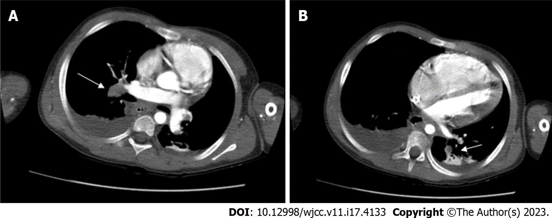
Figure 2 Pulmonary artery embolism.
The intraluminal filling defects are noted in A: The right interlobar; B: The left truncus. Bilateral posterior pleural effusions are noted with posterior passive atelectasis of both lungs.
- Citation: Lo CY, Chen KB, Chen LK, Chiou CS. Massive pulmonary embolism in Klippel-Trenaunay syndrome after leg raising: A case report. World J Clin Cases 2023; 11(17): 4133-4141
- URL: https://www.wjgnet.com/2307-8960/full/v11/i17/4133.htm
- DOI: https://dx.doi.org/10.12998/wjcc.v11.i17.4133


