Copyright
©The Author(s) 2023.
World J Clin Cases. May 26, 2023; 11(15): 3481-3490
Published online May 26, 2023. doi: 10.12998/wjcc.v11.i15.3481
Published online May 26, 2023. doi: 10.12998/wjcc.v11.i15.3481
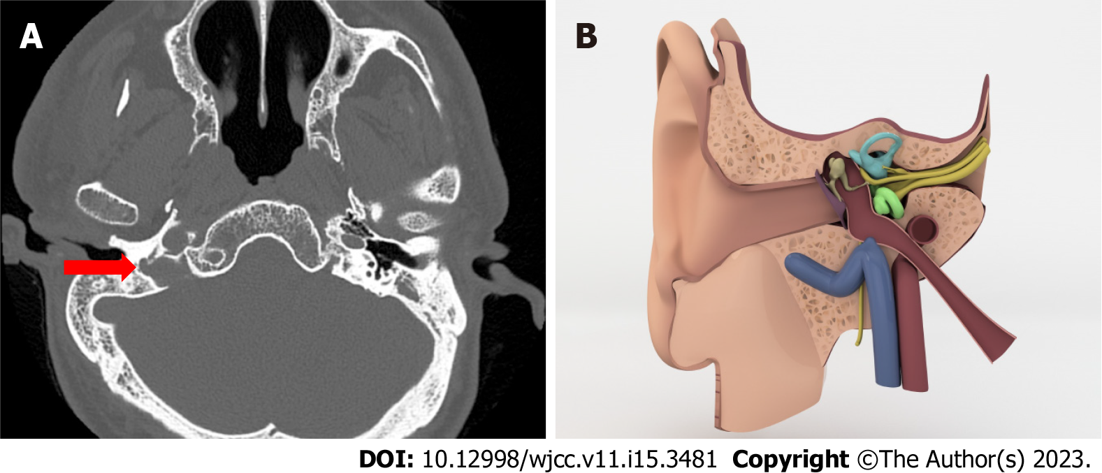
Figure 4 Jugular bulb diverticulum.
A: Axial computed tomography image shows jugular bulb diverticulum as an outpouching from tiny bulbus (red arrow); B: Illustration draw of jugular bulb diverticulum which out pouches to middle ear cavity.
- Citation: Gökharman FD, Şenbil DC, Aydin S, Karavaş E, Özdemir Ö, Yalçın AG, Koşar PN. Chronic otitis media and middle ear variants: Is there relation? World J Clin Cases 2023; 11(15): 3481-3490
- URL: https://www.wjgnet.com/2307-8960/full/v11/i15/3481.htm
- DOI: https://dx.doi.org/10.12998/wjcc.v11.i15.3481


