Copyright
©The Author(s) 2023.
World J Clin Cases. Jan 6, 2023; 11(1): 210-217
Published online Jan 6, 2023. doi: 10.12998/wjcc.v11.i1.210
Published online Jan 6, 2023. doi: 10.12998/wjcc.v11.i1.210
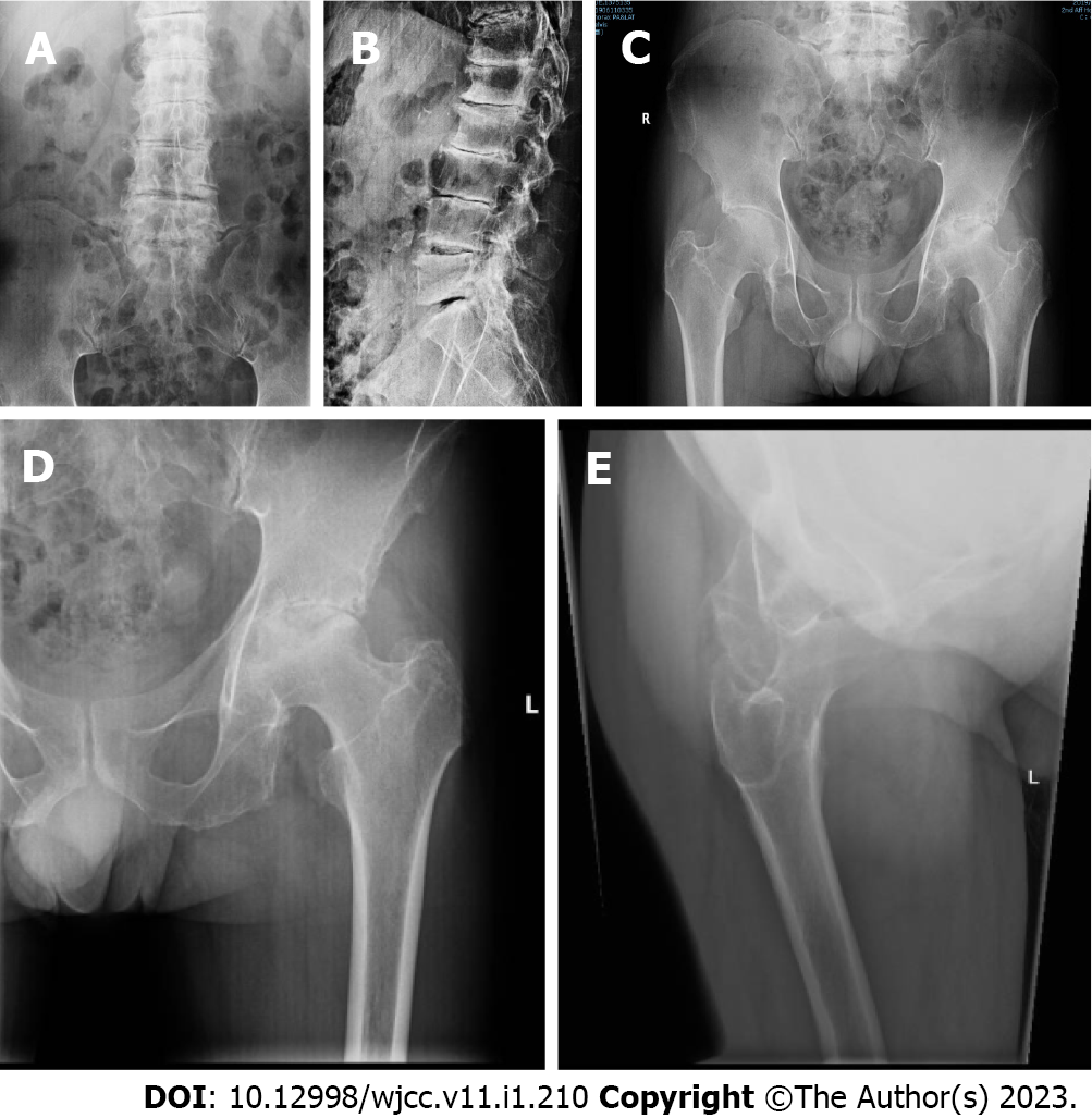
Figure 1 X-rays of lumbar spine and left hip.
A and B: Degenerative changes, lumbar vertebra spondylitis of lumbar spine; C: Pelvis indicated bilateral hip arthritis, aseptic necrosis of left hip joint of lumbar spine; D: Anteroposterior view of left hip; E: Lateral view showed disappearance of hip space and aseptic necrosis (stage 4) of left femoral head of left hip.
- Citation: Yap San Min N, Rafi U, Wang J, He B, Fan L. Ochronotic arthropathy of bilateral hip joints: A case report. World J Clin Cases 2023; 11(1): 210-217
- URL: https://www.wjgnet.com/2307-8960/full/v11/i1/210.htm
- DOI: https://dx.doi.org/10.12998/wjcc.v11.i1.210


