Published online Mar 6, 2022. doi: 10.12998/wjcc.v10.i7.2087
Peer-review started: June 25, 2021
First decision: July 26, 2021
Revised: August 6, 2021
Accepted: January 20, 2022
Article in press: January 20, 2022
Published online: March 6, 2022
Processing time: 250 Days and 1.4 Hours
Acromioclavicular joint (ACJ) space narrowing has been considered to be an important diagnostic image parameter of ACJ osteoarthritis (ACJO). However, the morphology of the ACJ space is irregular because of osteophyte formation, subchondral irregularity, capsular distention, sclerosis, and erosion. Therefore, we created the ACJ cross-sectional area (ACJCSA) as a new diagnostic image parameter to assess the irregular morphologic changes of the ACJ.
To hypothesize that the ACJCSA is a new diagnostic image parameter for ACJO.
ACJ samples were obtained from 35 patients with ACJO and 30 healthy individuals who underwent shoulder magnetic resonance (S-MR) imaging that revealed no evidence of ACJO. Oblique coronal, T2-weighted, fat-suppressed S-MR images were acquired at the ACJ level from the two groups. We measured the ACJCSA and the ACJ space width (ACJSW) at the ACJ on the S-MR images using our imaging analysis program. The ACJCSA was measured as the cross-sectional area of the ACJ. The ACJSW was measured as the narrowest point between the acromion and the clavicle.
The average ACJCSA was 39.88 ± 10.60 mm2 in the normal group and 18.80 ± 5.13 mm2 in the ACJO group. The mean ACJSW was 3.51 ± 0.58 mm in the normal group and 2.02 ± 0.48 mm in the ACJO group. ACJO individuals had significantly lower ACJCSA and ACJSW than the healthy individuals. Receiver operating characteristic curve analyses demonstrated that the most suitable ACJCSA cutoff score was 26.14 mm2, with 91.4% sensitivity and 90.0% specificity.
The optimal ACJSW cutoff score was 2.37 mm, with 88.6% sensitivity and 96.7% specificity. Even though both the ACJCSA and ACJSW were significantly associated with ACJO, the ACJCSA was a more sensitive diagnostic image parameter.
Core Tip: An acromioclavicular joint (ACJ) space narrowing has been considered to be an important diagnostic image parameter of ACJ osteoarthritis. However, the morphology of ACJ space is irregular, because of osteophyte formation, subchondral irregularity, capsular distention, sclerosis, and erosions. Therefore, we created the ACJ cross-sectional area as a new diagnostic image parameter to assess the irregular morphologic change of ACJ.
- Citation: Joo Y, Moon JY, Han JY, Bang YS, Kang KN, Lim YS, Choi YS, Kim YU. Usefulness of the acromioclavicular joint cross-sectional area as a diagnostic image parameter of acromioclavicular osteoarthritis. World J Clin Cases 2022; 10(7): 2087-2094
- URL: https://www.wjgnet.com/2307-8960/full/v10/i7/2087.htm
- DOI: https://dx.doi.org/10.12998/wjcc.v10.i7.2087
Acromioclavicular joint osteoarthritis (ACJO) is frequently diagnosed in patients older than the fifth decade[1-4]. ACJO is the main cause of shoulder pain relating to the acromioclavicular joint (ACJ). Clinically, the relevance of ACJ abnormalities is tested by the body cross-test and palpation. The body cross-test is performed by elevating the affected arm on the same side. The physician adducts the arm across the body and takes the patient’s elbow. Positive results on this test reproduce pain around the ACJ. Some pathologic and radiographic studies also have been performed to evaluate symptomatic ACJO. However, investigations using shoulder magnetic resonance (S-MR) scans have usually focused on disorders of the labrum and rotator cuff tears but rarely on the ACJ[1,2,5,6]. Only a few studies have been conducted to assess S-MR findings in symptomatic ACJO. However, the research findings varied. Shubin Stein et al[6] reported that bone edema at the distal clavicle or acromion was related to symptomatic ACJO whereas. In another study, S-MR findings were not related to symptomatic ACJO[7]. We think this discrepancy may be because the previous studies assessed ACJ space narrowing using only a single measurement called the ACJ space width (ACJSW) at the approximate halfway point of the ACJ[8]. However, partial narrowing and irregular osteophyte formation could occur anywhere. Thus, measurement mistakes can occur at any time. We think that it may be worthwhile to reconsider the morphological value of S-MR findings in the diagnosis of symptomatic ACJO.
Thus, to assess irregular narrowing of the ACJ, we devised the ACJ cross-sectional area (ACJCSA) as a new diagnostic image parameter. Contrast with the ACJSW, the ACJCSA does not influenced by measurement mistakes because the ACJCSA measures the entire irregular area of the ACJ. We hypothesized that the ACJCSA is an important diagnostic image parameter in ACJO diagnosis. Therefore, we used S-MR images to compare the ACJCSA and ACJSW between patients with ACJO and normal controls.
This original study was approved by the Catholic Kwandong University (Incheon, South Korea) Institutional Research Board (CKUIRB). The retrospective data used to support the findings of this research may be released upon application to the CKUIRB. A total of 35 patients with radiologically confirmed ACJO from January 2015 to October 2019 were enrolled in the study. The inclusion criteria of the ACJO group were as follows: (1) A history of pain and tenderness in the front of the shoulder around the ACJ; (2) A positive cross-arm adduction test; or (3) A positive active compression test. We excluded subjects if they had the following disorders: (1) history of shoulder infection; (2) inflammatory arthritis; (3) acute clavicle fracture; (4) humerus bone fracture, or (5) any history of shoulder surgery.
There were 14 (40.0%) men and 21 (60.0%) women with an average age of 60.60 ± 9.31 years (range, 45 to 80 years) in the study (Table 1). We enrolled normal individuals to compare to the ACJO patients. The normal group was people who voluntary wanted to undergo S-MR imaging for an exact diagnosis of shoulder pain but no evidence of ACJO. In the normal group, 30 subjects (9 males and 21 females) were enrolled with an average age of 57.30 ± 7.56 years (range, 40 to 69 years).
| Variable | Control group, n = 30 | ACJO group, n = 35 | Statistical significance |
| Gender (male/female) | 9/21 | 14/21 | NS |
| Age (yr) | 57.30 ± 7.56 | 60.60 ± 9.31 | NS |
| ACJSW (mm) | 3.51 ± 0.58 | 2.02 ± 0.48 | P < 0.001 |
| ACJCSA (mm2) | 39.88 ± 10.60 | 18.80 ± 5.13 | P < 0.001 |
| Location (Rt/Lt) | 13/17 | 22/13 | NS |
S-MR analysis was performed using a 3T magnetic resonance imaging Magnetom system (Siemens Medical care, Skyra, Germany) and 3T scanners (Philips, Healthcare, Angina, Netherlands). For all S-MR images, we acquired oblique coronal T2-weighted fat-suppressed turbo spin-echo imaging with a layer thickness of 3 mm, an intersection gap of 0.9 mm, a repetition time of 4010 ms, an echo time of 76 ms, a 512 × 256 matrix, a 150 cm × 150 cm field of view, and > 3 echo train length.
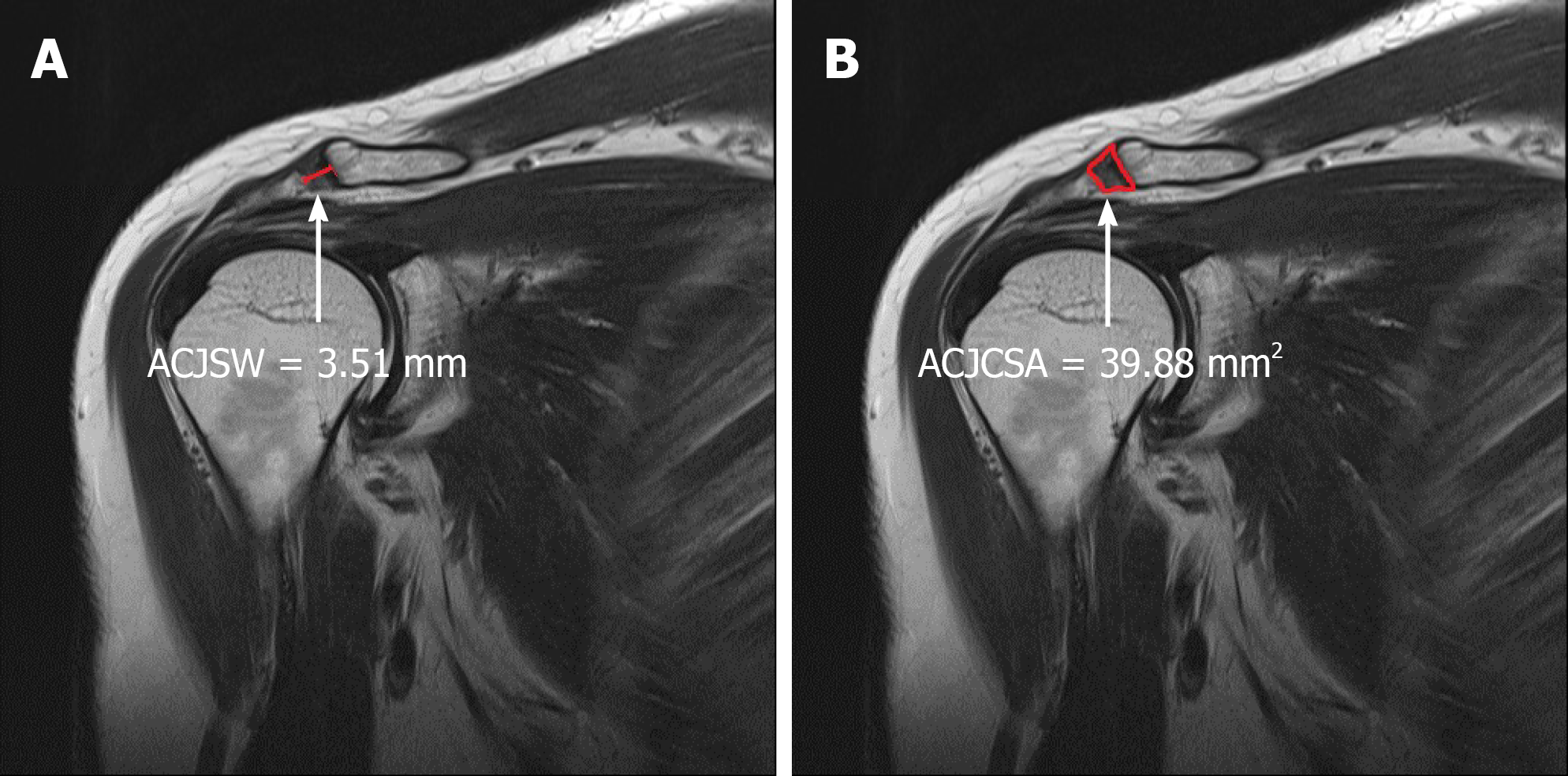
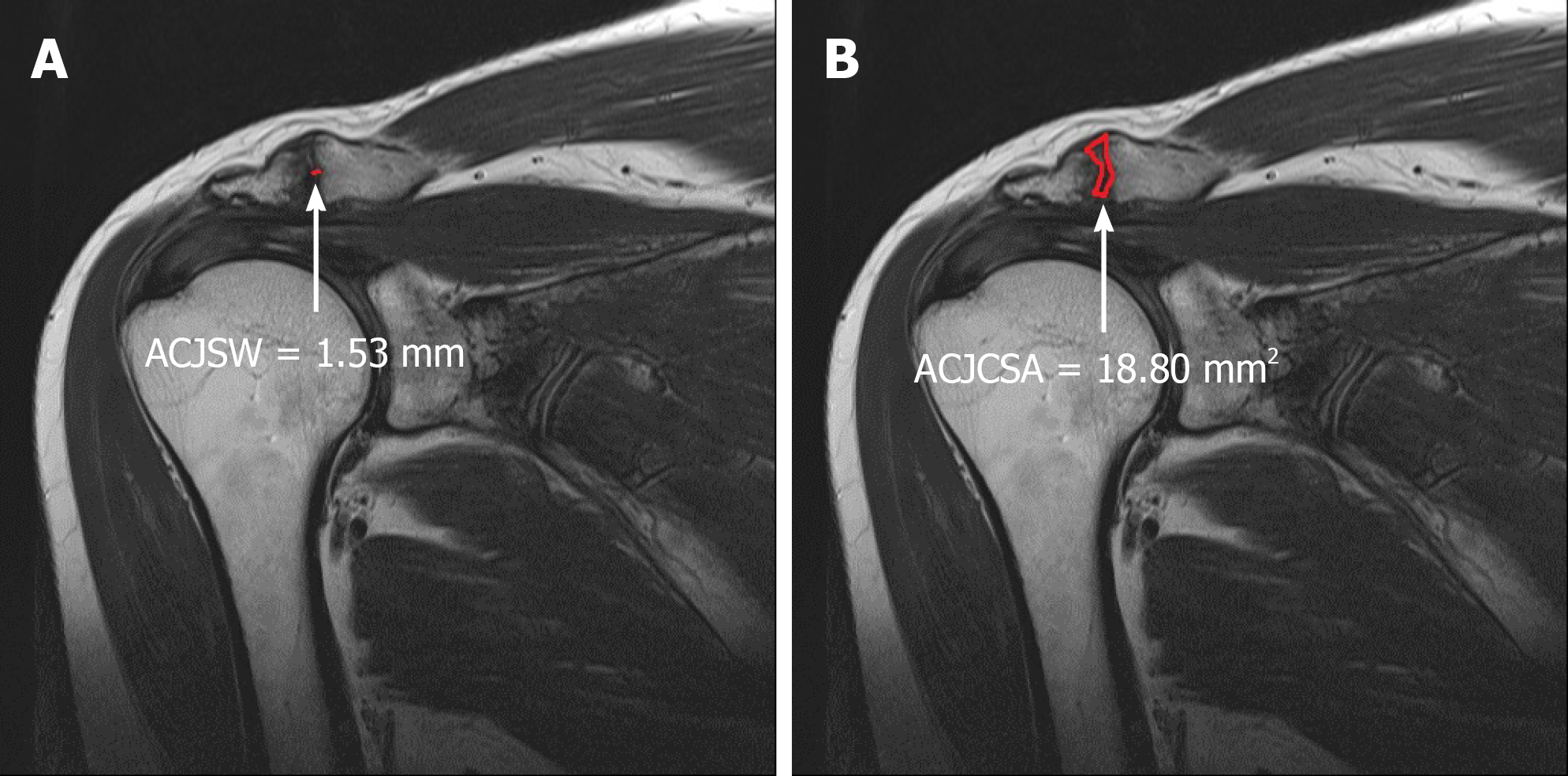
The ACJCSA and ACJSW data were acquired by the corresponding author who was blinded to the group of shoulder images. We obtained oblique coronal T2-weighted S-MR images at the narrowest visualization of the ACJ. We examined the ACJCSA and ACJSW on S-MR images using an image analysis program (INFINITT PACS; Incheon, South Korea) (Figures 1 and 2). We measured the ACJSW at the narrowest ACJ using the PACS system. The ACJCSA was examined as the cross-sectional whole area of the ACJ at the same point of the ACJSW.
We compared the ACJCSA and ACJSW between the ACJO and the normal group using unpaired t-tests. The predictive value of the ACJCSA and ACJSW in the diagnosis of ACJO was estimated by receiver operating characteristic (ROC) analysis. The area under the curve (AUC), sensitivity, and specificity were calculated. Statistical Package for Social Sciences (SPSS) version 22.0 software (SPSS Inc., Chicago, IL, USA) was used. P values of < 0.05 were considered statistically significant. All values are presented as the mean and standard deviation.
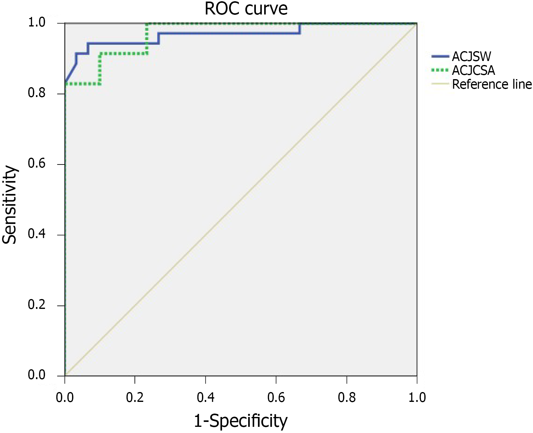
The mean ACJCSA was 39.88 ± 10.60 mm2 in the normal group and 18.80 ± 5.13 mm2 in the ACJO group. The mean ACJSW was 3.51 ± 0.58 mm in the normal group and 2.02 ± 0.48 mm in the ACJO group. The ACJO patients had significantly lower ACJCSA and ACJSW than the normal individuals (Table 1). The ROC analysis demonstrated that the most suitable ACJCSA cutoff value was 26.14 mm2, with an AUC of 0.98 (95%CI: 0.94-1.00), 91.4% sensitivity, and 90.0% specificity (Table 2 and Figure 3). The best ACJSW cutoff score was 2.37 mm, with an AUC of 0.97 (95%CI: 0.92-1.00), 88.6% sensitivity, and 96.7% specificity (Table 3 and Figure 3).
ACJO is a disabling and painful disorder in association with the more common diagnosis of shoulder impingement syndrome[1,7,9,10]. Inferiorly protruding osteophytes as well as soft tissue hypertrophy of the ACJ accelerates narrowing of the supraspinatus outlet[11-13]. Narrowing of the outlet space, whose borders are formed by the coracoacromial ligament, coracoid process, anterior aspect of the acromion, and ACJ, has been reported as the primary cause for the development of rotator cuff tears and subsequent impingement syndrome[14-24]. Thus, positive associations regarding the incidence of rotator cuff tears and the severity of ACJ degeneration have been demonstrated. Clinically, the possibility of ACJ abnormalities is examined using the body cross-test and palpation[4,9,25]. The body cross-test is performed by elevating the affected arm on the same side. The physician adducts the arm across the body and takes the patient’s elbow. Positive results on this test reproduce pain around the ACJ[2]. S-MR imaging and plain X-rays have been used to assess the severity and presence of ACJO[6]. Although plain shoulder X-rays are the first-choice imaging modality for the diagnosis of ACJ pathology, an exact diagnosis is impossible. The severity of ACJO has frequently been judged differently, with S-MR imaging compared to conventional radiography[1,11,12,26]. In S-MR imaging, the excellent soft tissue contrast and the associated benefits of multiplanar acquisition have optimized the assessment of ACJO. Subchondral bone marrow edema, osteophytes, sclerosis, subchondral cysts, and soft-tissue abnormalities (joint effusion and capsular hypertrophy) and may also be seen on S-MR images[1]. However, only a few studies have been performed to assess the predictability of S-MR findings in diagnosing symptomatic ACJO. Moreover, the previous conclusions of these studies varied. Gordon et al[8] insisted that ACJO may mimic the clinical symptoms of rotator cuff disorder. Several S-MR features are common to distal clavicle osteolysis, os acromiale. Shubin Stein et al[6] reported that bone edema at the distal clavicle or acromion was related to symptomatic ACJO. Hawkins et al[7] insisted that any S-MR findings were not related to symptomatic ACJO. Moreover, previous studies only investigated ACJ space narrowing using a single measurement called the ACJSW at the approximate halfway point of the ACJ. However, partial narrowing and irregular osteophyte formation can occur at any time. Thus, measurement mistakes could occur at any time.
We think it can be worthwhile to reconsider the morphological value of S-MR findings in the diagnosis of symptomatic ACJO. Thus, to evaluate the irregular narrowing of the ACJ, we devised the ACJCSA as a new morphological parameter. Compared to the ACJSW, the ACJCSA does not influenced by these measurement biases because the ACJCSA measures the entire cross-sectional area of the ACJ. Eventually, we concluded that the ACJCSA was better than the ACJSW as a diagnostic image parameter of ACJO. In this research, we demonstrated that the ACJCSA had 91.4% sensitivity, and an AUC of 0.98 to evaluate ACJO. The ACJSW had 88.6% sensitivity, and an AUC of 0.97. Our results suggest that the ACJCSA was a better morphological parameter of ACJO than the ACJSW. We hope our results will help to improve the quality of ACJO diagnosis.
The current research had several limitations. There are several isolated ACJ pathologies in symptomatic shoulders such as distal clavicle osteolysis, acromiale syndrome, and ACJO. However, we only focused on ACJO because the ACJ is the most commonly damaged area. Second, some different methods to assess ACJO, such as subchondral bone marrow edema, osteophytes, subchondral cysts, sclerosis, and soft-tissue abnormalities, have been reported to be effective in discriminating ACJO. However, in this research, we only analyzed the ACJCSA and ACJSW measurements on S-MR images. Third, we enrolled a relatively small sample. Fifth, this study was retrospective in nature.
We demonstrated the optimal ACJCSA cutoff value as 26.14 mm2, with 91.4% sensitivity and 90.0% specificity. The best ACJSW cutoff value was 2.37 mm, with 88.6% sensitivity and 96.7% specificity. When evaluating patients with ACJO, physicians should carefully assess the ACJCSA rather than the ACJSW.
Acromioclavicular joint (ACJ) space narrowing has been considered to be an important diagnostic image parameter of ACJ osteoarthritis (ACJO).
The morphology of the ACJ space is irregular because of osteophyte formation, subchondral irregularity, capsular distention, sclerosis, and erosion. Therefore, we created the ACJ cross-sectional area (ACJCSA) as a new diagnostic image parameter to assess the irregular morphologic changes of the ACJ.
To hypothesize that the ACJCSA is a new diagnostic image parameter for ACJO.
ACJ samples were obtained from 35 patients with ACJO and 30 healthy individuals who underwent shoulder magnetic resonance (S-MR) imaging that revealed no evidence of ACJO. Oblique coronal, T2-weighted, fat-suppressed S-MR images were acquired at the ACJ level from the two groups. We measured the ACJCSA and ACJ space width (ACJSW) at the ACJ on S-MR images using our imaging analysis program. The ACJCSA was measured as the cross-sectional area of the ACJ. The ACJSW was measured as the narrowest point between the acromion and the clavicle.
The average ACJCSA was 39.88 ± 10.60 mm2 in the normal group and 18.80 ± 5.13 mm2 in the ACJO group. The mean ACJSW was 3.51 ± 0.58 mm in the normal group and 2.02 ± 0.48 mm in the ACJO group. ACJO individuals had significantly lower ACJCSA and ACJSW than the healthy individuals. Receiver operating characteristic curve analysis demonstrated that the most suitable ACJCSA cutoff score was 26.14 mm2, with 91.4% sensitivity and 90.0% specificity.
The optimal ACJSW cutoff score was 2.37 mm, with 88.6% sensitivity and 96.7% specificity. Even though both the ACJCSA and ACJSW were significantly associated with ACJO, the ACJCSA was a more sensitive diagnostic image parameter.
We enrolled a relatively small sample.
All authors thank International St. Mary’s Hospital.
Provenance and peer review: Unsolicited article; Externally peer reviewed.
Peer-review model: Single blind
Specialty type: Medicine, research and experimental
Country/Territory of origin: South Korea
Peer-review report’s scientific quality classification
Grade A (Excellent): A
Grade B (Very good): 0
Grade C (Good): C
Grade D (Fair): 0
Grade E (Poor): 0
P-Reviewer: Kim YU, Naserian S S-Editor: Chang KL L-Editor: A P-Editor: Chang KL
| 1. | de Abreu MR, Chung CB, Wesselly M, Jin-Kim H, Resnick D. Acromioclavicular joint osteoarthritis: comparison of findings derived from MR imaging and conventional radiography. Clin Imaging. 2005;29:273-277. [RCA] [PubMed] [DOI] [Full Text] [Cited by in Crossref: 37] [Cited by in RCA: 29] [Article Influence: 1.5] [Reference Citation Analysis (0)] |
| 2. | Strobel K, Pfirrmann CW, Zanetti M, Nagy L, Hodler J. MRI features of the acromioclavicular joint that predict pain relief from intraarticular injection. AJR Am J Roentgenol. 2003;181:755-760. [RCA] [PubMed] [DOI] [Full Text] [Cited by in Crossref: 51] [Cited by in RCA: 43] [Article Influence: 2.0] [Reference Citation Analysis (0)] |
| 3. | Widman DS, Craig JG, van Holsbeeck MT. Sonographic detection, evaluation and aspiration of infected acromioclavicular joints. Skeletal Radiol. 2001;30:388-392. [RCA] [PubMed] [DOI] [Full Text] [Cited by in Crossref: 48] [Cited by in RCA: 50] [Article Influence: 2.1] [Reference Citation Analysis (0)] |
| 4. | Worcester JN, Jr. , Green DP. Osteoarthritis of the acromioclavicular joint. Clin Orthop Relat Res. 1968;58. [DOI] [Full Text] |
| 5. | Nemec U, Oberleitner G, Nemec SF, Gruber M, Weber M, Czerny C, Krestan CR. MRI vs radiography of acromioclavicular joint dislocation. AJR Am J Roentgenol. 2011;197:968-973. [RCA] [PubMed] [DOI] [Full Text] [Cited by in Crossref: 51] [Cited by in RCA: 53] [Article Influence: 3.8] [Reference Citation Analysis (0)] |
| 6. | Shubin Stein BE, Ahmad CS, Pfaff CH, Bigliani LU, Levine WN. A comparison of magnetic resonance imaging findings of the acromioclavicular joint in symptomatic vs asymptomatic patients. J Shoulder Elbow Surg. 2006;15:56-59. [RCA] [PubMed] [DOI] [Full Text] [Cited by in Crossref: 60] [Cited by in RCA: 42] [Article Influence: 2.2] [Reference Citation Analysis (0)] |
| 7. | Hawkins BJ, Covey DC, Thiel BG. Distal clavicle osteolysis unrelated to trauma, overuse, or metabolic disease. Clin Orthop Relat Res. 2000;208-211. [RCA] [PubMed] [DOI] [Full Text] [Cited by in Crossref: 9] [Cited by in RCA: 11] [Article Influence: 0.4] [Reference Citation Analysis (0)] |
| 8. | Gordon BH, Chew FS. Isolated acromioclavicular joint pathology in the symptomatic shoulder on magnetic resonance imaging: a pictorial essay. J Comput Assist Tomogr. 2004;28:215-222. [RCA] [PubMed] [DOI] [Full Text] [Cited by in Crossref: 20] [Cited by in RCA: 22] [Article Influence: 1.0] [Reference Citation Analysis (0)] |
| 9. | Apivatgaroon A, Sanguanjit P. Arthroscopic Distal Clavicle and Medial Border of Acromion Resection for Symptomatic Acromioclavicular Joint Osteoarthritis. Arthrosc Tech. 2017;6:e25-e29. [RCA] [PubMed] [DOI] [Full Text] [Cited by in Crossref: 6] [Cited by in RCA: 6] [Article Influence: 0.8] [Reference Citation Analysis (0)] |
| 10. | Garretson RB, 3rd, Williams GR, Jr. Clinical evaluation of injuries to the acromioclavicular and sternoclavicular joints. Clin Sports Med. 2003;22:239-254. [RCA] [DOI] [Full Text] [Cited by in Crossref: 45] [Cited by in RCA: 37] [Article Influence: 1.7] [Reference Citation Analysis (0)] |
| 11. | Docimo S, Jr. , Kornitsky D, Futterman B, Elkowitz DE. Surgical treatment for acromioclavicular joint osteoarthritis: patient selection, surgical options, complications, and outcome. Curr Rev Musculoskelet Med. 2008;1:154-160. [RCA] [PubMed] [DOI] [Full Text] [Full Text (PDF)] [Cited by in Crossref: 29] [Cited by in RCA: 22] [Article Influence: 1.5] [Reference Citation Analysis (0)] |
| 12. | Ernberg LA, Potter HG. Radiographic evaluation of the acromioclavicular and sternoclavicular joints. Clin Sports Med. 2003;22:255-275. [RCA] [DOI] [Full Text] [Cited by in Crossref: 91] [Cited by in RCA: 62] [Article Influence: 2.8] [Reference Citation Analysis (0)] |
| 13. | Lawrence RL, Braman JP, Laprade RF, Ludewig PM. Comparison of 3-dimensional shoulder complex kinematics in individuals with and without shoulder pain, part 1: sternoclavicular, acromioclavicular, and scapulothoracic joints. J Orthop Sports Phys Ther. 2014;44:636-645. [RCA] [PubMed] [DOI] [Full Text] [Cited by in Crossref: 97] [Cited by in RCA: 115] [Article Influence: 10.5] [Reference Citation Analysis (0)] |
| 14. | Freitag J, Wickham J, Shah K, Tenen A. Effect of autologous adipose-derived mesenchymal stem cell therapy in the treatment of acromioclavicular joint osteoarthritis. BMJ Case Rep. 2019;12. [RCA] [PubMed] [DOI] [Full Text] [Full Text (PDF)] [Cited by in Crossref: 7] [Cited by in RCA: 12] [Article Influence: 2.0] [Reference Citation Analysis (0)] |
| 15. | Gokkus K, Saylik M, Atmaca H, Sagtas E, Aydin AT. Limited distal clavicle excision of acromioclavicular joint osteoarthritis. Orthop Traumatol Surg Res. 2016;102:311-318. [RCA] [PubMed] [DOI] [Full Text] [Cited by in Crossref: 14] [Cited by in RCA: 18] [Article Influence: 2.0] [Reference Citation Analysis (0)] |
| 16. | Lee JH, Heo HJ, Kim YY, Baek SM, Kim KM, Jung DW. The effect of interscalene brachial plexus block with propofol sedation on preventing perioperative hypothermia during arthroscopic shoulder surgery. Korean J Anesthesiol. 2021;74:53-58. [RCA] [PubMed] [DOI] [Full Text] [Full Text (PDF)] [Cited by in Crossref: 5] [Cited by in RCA: 9] [Article Influence: 1.8] [Reference Citation Analysis (0)] |
| 17. | Lee SC, Jeong JH, Jeong SY, Kim SW, Chung CJ, Choi SR, Kim JH, Park SY. Comparison between two different concentrations of a fixed dose of ropivacaine in interscalene brachial plexus block for pain management after arthroscopic shoulder surgery: a randomized clinical trial. Korean J Anesthesiol. 2021;74:226-233. [RCA] [PubMed] [DOI] [Full Text] [Full Text (PDF)] [Cited by in Crossref: 2] [Cited by in RCA: 4] [Article Influence: 0.8] [Reference Citation Analysis (0)] |
| 18. | Lim YC, Koo ZK, Ho VW, Chang SS, Manohara S, Tong QJ. Randomized, controlled trial comparing respiratory and analgesic effects of interscalene, anterior suprascapular, and posterior suprascapular nerve blocks for arthroscopic shoulder surgery. Korean J Anesthesiol. 2020;73:408-416. [RCA] [PubMed] [DOI] [Full Text] [Full Text (PDF)] [Cited by in Crossref: 11] [Cited by in RCA: 16] [Article Influence: 3.2] [Reference Citation Analysis (0)] |
| 19. | Mustafaoglu R, Yasaci Z, Zirek E, Griffiths MD, Ozdincler AR. The relationship between smartphone addiction and musculoskeletal pain prevalence among young population: a cross-sectional study. Korean J Pain. 2021;34:72-81. [RCA] [PubMed] [DOI] [Full Text] [Full Text (PDF)] [Cited by in Crossref: 17] [Cited by in RCA: 61] [Article Influence: 15.3] [Reference Citation Analysis (0)] |
| 20. | Oh C, Noh C, Eom H, Lee S, Park S, Shin YS, Ko Y, Chung W, Hong B. Costoclavicular brachial plexus block reduces hemidiaphragmatic paralysis more than supraclavicular brachial plexus block: retrospective, propensity score matched cohort study. Korean J Pain. 2020;33:144-152. [RCA] [PubMed] [DOI] [Full Text] [Full Text (PDF)] [Cited by in Crossref: 12] [Cited by in RCA: 21] [Article Influence: 4.2] [Reference Citation Analysis (0)] |
| 21. | Ryu T, Kim BJ, Woo SJ, Lee SY, Lim JA, Kwak SG, Roh WS. Retrospective analysis of risk factors of hypotensive bradycardic events during shoulder arthroscopic surgery under interscalene blockade in the sitting position. Korean J Anesthesiol. 2020;73:542-549. [RCA] [PubMed] [DOI] [Full Text] [Full Text (PDF)] [Cited by in Crossref: 3] [Cited by in RCA: 4] [Article Influence: 0.8] [Reference Citation Analysis (0)] |
| 22. | Gharaei H, Imani F, Vakily M. Radiofrequency thermal ablation in painful myeloma of the clavicle. Korean J Pain. 2014;27:72-76. [RCA] [PubMed] [DOI] [Full Text] [Full Text (PDF)] [Cited by in Crossref: 3] [Cited by in RCA: 2] [Article Influence: 0.2] [Reference Citation Analysis (0)] |
| 23. | Tantawy SA, Abdul Rahman A, Abdul Ameer M. The relationship between the development of musculoskeletal disorders, body mass index, and academic stress in Bahraini University students. Korean J Pain. 2017;30:126-133. [RCA] [PubMed] [DOI] [Full Text] [Full Text (PDF)] [Cited by in Crossref: 16] [Cited by in RCA: 34] [Article Influence: 4.3] [Reference Citation Analysis (0)] |
| 24. | Baran O, Kır B, Ateş İ, Şahin A, Üztürk A. Combined supraclavicular and superficial cervical plexus block for clavicle surgery. Korean J Anesthesiol. 2020;73:67-70. [RCA] [PubMed] [DOI] [Full Text] [Full Text (PDF)] [Cited by in Crossref: 5] [Cited by in RCA: 11] [Article Influence: 1.6] [Reference Citation Analysis (0)] |
| 25. | Schuh A, Eibl P, Pfeiffer M, Manolikakis G. [Impingement syndrome of the shoulder]. MMW Fortschr Med. 2011;153:29-31. [RCA] [PubMed] [DOI] [Full Text] [Cited by in Crossref: 1] [Cited by in RCA: 1] [Article Influence: 0.1] [Reference Citation Analysis (0)] |
| 26. | Ferri M, Finlay K, Popowich T, Jurriaans E, Friedman L. Sonographic examination of the acromioclavicular and sternoclavicular joints. J Clin Ultrasound. 2005;33:345-355. [RCA] [PubMed] [DOI] [Full Text] [Cited by in Crossref: 32] [Cited by in RCA: 27] [Article Influence: 1.4] [Reference Citation Analysis (0)] |


